Downloaded 158 times

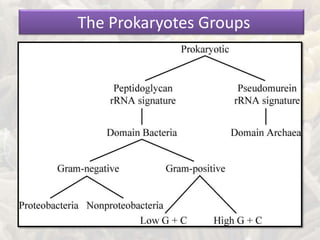


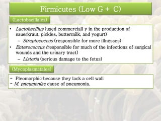
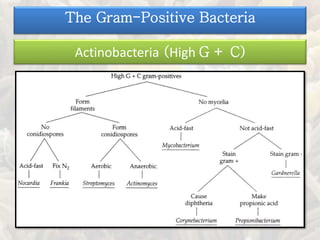
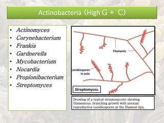
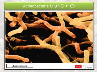
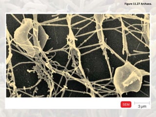


This document summarizes key information about prokaryotes from several domains. It describes the major groups within the domains of bacteria (including Proteobacteria, Firmicutes, Actinobacteria) and Archaea (including extremophiles). It also discusses the diversity of prokaryotes, noting that many have not been isolated or identified due to difficulties in culturing them.