Purpose of taxonomy
HistoricalBackground and Naming Conventions
Species Characteristics and Strains
Linnaean Taxonomic Hierarchy
Challenges in the Taxonomic Hierarchy
Evolution of Classification Systems
Bacterial Classification Challenges
Archaeal Diversity
Three-Domain System
Gram-Negative Bacteria
Gram-Positive Bacteria
Bacterial Classification Methods
Key Microbial Groups and Their Importance
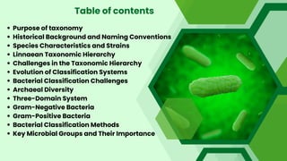
Table of contents
3.
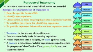
Purpose of taxonomy
Inscience, accurate and standardized names are essential.
Biologists use characteristics of organisms to :
Describe specific forms of life.
Identify new organisms.
Classification is based on grouping related organisms together.
To establish the criteria for identifying organisms.
To provide important information on how organisms evolved.
Taxonomy is the science of classification.
Provides an orderly basis for naming organisms.
Places organisms into a category, or taxon (plural: taxa).
A taxon is a collection of related organisms grouped together
for purposes of classification.Thus, genus, family, etc., are
taxonomic levels .
4.
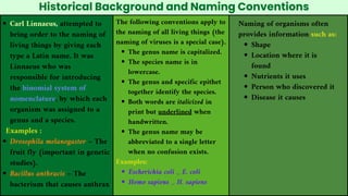
Historical Background andNaming Conventions
Carl Linnaeus, attempted to
bring order to the naming of
living things by giving each
type a Latin name. It was
Linnaeus who was
responsible for introducing
the binomial system of
nomenclature, by which each
organism was assigned to a
genus and a species.
Examples :
Drosophila melanogaster – The
fruit fly (important in genetic
studies).
Bacillus anthracis – The
bacterium that causes anthrax
The following conventions apply to
the naming of all living things (the
naming of viruses is a special case).
The genus name is capitalized.
The species name is in
lowercase.
The genus and specific epithet
together identify the species.
Both words are italicized in
print but underlined when
handwritten.
The genus name may be
abbreviated to a single letter
when no confusion exists.
Examples:
Escherichia coli _ E. coli
Homo sapiens _ H. sapiens
Naming of organisms often
provides information such as:
Shape
Location where it is
found
Nutrients it uses
Person who discovered it
Disease it causes
5.
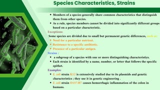
Species Characteristics, Strains
Membersof a species generally share common characteristics that distinguish
them from other species.
As a rule, species members cannot be divided into significantly different groups
based on a particular characteristic.
Exceptions:
Some species are divided due to small but permanent genetic differences, such as:
Need for a particular nutrient.
Resistance to a specific antibiotic.
Presence of a particular antigen.
Strains :
a subgroup of a species with one or more distinguishing characteristics.
Each strain is identified by a name, number, or letter that follows the specific
epithet.
Examples:
E. coli strain K12 is extensively studied due to its plasmids and genetic
characteristics ; they use it in genetic engineering .
E. coli strain O157:H7 causes hemorrhagic inflammation of the colon in
humans.
6.
Species Characteristics, Strains
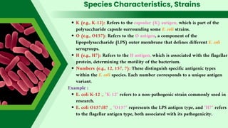
K(e.g., K-12): Refers to the capsular (K) antigen, which is part of the
polysaccharide capsule surrounding some E. coli strains.
O (e.g., O157): Refers to the O antigen, a component of the
lipopolysaccharide (LPS) outer membrane that defines different E. coli
serogroups.
H (e.g., H7): Refers to the H antigen, which is associated with the flagellar
protein, determining the motility of the bacterium.
Numbers (e.g., 12, 157, 7): These distinguish specific antigenic types
within the E. coli species. Each number corresponds to a unique antigen
variant.
Example :
E. coli K-12 _ "K-12" refers to a non-pathogenic strain commonly used in
research.
E. coli O157:H7 _ "O157" represents the LPS antigen type, and "H7" refers
to the flagellar antigen type, both associated with its pathogenicity.
7.
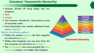
Linnaeus’ Taxonomic Hierarchy
Linnaeusdivided all living things into two
kingdoms:
Plant
Animal
Carl Linnaeus introduced a hierarchical system
of taxonomic ranks .
Some hierarchies now include additional levels,
such as subphyla.
Modern classification updates:
Within the animal kingdom, the first categories
are referred to as phyla.
Within other kingdoms (we now have five), the
first categories are referred to as divisions.
The five kingdoms have been grouped into three
domains, a category even higher than kingdom.
Taxonomic hierarchy
8.
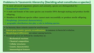
Problems in TaxonomicHierarchy (Deciding what constitutes a species)
In bacteria, these criteria cannot be used because:
Lateral gene transfer (genetic recombination) is common in bacterial evolution.
Morphological differences are minor among bacteria.
Bacterial species are defined by similarities in:
Biochemical reactions
Chemical composition
Cellular structures
Genetic characteristics
Immunological features
In most advanced organisms (plants and animals), species are distinguished by:
Reproductive capabilities:
1.
A male and female of the same species can transfer DNA through mating and produce fertile
offspring.
Members of different species either cannot mate successfully or produce sterile offspring.
Morphology (structural characteristics).
2.
geographic distribution also play a role in defining species.
3.
9.
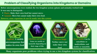
Before microorganisms werestudied, the two-kingdom system (plants and animals) worked well.
It was easy to distinguish:
Plants: Make their own food but cannot move.
Animals: Move but cannot make their own food.
However, some organisms do not fit neatly into this system:
Problem of Classifying Organisms into Kingdoms or Domains
Euglena: A mobile
microorganism that
makes its own food
Many organisms pose problems when trying to use a two-kingdom system for classification.
Jellyfishes and sponges:
Some are motile or
immotile depending on
their stage of life.
Colorless fungi:
Neither move nor
make their own
food
Slime molds: Can be
unicellular or
multicellular, and
mobile or immobile.
10.
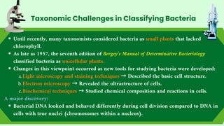
Until recently, manytaxonomists considered bacteria as small plants that lacked
chlorophyll.
As late as 1957, the seventh edition of Bergey’s Manual of Determinative Bacteriology
classified bacteria as unicellular plants.
Changes in this viewpoint occurred as new tools for studying bacteria were developed:
Light microscopy and staining techniques → Described the basic cell structure.
a.
Electron microscopy → Revealed the ultrastructure of cells.
b.
Biochemical techniques → Studied chemical composition and reactions in cells.
c.
A major discovery:
Bacterial DNA looked and behaved differently during cell division compared to DNA in
cells with true nuclei (chromosomes within a nucleus).
Taxonomic Challenges in Classifying Bacteria
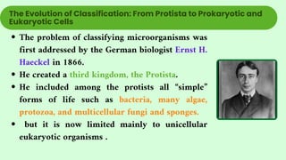
11.
The problem ofclassifying microorganisms was
first addressed by the German biologist Ernst H.
Haeckel in 1866.
He created a third kingdom, the Protista.
He included among the protists all “simple”
forms of life such as bacteria, many algae,
protozoa, and multicellular fungi and sponges.
but it is now limited mainly to unicellular
eukaryotic organisms .
The Evolution of Classification: From Protista to Prokaryotic and
Eukaryotic Cells
12.
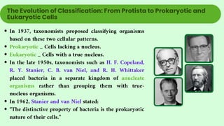
In 1937, taxonomistsproposed classifying organisms
based on these two cellular patterns.
Prokaryotic _ Cells lacking a nucleus.
Eukaryotic _ Cells with a true nucleus.
In the late 1950s, taxonomists such as H. F. Copeland,
R. Y. Stanier, C. B. van Niel, and R. H. Whittaker
placed bacteria in a separate kingdom of anucleate
organisms rather than grouping them with true-
nucleus organisms.
In 1962, Stanier and van Niel stated:
“The distinctive property of bacteria is the prokaryotic
nature of their cells.”
The Evolution of Classification: From Protista to Prokaryotic and
Eukaryotic Cells
13.
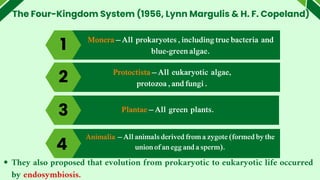
The Four-Kingdom System(1956, Lynn Margulis & H. F. Copeland)
1
4
Monera – All prokaryotes , including true bacteria and
blue-green algae.
Protoctista – All eukaryotic algae,
protozoa , and fungi .
Plantae – All green plants.
3
Animalia – All animals derived from a zygote (formed by the
union of an egg and a sperm).
4
They also proposed that evolution from prokaryotic to eukaryotic life occurred
by endosymbiosis.
2
15.
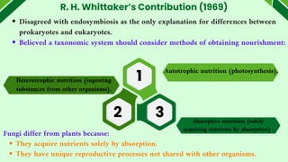
R. H. Whittaker’sContribution (1969)
Disagreed with endosymbiosis as the only explanation for differences between
prokaryotes and eukaryotes.
Believed a taxonomic system should consider methods of obtaining nourishment:
Autotrophic nutrition (photosynthesis).
Fungi differ from plants because:
They acquire nutrients solely by absorption.
They have unique reproductive processes not shared with other organisms.
Heterotrophic nutrition (ingesting
substances from other organisms).
Absorptive nutrition (solely
acquiring nutrients by absorption) .
1
2 3
16.
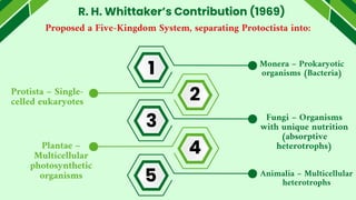
R. H. Whittaker’sContribution (1969)
Proposed a Five-Kingdom System, separating Protoctista into:
Monera – Prokaryotic
organisms (Bacteria)
Protista – Single-
celled eukaryotes
Fungi – Organisms
with unique nutrition
(absorptive
heterotrophs)
Plantae –
Multicellular
photosynthetic
organisms Animalia – Multicellular
heterotrophs
1
2
3
4
5
17.
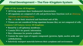
Final Development –The Five-Kingdom System
Unity of Life Across All Kingdoms:
All living organisms share fundamental characteristics.
All organisms are composed of cells and perform basic functions such as obtaining
nutrients and excreting waste.
The cell is the basic structural and functional unit of life.
Viruses are not considered living organisms because they are not composed of cells.
Common Cell Features Across All Kingdoms:
Bound by a cell (plasma) membrane.
Contain DNA for genetic information.
Have ribosomes for protein synthesis.
Contain the same types of organic compounds (proteins, lipids, nucleic acids, and
carbohydrates).
Selectively transport materials between their cytoplasm and environment.
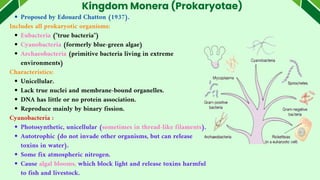
Kingdom Monera (Prokaryotae)
Proposedby Edouard Chatton (1937).
Includes all prokaryotic organisms:
Eubacteria ("true bacteria")
Cyanobacteria (formerly blue-green algae)
Archaeobacteria (primitive bacteria living in extreme
environments)
Characteristics:
Unicellular.
Lack true nuclei and membrane-bound organelles.
DNA has little or no protein association.
Reproduce mainly by binary fission.
Cyanobacteria :
Photosynthetic, unicellular (sometimes in thread-like filaments).
Autotrophic (do not invade other organisms, but can release
toxins in water).
Some fix atmospheric nitrogen.
Cause algal blooms, which block light and release toxins harmful
to fish and livestock.
20.
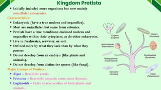
Kingdom Protista
Initially includedmore organisms but now mainly
unicellular eukaryotes.
Characteristics:
Eukaryotic (have a true nucleus and organelles).
Most are unicellular, but some form colonies.
Protists have a true membrane-enclosed nucleus and
organelles within their cytoplasm, as do other eukaryotes.
Live in freshwater, seawater, or soil.
Defined more by what they lack than by what they
possess
Do not develop from an embryo (like plants and
animals).
Do not develop from distinctive spores (like fungi).
Major Groups of Protists :
Algae – Resemble plants.
Protozoa – Resemble animals; some cause diseases.
Euglenoids – Share characteristics of both plants and
animals .
21.
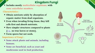
Includes mostly multicellularorganisms, with
some unicellular members.
Characteristics :
Obtain nutrients solely by absorption of
organic matter from dead organisms.
Even when invading living tissue, they kill
cells first and absorb nutrients.
Have simpler structures compared to plants
(e.g., no true leaves or stems).
Form spores but not seeds.
Importance of Fungi :
Some attack plants and animals, including
humans.
Some are beneficial, such as yeast and
mushrooms used in food production.
Kingdom Fungi
22.
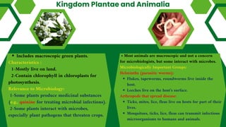
Kingdom Plantae andAnimalia
Includes macroscopic green plants.
Characteristics :
1-Mostly live on land.
2-Contain chlorophyll in chloroplasts for
photosynthesis.
Relevance to Microbiology:
1-Some plants produce medicinal substances
(e.g., quinine for treating microbial infections).
2-Some plants interact with microbes,
especially plant pathogens that threaten crops.
• Most animals are macroscopic and not a concern
for microbiologists, but some interact with microbes.
Microbiologically Important Groups:
Helminths (parasitic worms):
Flukes, tapeworms, roundworms live inside the
host.
Leeches live on the host’s surface.
Arthropods that spread disease:
Ticks, mites, lice, fleas live on hosts for part of their
lives.
Mosquitoes, ticks, lice, fleas can transmit infectious
microorganisms to humans and animals.
23.
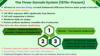
The Three-Domain System(1970s–Present)
Advances in molecular biology revealed fundamental differences between major groups of microbes.
Key discoveries about Archaea:
16S rRNA sequences differ significantly from Bacteria.
Cell wall composition is distinct.
Membrane lipids are unique.
Protein synthesis machinery resembles that of eukaryotes.
This led to the three-domain classification:
1
2 3
Domains represent a higher taxonomic level than kingdoms.
Interestingly, Archaea share some traits with Eukarya .
Traditional Kingdom Monera was split into two domains: Bacteria and Archaea.
Bacteria – True bacteria (Eubacteria)
Archaea – Ancient prokaryotic
microbes. Contains archaeobacteria
Eukarya – All eukaryotic organisms.
, Contains all eukaryotic kingdoms
(Animals, Plants, Fungi, Protists).
24.
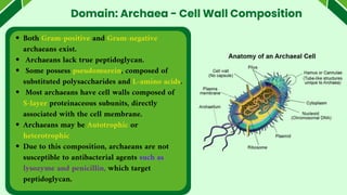
Domain: Archaea -Cell Wall Composition
Both Gram-positive and Gram-negative
archaeans exist.
Archaeans lack true peptidoglycan.
Some possess pseudomurein, composed of
substituted polysaccharides and L-amino acids.
Most archaeans have cell walls composed of
S-layer proteinaceous subunits, directly
associated with the cell membrane.
Archaeans may be Autotrophic or
heterotrophic
Due to this composition, archaeans are not
susceptible to antibacterial agents such as
lysozyme and penicillin, which target
peptidoglycan.
25.
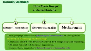
Domain: Archaea
Three MajorGroups
of Archaeobacteria
These groupings are based on physiological characteristics of the organisms .
Diversity in Morphology and Physiology:
• Archaeans exhibit considerable diversity in both morphology and physiology.
• All main bacterial cell shapes are represented.
• Some archaeal species have flattened square or triangular cells.
1
2
3
Methanogens
Extreme Halophiles
Extreme Thermophiles
26.
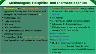
Methanogens, Halophiles, andThermoacidophiles
Methanogens are strictly anaerobic
organisms, having been isolated from such
divergent anaerobic environments as :
Waterlogged soils
Lake sediments
Marshes
Marine sediments
The gastrointestinal tracts of animals,
including humans
As members of the anaerobic food chain,
they degrade organic molecules to methane.
Extreme Thermoacidophiles occupy unique
niches where bacteria are very rarely found,
such as:
Hot springs
Geothermally heated marine sediments
Submarine hydrothermal vents
With optimum temperatures usually in
excess of 80°C, they may be:
• Obligate aerobes
• Facultative aerobes
• Obligate anaerobes
The heat-stable enzymes known as
extremozymes that are found in these
organisms have become of special interest to
scientists.
27.
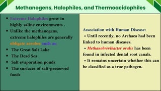
Methanogens, Halophiles, andThermoacidophiles
Extreme Halophiles grow in
highly saline environments .
Unlike the methanogens,
extreme halophiles are generally
obligate aerobes such as:
The Great Salt Lake
The Dead Sea
Salt evaporation ponds
The surfaces of salt-preserved
foods
Association with Human Disease:
• Until recently, no Archaea had been
linked to human diseases.
• Methanobrevibacter oralis has been
found in infected dental root canals.
• It remains uncertain whether this can
be classified as a true pathogen.
Classification Methods andExploring Evolutionary
Relationships in Prokaryotes
Since morphology and fossil records are insufficient, classification relies on:
Metabolic reactions.
Genetic relationships.
specialized biochemical traits.
These properties help health scientists identify pathogenic bacteria, but they
do not always reflect evolutionary history.
Various alternative methods help determine prokaryotic evolutionary
relationships.
While initially developed for eukaryotes, many of these methods are also
applicable to bacteria.
30.
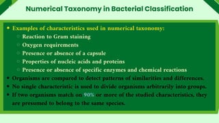
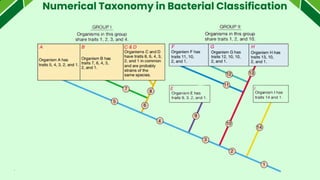
Numerical taxonomy isbased on the idea that observing more
characteristics increases accuracy in detecting similarities among organisms.
If characteristics are genetically determined, the more traits two organisms
share, the closer their evolutionary relationship.
Although developed before computers, computers now allow rapid
comparisons of large numbers of organisms based on multiple traits.
Each characteristic is assigned a value: 1 if present, 0 if absent.
Numerical Taxonomy in Bacterial Classification
31.
Examples of characteristicsused in numerical taxonomy:
Reaction to Gram staining
Oxygen requirements
Presence or absence of a capsule
Properties of nucleic acids and proteins
Presence or absence of specific enzymes and chemical reactions
Organisms are compared to detect patterns of similarities and differences.
No single characteristic is used to divide organisms arbitrarily into groups.
If two organisms match on 90% or more of the studied characteristics, they
are presumed to belong to the same species.
Numerical Taxonomy in Bacterial Classification
Genetic homology refersto the similarity of DNA sequences among
organisms.
The discovery of DNA structure by Watson and Crick (1953) led to its
application in studying taxonomic relationships and evolution.
Ideally, sequencing entire genomes would allow direct comparisons, but this is
currently impractical due to time and effort.
Several faster techniques for estimating genetic homology include:
Determining DNA base composition.
Sequencing portions of DNA or RNA.
Using DNA hybridization.
Since proteins are determined by DNA, genetic homology can also be
estimated indirectly by:
Preparing protein profiles and analyzing amino acid sequences in protein
Genetic Homology in Taxonomy
34.
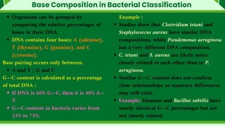
Base Composition inBacterial Classification
Organisms can be grouped by
comparing the relative percentages of
bases in their DNA.
DNA contains four bases: A (adenine),
T (thymine), G (guanine), and C
(cytosine).
Base pairing occurs only between:
A and T / G and C
G—C content is calculated as a percentage
of total DNA :
If DNA is 60% G—C, then it is 40% A—
T.
G—C content in bacteria varies from
23% to 75%.
Example :
Studies show that Clostridium tetani and
Staphylococcus aureus have similar DNA
compositions, while Pseudomonas aeruginosa
has a very different DNA composition.
C. tetani and S. aureus are likely more
closely related to each other than to P.
aeruginosa.
Similar G—C content does not confirm
close relationships, as sequence differences
may still exist.
Example: Humans and Bacillus subtilis have
nearly identical G—C percentages but are
not closely related.
35.
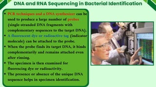
DNA and RNASequencing in Bacterial Identification
PCR techniques and a DNA synthesizer can be
used to produce a large number of probes
(single-stranded DNA fragments with
complementary sequences to the target DNA).
A fluorescent dye or radioactive tag (indicator
molecule) can be attached to the probe.
When the probe finds its target DNA, it binds
complementarily and remains attached even
after rinsing.
The specimen is then examined for
fluorescing dye or radioactivity.
The presence or absence of the unique DNA
sequence helps in specimen identification.
36.
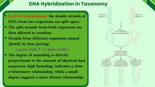
DNA Hybridization inTaxonomy
In DNA hybridization, the double strands of
DNA from two organisms are split apart.
The split strands from both organisms are
then allowed to combine.
Strands from different organisms anneal
(bond) by base pairing:
A pairs with T / G pairs with C
The degree of annealing is directly
proportional to the amount of identical base
sequences; high homology indicates a close
evolutionary relationship, while a small
degree suggests a more distant relationship.
37.
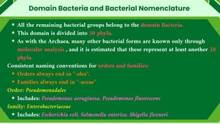
All the remainingbacterial groups belong to the domain Bacteria.
This domain is divided into 30 phyla.
As with the Archaea, many other bacterial forms are known only through
molecular analysis , and it is estimated that these represent at least another 20
phyla.
Consistent naming conventions for orders and families:
Orders always end in "-ales".
Families always end in "-aceae"
Order: Pseudomonadales
Includes: Pseudomonas aeruginosa, Pseudomonas fluorescens
family: Enterobacteriaceae
Includes: Escherichia coli, Salmonella enterica, Shigella flexneri
Domain Bacteria and Bacterial Nomenclature
38.
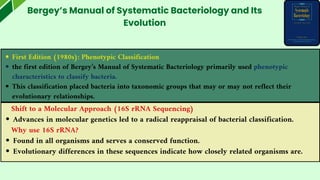
First Edition (1980s):Phenotypic Classification
the first edition of Bergey’s Manual of Systematic Bacteriology primarily used phenotypic
characteristics to classify bacteria.
This classification placed bacteria into taxonomic groups that may or may not reflect their
evolutionary relationships.
Bergey’s Manual of Systematic Bacteriology and Its
Evolution
Shift to a Molecular Approach (16S rRNA Sequencing)
Advances in molecular genetics led to a radical reappraisal of bacterial classification.
Why use 16S rRNA?
Found in all organisms and serves a conserved function.
Evolutionary differences in these sequences indicate how closely related organisms are.
39.
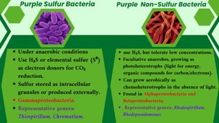
Bergey’s Manual ofSystematic Bacteriology and Its
Evolution
Bergey’s Second Edition (2001–2012) - Molecular Approach
Reflects the shift from phenotypic to phylogenetic classification.
Many bacteria were reassigned based on molecular evidence, especially 16S
rRNA sequencing.
Example: The genus Pseudomonas previously contained ~70 species classified
based on phenotypic similarities, but in the second edition, many species were
reassigned to newly created genera based on molecular data.
40.
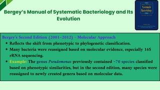
Phylum : Proteobacteria
01
Thelargest single phylum, representing about one-third of all known bacterial species.
Occupies the entire Volume 2 in the second edition of Bergey.
Highly diverse in both morphology and physiology, encompassing most forms of metabolism.
Assigned to a single taxonomic group based on 16S rRNA sequences.
Includes many well-known Gram-negative bacteria of medical, industrial, and agricultural importance.
For taxonomic purposes, Proteobacteria are divided into six classes :
Alphaproteobacteria Betaproteobacteria 03
02
04
Gammaproteobacteria
Deltaproteobacteria Epsilonproteobacteria 06
05 Zetaproteobacteria
(added in 2010, includes a single
iron-oxidizing deep-sea species).
Shared traits often appear in multiple classes.
Example: Nitrifying bacteria occur in Alpha-, Beta-, Gamma-, and
Deltaproteobacteria.
41.
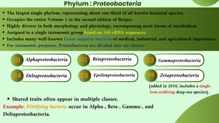
Only the purplesulfur and purple non-sulfur bacteria retain photosynthetic
ability, include rods, cocci and spiral forms.
Differences from Plant Photosynthesis:
Anoxygenic (does not produce oxygen).
Uses bacteriochlorophylls a and/or b instead of chlorophyll a/b.
H₂S or elemental sulfur as the electron donor (not water).
Operates with a single photosystem.
Similarity to Oxygenic Photosynthesis:
Uses the Calvin cycle to fix CO₂.
Typically found in stagnant lakes and salt marshes, forming colored blooms .
Light Utilization & Pigmentation
Bacteriochlorophylls absorb light in the infrared region, allowing deep water
penetration.
Colors range from orange/brown to purple, masked by carotenoids (e.g.,
lycopene, spirillixanthin).
Photosynthetic pigments are located on highly folded plasma membrane
extensions.
Photosynthetic Proteobacteria
42.
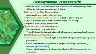
Purple Sulfur Bacteria
Underanaerobic conditions
Use H₂S or elemental sulfur (S⁰)
as electron donors for CO₂
reduction.
Sulfur stored as intracellular
granules or produced externally.
Gammaproteobacteria.
Representative genera:
Thiospirillum, Chromatium.
use H₂S, but tolerate low concentrations.
Facultative anaerobes, growing as
photoheterotrophs (light for energy,
organic compounds for carbon/electrons).
Can grow aerobically as
chemoheterotrophs in the absence of light.
Found in Alphaproteobacteria and
Betaproteobacteria.
Representative genera: Rhodospirillum,
Rhodopseudomonas.
Purple Non-Sulfur Bacteria
43.
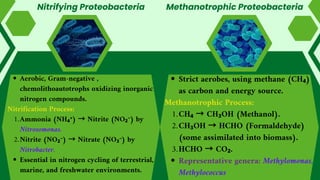
Nitrifying Proteobacteria
Strict aerobes,using methane (CH₄)
as carbon and energy source.
Methanotrophic Process:
CH₄ → CH₃OH (Methanol).
1.
CH₃OH → HCHO (Formaldehyde)
(some assimilated into biomass).
2.
HCHO → CO₂.
3.
Representative genera: Methylomonas,
Methylococcus
Aerobic, Gram-negative ,
chemolithoautotrophs oxidizing inorganic
nitrogen compounds.
Nitrification Process:
Ammonia (NH₄⁺) → Nitrite (NO₂⁻) by
Nitrosomonas.
1.
Nitrite (NO₂⁻) → Nitrate (NO₃⁻) by
Nitrobacter.
2.
Essential in nitrogen cycling of terrestrial,
marine, and freshwater environments.
Methanotrophic Proteobacteria
44.
Two groups ofchemolithoautotrophs oxidizing iron and sulfur for energy.
Iron- and Sulfur-Oxidizing Proteobacteria
Sulfur-Oxidizing Proteobacteria
Best-studied genus: Acidithiobacillus, extreme
acidophile (growth at pH ~1.0).
Uses S⁰, H₂S, metal sulfides, and thiosulfate.
Sulfur oxidation reactions produce sulfuric acid,
contributing to acid mine drainage and toxic
metal release.
Used in bioleaching.
Representative genera: Acidithiobacillus,
Beggiatoa.
Iron-Oxidizing Proteobacteria
Convert Fe²⁺ → Fe³⁺ under acidic
conditions.
Acidithiobacillus ferrooxidans oxidizes Fe²⁺
at pH ~2.
Gallionella ferruginea deposits iron
hydroxide in oxygen-poor environments.
Representative genera: Leptospirillum,
Gallionella.
45.
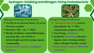
Hydrogen-Oxidizing and Nitrogen-FixingProteobacteria
Hydrogen-Oxidizing Proteobacteria
Use H₂ as an electron donor, O₂ as an
electron acceptor.
Reaction: 2H₂ + O₂ → 2H₂O.
Mostly facultative chemolithotrophs,
meaning they can switch to
heterotrophic growth (using organic
compounds).
Representative genera: Alcaligenes,
Ralstonia.
Nitrogen-Fixing Proteobacteria
Alphaproteobacteria convert
atmospheric N₂ → NH₄⁺
Ammonium (requires ATP).
Free-living: Azotobacter.
Symbiotic: Rhizobium colonizes
legume roots, forming nodules
where nitrogen fixation occurs.
Representative genera: Rhizobium,
Azotobacter.
46.
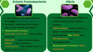
Enteric Proteobacteria
Facultative anaerobes,similar to
enteric bacteria but oxidase-
positive.
Vibrio cholerae causes cholera, a
major waterborne disease.
Some species, like Vibrio and
Photobacterium, are
bioluminescent.
Representative Genera: Vibrio ,
Aeromonas
Gram-negative, rod-shaped bacteria
belonging to Gammaproteobacteria.
Facultative anaerobes that ferment
glucose into various byproducts.
Representative Genera:
Escherichia coli (E. coli) – Most studied
bacterium.
Salmonella – Causes food poisoning.
Shigella – Causes dysentery.
Yersinia pestis – The causative agent of
plague. البراغيث لدغات عبر
Vibrio
47.
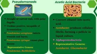
Pseudomonads
Straight or curvedrods, with polar
flagella.
Oxidase-positive, incapable of
fermentation.
Pseudomonas aeruginosa infects
wounds and burns.
Pseudomonas syringae causes plant
disease.
Representative Genera:
Pseudomonas, Burkholderia
Convert ethanol into acetic
acid.
Acetobacter produces cellulose
fibrils, forming a pellicle in
liquid culture.
Alphaproteobacteria
Representative Genera:
Acetobacter, Gluconobacter
Acetic Acid Bacteria
48.
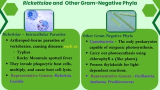
Rickettsiae and OtherGram-Negative Phyla
Rickettsiae – Intracellular Parasites
Arthropod-borne parasites of
vertebrates, causing diseases such as:
Typhus
Rocky Mountain spotted fever
They invade phagocytic host cells,
multiply, and cause host cell lysis.
Representative Genera: Rickettsia,
Coxiella
Other Gram-Negative Phyla
Cyanobacteria – The only prokaryotes
capable of oxygenic photosynthesis.
Carry out photosynthesis using
chlorophyll a (like plants).
Possess thylakoids for light-
dependent reactions.
Representative Genera : Oscillatoria,
Anabaena, Prochlorococcus
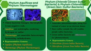
49.
Anaerobic photolithotrophs, usingH₂S
instead of water as an electron donor.
Store sulfur outside the cell (unlike
purple sulfur bacteria).
Green sulfur bacteria use chlorosomes
for photosynthesis.
Representative Genera:
Chlorobium (Green Sulfur Bacteria)
Chloroflexus (Green Non-Sulfur Bacteria)
Phylum Chlorobi (Green Sulfur
Bacteria) & Phylum Chloroflexi
(Green Non-Sulfur Bacteria)
Phylum Aquificae and
Phylum Thermotogae
Oldest bacterial lineages, thermophilic
Gram-negative rods.
Aquificae are autotrophs, oxidizing
hydrogen or sulfur.
Thermotogae are anaerobic heterotrophs,
fermenting carbohydrates.
Representative Genera:
Aquifex (Phylum Aquificae)
Thermotoga (Phylum Thermotogae)
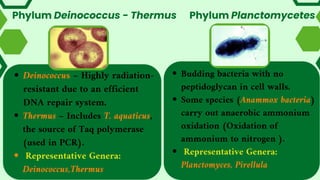
50.
Phylum Deinococcus -Thermus
Budding bacteria with no
peptidoglycan in cell walls.
Some species (Anammox bacteria)
carry out anaerobic ammonium
oxidation (Oxidation of
ammonium to nitrogen ).
Representative Genera:
Planctomyces, Pirellula
Deinococcus – Highly radiation-
resistant due to an efficient
DNA repair system.
Thermus – Includes T. aquaticus,
the source of Taq polymerase
(used in PCR).
Representative Genera:
Deinococcus,Thermus
Phylum Planctomycetes
51.
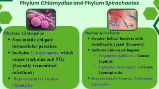
Phylum Chlamydiae andPhylum Spirochaetes
Phylum Chlamydiae
Non-motile obligate
intracellular parasites.
Includes C. trachomatis, which
causes trachoma and STIs
(Sexually transmitted
infections)
Representative Genus:
Chlamydia
Phylum Spirochaetes
Slender, helical bacteria with
endoflagella (axial filaments).
Includes human pathogens:
Treponema pallidum – Causes
Syphilis
Leptospira interrogans – Causes
Leptospirosis
Representative Genera: Treponema,
Leptospira
52.
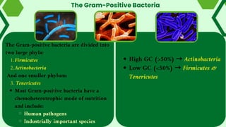
The Gram-Positive Bacteria
HighGC (>50%) → Actinobacteria
Low GC (<50%) → Firmicutes &
Tenericutes
The Gram-positive bacteria are divided into
two large phyla:
Firmicutes
1.
Actinobacteria
2.
And one smaller phylum:
3. Tenericutes
Most Gram-positive bacteria have a
chemoheterotrophic mode of nutrition
and include:
Human pathogens
Industrially important species
53.
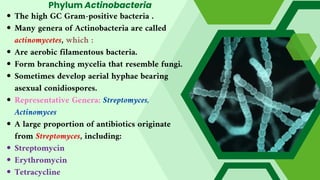
Phylum Actinobacteria
The highGC Gram-positive bacteria .
Many genera of Actinobacteria are called
actinomycetes, which :
Are aerobic filamentous bacteria.
Form branching mycelia that resemble fungi.
Sometimes develop aerial hyphae bearing
asexual conidiospores.
Representative Genera: Streptomyces,
Actinomyces
A large proportion of antibiotics originate
from Streptomyces, including:
Streptomycin
Erythromycin
Tetracycline
54.
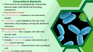
Coryneform Bacteria
These bacteriaare morphologically intermediate
between single-celled bacilli and branching
actinomycetes.
Representative Genera:
Corynebacterium – Common in soil and animal
mouths.
C. diphtheriae causes diphtheria, but only when
infected by a bacteriophage carrying the diphtheria
toxin gene.
Other Coryneform Bacteria :
Mycobacterium – Have complex cell walls with
mycolic acids.
Stain positive in the acid-fast test.
Includes M. tuberculosis (causes tuberculosis) and M.
leprae (causes leprosy).
Propionibacterium – Ferments lactic acid to propionic
acid.
P. acnes is a major cause of acne.
55.
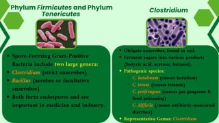
Phylum Firmicutes andPhylum
Tenericutes
Obligate anaerobes, found in soil.
Ferment sugars into various products
(butyric acid, acetone, butanol).
Pathogenic species:
C. botulinum (causes botulism)
C. tetani (causes tetanus)
C. perfringens (causes gas gangrene &
food poisoning)
C. difficile (causes antibiotic-associated
diarrhea)
Representative Genus: Clostridium
Clostridium
Spore-Forming Gram-Positive
Bacteria include two large genera:
Clostridium (strict anaerobes)
Bacillus (aerobes or facultative
anaerobes)
Both form endospores and are
important in medicine and industry.
56.
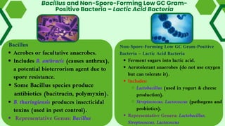
Bacillus
Aerobes or facultativeanaerobes.
Includes B. anthracis (causes anthrax),
a potential bioterrorism agent due to
spore resistance.
Some Bacillus species produce
antibiotics (bacitracin, polymyxin).
B. thuringiensis produces insecticidal
toxins (used in pest control).
Representative Genus: Bacillus
Bacillus and Non-Spore-Forming Low GC Gram-
Positive Bacteria – Lactic Acid Bacteria
Non-Spore-Forming Low GC Gram-Positive
Bacteria – Lactic Acid Bacteria
Ferment sugars into lactic acid.
Aerotolerant anaerobes (do not use oxygen
but can tolerate it).
Includes:
Lactobacillus (used in yogurt & cheese
production).
Streptococcus, Lactococcus (pathogens and
probiotics).
Representative Genera: Lactobacillus,
Streptococcus, Lactococcus
57.
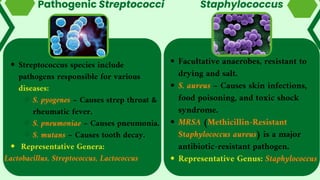
Facultative anaerobes, resistantto
drying and salt.
S. aureus – Causes skin infections,
food poisoning, and toxic shock
syndrome.
MRSA (Methicillin-Resistant
Staphylococcus aureus) is a major
antibiotic-resistant pathogen.
Representative Genus: Staphylococcus
Pathogenic Streptococci
Streptococcus species include
pathogens responsible for various
diseases:
S. pyogenes – Causes strep throat &
rheumatic fever.
S. pneumoniae – Causes pneumonia.
S. mutans – Causes tooth decay.
Representative Genera:
Lactobacillus, Streptococcus, Lactococcus
Staphylococcus
58.
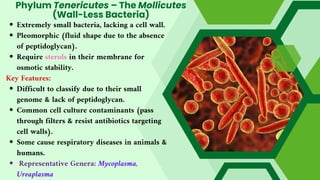
Phylum Tenericutes –The Mollicutes
(Wall-Less Bacteria)
Extremely small bacteria, lacking a cell wall.
Pleomorphic (fluid shape due to the absence
of peptidoglycan).
Require sterols in their membrane for
osmotic stability.
Key Features:
Difficult to classify due to their small
genome & lack of peptidoglycan.
Common cell culture contaminants (pass
through filters & resist antibiotics targeting
cell walls).
Some cause respiratory diseases in animals &
humans.
Representative Genera: Mycoplasma,
Ureaplasma