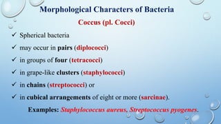
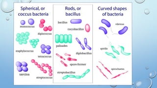
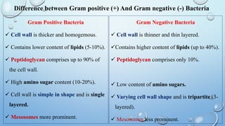
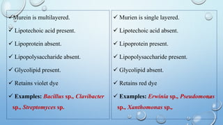
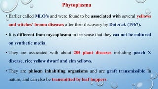
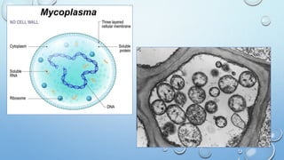
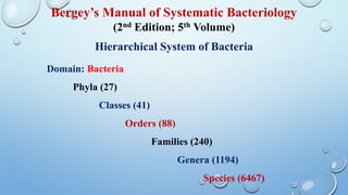
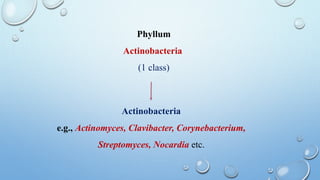
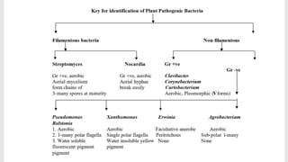
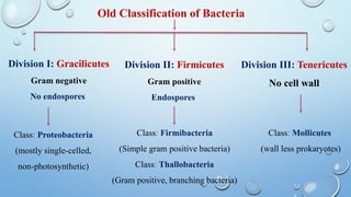
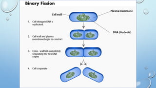
Bacteria
Definition
Bacteria are extremely minute, rigid, essentially unicellular
organisms (actinomycetes are filamentous), devoid of
chlorophyll, most commonly reproduce by transverse binary
fission and the resulting cells are identical in size and
morphology.
General characters of Bacteria
 Bacteria and mollicutes are prokaryotes.
 These are generally single-celled microorganisms whose
genetic material (DNA) is not bound by a membrane and
therefore is not organized into a nucleus.
 Plant pathogenic bacteria have been known since 1882.
 About 1,600 bacterial species are known.
 Most are strictly saprophytic and as such are beneficial to
humans because they help decompose the enormous quantities
of organic matter.
 Several species cause diseases in humans, including
tuberculosis, pneumonia and typhoid fever and in animals,
such as brucellosis and anthrax.
 About 100 species of bacteria cause diseases in plants.
 They are by far the largest group of plant pathogenic prokaryotes,
cause a variety of plant disease symptoms and are the best
understood prokaryotic pathogens of plants.
 Most plant pathogenic bacteria are facultative saprophytes and
can be grown artificially on nutrient media.
 Bacteria may be rod shaped, spherical, spiral or filamentous
(threadlike).
Some bacteria can move by means of flagella, whereas others have
no flagella and cannot move themselves.
Some can transform themselves into spores and the filamentous
bacteria.
Streptomyces can produce spores, called conidia, at the end of the
filament.
Other bacteria, however, do not produce any spores.
 Bacteria multiply with astonishing rapidity.
Structure of Bacterial Cell
 A bacterium has a thin, relatively tough, rigid cell wall, and a
distinct three layered but thin cytoplasmic membrane.
 Most bacteria have a slime layer made up of viscous gummy
material. Slime layer has bacterial immunological property.
 When the layer is thick and firm, it is called capsule.
 Generally plant pathogenic bacteria lack capsule but some of
them like Pseudomonas and Xanthomonas produce slime.
A. Surface appendages: flagella, pili.
B. Cell envelop: cytoplasmic membrane, cell wall, capsule or
slime layer.
C. Cytoplasmic organelles: genophore, plasmid, ribosome,
mesosome.
 Slime layer is mostly composed of polysaccharides but may
rarely contain amino sugars, sugar acids etc.
Flagella
 Most of the plant pathogenic bacteria have delicate thread
like flagella, which are usually longer than the cell.
 They are the organs of locomotion.
 The arrangement of flagella on bacterial cell is an
important taxonomic character that aid in
bacterial classification.
 This arrangement may be:
1. Atrichous- bacteria lacking flagella,
e.g., Lactobacillus sp.
2. Monotrichous- with single polar
flagellum at one end only, e.g.,
Xanthomonas sp.
3. Amphitrichous- with single polar
flagellum at both polar ends, e.g.,
Pseudomonas spp.
4. Cephalotrichous- with tuft
(several) flagella at one end, e.g.,
Pseudomonas fluorescens
5. Lophotrichous- tuft of flagella at
both end, e.g., Spirillum sp.
6. Peritrichous- distributed all
around the cell or surface, e.g.,
Erwinia sp.
Pili (sing. Pilus)
 Short hair like structure.
 Shorter and stiffer than flagella.
 Composed of protein pilin.
 Involved in attachment.
 Helps in conjugation.
 Mostly gram –ve bacteria have pili.
Morphological Characters of Bacteria
Coccus (pl. Cocci)
 Spherical bacteria
 may occur in pairs (diplococci)
 in groups of four (tetracocci)
 in grape-like clusters (staphylococci)
 in chains (streptococci) or
 in cubical arrangements of eight or more (sarcinae).
Examples: Staphylococcus aureus, Streptococcus pyogenes.
Rod-shaped bacteria; generally occur singly, but may
occasionally be found in pairs (diplo-bacilli) or chains
(streptobacilli).
Examples: Bacillus cereus, Clostridium tetani.
Bacillus (pl. Bacilli)
Coccobacilli
Elongated spherical or ovoid form, e.g., Haemophilus
influenzae
Filamentous
Bacilli that occur in long chains or threads, e.g., Streptomyces
scabies
Fusiform
Bacilli with tapered ends, e.g., Fusobacterium necrophorum
 These are small, regularly coiled, rigid organisms
measuring about 3–4 um in length.
 Each coil measures about 1 um.
 Spirilla are motile with groups of flagella at both ends.
Example: Spirillum minus.
Spirillum (pl. Spirilla)
 These are small slightly curved rods measuring 3–4 um in
length by 0.5 um in width.
 Most vibrios are motile with a single flagellum at one end.
 They show rapid darting motility, e.g., Vibrio cholerae.
Vibrios
These are flexible, coiled, motile organisms. They progress by
rapid body movements.
Treponemes, which are thin delicate spirochaetes with regular
tight coils, e.g., Treponema pallidum.
Borreliae, which are large spirochaetes with irregular open coils,
e.g., Borrelia duttoni.
Leptospires, which are thin spirochaetes with many tightly
packed coils, e.g., Leptospira interrogans.
Spirochaetes
Difference between Gram positive (+) And Gram negative (-) Bacteria
Gram Positive Bacteria
 Cell wall is thicker and homogemous.
 Contains lower content of lipids (5-10%).
 Peptidoglycan comprises up to 90% of
the cell wall.
 High amino sugar content (10-20%).
 Cell wall is simple in shape and is single
layered.
 Mesosomes more prominent.
Gram Negative Bacteria
 Cell wall is thinner and thin layered.
Contains higher content of lipids (up to 40%).
 Peptidoglycan comprises only 10%.
 Low content of amino sugars.
 Varying cell wall shape and is tripartite (3-
layered).
 Mesosomes less prominent.
Murein is multilayered.
 Lipotechoic acid present.
 Lipoprotein absent.
 Lipopolysaccharide absent.
 Glycolipid present.
 Retains violet dye
 Examples: Bacillus sp., Clavibacter
sp., Streptomyces sp.
 Murien is single layered.
 Lipotechoic acid absent.
 Lipoprotein present.
 Lipopolysaccharide present.
 Glycolipid absent.
 Retains red dye
 Examples: Erwinia sp., Pseudomonas
sp., Xanthomonas sp.,
Gram’s Staining
Mycoplasma
Earlier known as ‘Pleuro Pneumonia Like Organisms’ (PPLO’s) were
discovered to be associated with the disease Bovine Pleuro Pneumonia.
Placed in order Mycoplasmatales under Eubacteria.
It is a group of organisms that lack cell wall and contain a very small
genome.
Phylogenetically, they are closely related to Clostridia, the gram positive
bacteria.
On the basis of growth, it can be two types; require sterol
(mycoplasma and spiroplasma); and do not require sterols
(acholeplasma and thermoplasma).
 The mycoplasma cells are small, pleomorphic (of different
shapes) and divide by budding.
 The colonies of mycoplasma on agar exhibit a characteristic ‘fried
egg’ appearance.
 The growth of mycoplasma is not inhibited by penicillin or other
antibiotics that inhibit cell wall synthesis, but sensitive to
tetracycline.
 Spiroplasma citri causes citrus stubborn disease and corn stunt.
Phytoplasma
• Earlier called MLO’s and were found to be associated with several yellows
and witches’ broom diseases after their discovery by Doi et al. (1967).
• It is different from mycoplasma in the sense that they can not be cultured
on synthetic media.
• They are associated with about 200 plant diseases including peach X
disease, rice yellow dwarf and elm yellows.
• They are phloem inhabiting organisms and are graft transmissible in
nature, and can also be transmitted by leaf hoppers.
Classification of Bacteria
 Traditionally bacteria have been included in Plantae
kingdom under Thallophyta.
 Haeckel (1966) proposed the kingdom Protista to include
all unicellular organisms and placed various organisms of
Thallophyta plants and Protozoa animals in Protista.
 Chatton (1937) proposed the most appropriate conceptual
basis for taxa as prokaryotes and eukaryotes.
 Stanier (1969) considered prokaryotes as lower protists
including blue green algae, myxobacteria and eubacteria;
and eukaryotes as higher protists including algae, fungi and
protozoa.
 Prokaryotae was recognised a separate kingdom.
 Whittaker (1969) proposed five-kingdom classification
including 1. Plantae, 2. Animalia, 3. Fungi, 4. Protista and 4.
Monera (Prokaryotes)
Bergey’s Manual of Systematic Bacteriology
(2nd Edition; 5th Volume)
Hierarchical System of Bacteria
Domain: Bacteria
Phyla (27)
Classes (41)
Orders (88)
Families (240)
Genera (1194)
Species (6467)
Out of 27 phyla, three phyla Proteobacteria (2005; Vol. 2),
Fermicutes (2009; Vol. 3) and Actinobacteria (2010; Vol. 5)
mainly contains bacterial genera of both beneficial or
pathogenic to plants.
Phyllum
Proteobacteria
(5 classes)
Alphaproteobacteria, e.g., Azosprillum,
Rhizobium, Agrobacterium etc.
Betaproteobacteria, e.g., Ralstonia,
Burkholderia, Spirilum, Nitrosomonas etc.
Gammaproteobacteria, e.g., Xanthomonas,
Erwinia, Pseudomonas, Azotobactor etc.
Deltaproteobacteria, e.g., Bdellovibrio etc.
Epsilonproteobacteria, e.g., Helicobacter etc.
Gram -ve
Phyllum
Firmicutes
(3 classes)
Clostridia, e.g., Clostridium, Helicococcus,
Acetobacterium, Syntrophomonas etc.
Mollicutes, e.g., Mycoplasma, Spiroplasma,
Acholeplasma, Anaeroplasma etc.
Bacilli, e.g., Bacillus, Staphylococcus,
Lactobacillus, Streptococcus etc.
Gram +ve
Phyllum
Actinobacteria
(1 class)
Actinobacteria
e.g., Actinomyces, Clavibacter, Corynebacterium,
Streptomyces, Nocardia etc.
Old Classification of Bacteria
Division I: Gracilicutes
Gram negative
No endospores
Division II: Firmicutes
Gram positive
Endospores
Division III: Tenericutes
No cell wall
Class: Proteobacteria
(mostly single-celled,
non-photosynthetic)
Class: Firmibacteria
(Simple gram positive bacteria)
Class: Thallobacteria
(Gram positive, branching bacteria)
Class: Mollicutes
(wall less prokaryotes)
Reproduction in Bacteria
 Bacterial cell undergo reproduction both asexually and sexually.
 Rod shaped plant pathogenic bacteria reproduce by the asexual process
known as binary fission or fission.
 In addition to this, bacterial cells also produces endospore or exospore.
 Endospore: It is a structure formed by bacteria to survive under
unfavourable conditions, consisting of DNA and small amount of
cytoplasm.
 Exospore: It is an asexual spore that is separated from the mother cell
by the formation of a septum.
 Sexual reproduction takes place by 3 methods:
 Conjugation
 Transformation
 Transduction
Asexual reproduction
 As the cytoplasm and cell wall undergo division into two, the
nuclear material is organized into a circular chromosome like
structure which ultimately duplicates itself and gets distributed
equally into 2 newly formed cells.
 Similarly, plasmids also duplicate and come into 2 daughter cells.
 The duplication occurs rapidly, once every 20 minutes.
 Escherichia coli may produce 1 million bacteria in 10 hours.
Conjugation
 It was first observed by Lederberg and Tatum (1956) in Escherichia
coli.
 Conjugation occurs when two compatible bacteria come into contact
and part of the chromosomal or non-chromosomal genetic material of
one is transferred to the other.
 Incorporated into the genome through conjugal zygote formation and,
breakage and reunion.
Sexual reproduction
Transformation
 It was first observed by Griffith (1928) in Enterococcus pneumoniae.
 It occurs when the bacterium is genetically transformed by absorption
of genetic material from another compatible bacterium.
 The process of transformation can
transfer DNA regions of one to
tens of kilobases.
Transduction
 It was first discovered by Zinder and Lederberg (1952) in Salmonella.
 When genetic material from one bacterium is carried by virus to another
bacterium that it visits next and the later is genetically transformed.
 Two types of Transduction:
 Generalised Transduction:
Takes place during lytic cycle
 Specialised Transduction:
Lysogenic cycle
Growth phases of Bacteria
 Plant pathogenic bacteria causes a variety of symptoms like leaf spot,
blight soft rot, canker, gall and tumors, wilt and scab, in plant.
 The pathogen belonging to several genera causes same type of symptom.
1. Leaf spot
 Symptom include the appearance of water soaked, circular or irregular,
necrotic spot on leaves.
 Some time the spots are encircled with a yellow halo, e.g., angular leaf spot
in cotton: Xanthomonas campestris pv. malvacearum
Important Diseases
2. Leaf blight
Development of rapid and extensive necrosis of affected leaves resulting
in scorched appearance, e.g., bacterial blight in paddy (Xanthomonas
campestris pv. oryzae) and beans (Xanthomonas campestris pv. phaseoli).
3. Canker
Formation of corky outgrowth on the surface of leaves, swigs and fruits as
a result of necrosis of host tissues and reaction of undamaged tissues to
produce corky cells, e.g., citrus canker (Xanthomonas campestris pv.
citri)
4. Scab
The infected area become rough, corky (epidermal), slightly raised with
rusty surface and pitted due to the abnormal proliferation of tissues in
the epidermis, e.g., potato scab (Streptomyces scabies).
5. Galls and Tumours
Development of elongate or irregular large sized outgrowth on the
affected plant part due to the hypertrophy and hyperplasia of cell, e.g.,
crown gall in apple (Agrobacterium tumefaciens).
6. Wilt
Yellowing, dropping, wilting and death of the above ground parts of
the plant. In general, bacteria enter and multiply inside the xylem
vessels of host plant, e.g., bacterial wilt in solanaceous plants
(Ralstonia solanacearum).

UNIT 3 BACTERIA.pptx

  • 1.
  • 2.
    Definition Bacteria are extremelyminute, rigid, essentially unicellular organisms (actinomycetes are filamentous), devoid of chlorophyll, most commonly reproduce by transverse binary fission and the resulting cells are identical in size and morphology.
  • 3.
    General characters ofBacteria  Bacteria and mollicutes are prokaryotes.  These are generally single-celled microorganisms whose genetic material (DNA) is not bound by a membrane and therefore is not organized into a nucleus.  Plant pathogenic bacteria have been known since 1882.  About 1,600 bacterial species are known.
  • 4.
     Most arestrictly saprophytic and as such are beneficial to humans because they help decompose the enormous quantities of organic matter.  Several species cause diseases in humans, including tuberculosis, pneumonia and typhoid fever and in animals, such as brucellosis and anthrax.  About 100 species of bacteria cause diseases in plants.
  • 5.
     They areby far the largest group of plant pathogenic prokaryotes, cause a variety of plant disease symptoms and are the best understood prokaryotic pathogens of plants.  Most plant pathogenic bacteria are facultative saprophytes and can be grown artificially on nutrient media.  Bacteria may be rod shaped, spherical, spiral or filamentous (threadlike).
  • 6.
    Some bacteria canmove by means of flagella, whereas others have no flagella and cannot move themselves. Some can transform themselves into spores and the filamentous bacteria. Streptomyces can produce spores, called conidia, at the end of the filament. Other bacteria, however, do not produce any spores.  Bacteria multiply with astonishing rapidity.
  • 7.
    Structure of BacterialCell  A bacterium has a thin, relatively tough, rigid cell wall, and a distinct three layered but thin cytoplasmic membrane.  Most bacteria have a slime layer made up of viscous gummy material. Slime layer has bacterial immunological property.  When the layer is thick and firm, it is called capsule.  Generally plant pathogenic bacteria lack capsule but some of them like Pseudomonas and Xanthomonas produce slime.
  • 8.
    A. Surface appendages:flagella, pili. B. Cell envelop: cytoplasmic membrane, cell wall, capsule or slime layer. C. Cytoplasmic organelles: genophore, plasmid, ribosome, mesosome.  Slime layer is mostly composed of polysaccharides but may rarely contain amino sugars, sugar acids etc.
  • 10.
    Flagella  Most ofthe plant pathogenic bacteria have delicate thread like flagella, which are usually longer than the cell.  They are the organs of locomotion.  The arrangement of flagella on bacterial cell is an important taxonomic character that aid in bacterial classification.
  • 11.
     This arrangementmay be: 1. Atrichous- bacteria lacking flagella, e.g., Lactobacillus sp. 2. Monotrichous- with single polar flagellum at one end only, e.g., Xanthomonas sp. 3. Amphitrichous- with single polar flagellum at both polar ends, e.g., Pseudomonas spp.
  • 12.
    4. Cephalotrichous- withtuft (several) flagella at one end, e.g., Pseudomonas fluorescens 5. Lophotrichous- tuft of flagella at both end, e.g., Spirillum sp. 6. Peritrichous- distributed all around the cell or surface, e.g., Erwinia sp.
  • 13.
    Pili (sing. Pilus) Short hair like structure.  Shorter and stiffer than flagella.  Composed of protein pilin.  Involved in attachment.  Helps in conjugation.  Mostly gram –ve bacteria have pili.
  • 14.
    Morphological Characters ofBacteria Coccus (pl. Cocci)  Spherical bacteria  may occur in pairs (diplococci)  in groups of four (tetracocci)  in grape-like clusters (staphylococci)  in chains (streptococci) or  in cubical arrangements of eight or more (sarcinae). Examples: Staphylococcus aureus, Streptococcus pyogenes.
  • 15.
    Rod-shaped bacteria; generallyoccur singly, but may occasionally be found in pairs (diplo-bacilli) or chains (streptobacilli). Examples: Bacillus cereus, Clostridium tetani. Bacillus (pl. Bacilli)
  • 16.
    Coccobacilli Elongated spherical orovoid form, e.g., Haemophilus influenzae Filamentous Bacilli that occur in long chains or threads, e.g., Streptomyces scabies Fusiform Bacilli with tapered ends, e.g., Fusobacterium necrophorum
  • 17.
     These aresmall, regularly coiled, rigid organisms measuring about 3–4 um in length.  Each coil measures about 1 um.  Spirilla are motile with groups of flagella at both ends. Example: Spirillum minus. Spirillum (pl. Spirilla)
  • 18.
     These aresmall slightly curved rods measuring 3–4 um in length by 0.5 um in width.  Most vibrios are motile with a single flagellum at one end.  They show rapid darting motility, e.g., Vibrio cholerae. Vibrios
  • 19.
    These are flexible,coiled, motile organisms. They progress by rapid body movements. Treponemes, which are thin delicate spirochaetes with regular tight coils, e.g., Treponema pallidum. Borreliae, which are large spirochaetes with irregular open coils, e.g., Borrelia duttoni. Leptospires, which are thin spirochaetes with many tightly packed coils, e.g., Leptospira interrogans. Spirochaetes
  • 22.
    Difference between Grampositive (+) And Gram negative (-) Bacteria Gram Positive Bacteria  Cell wall is thicker and homogemous.  Contains lower content of lipids (5-10%).  Peptidoglycan comprises up to 90% of the cell wall.  High amino sugar content (10-20%).  Cell wall is simple in shape and is single layered.  Mesosomes more prominent. Gram Negative Bacteria  Cell wall is thinner and thin layered. Contains higher content of lipids (up to 40%).  Peptidoglycan comprises only 10%.  Low content of amino sugars.  Varying cell wall shape and is tripartite (3- layered).  Mesosomes less prominent.
  • 23.
    Murein is multilayered. Lipotechoic acid present.  Lipoprotein absent.  Lipopolysaccharide absent.  Glycolipid present.  Retains violet dye  Examples: Bacillus sp., Clavibacter sp., Streptomyces sp.  Murien is single layered.  Lipotechoic acid absent.  Lipoprotein present.  Lipopolysaccharide present.  Glycolipid absent.  Retains red dye  Examples: Erwinia sp., Pseudomonas sp., Xanthomonas sp.,
  • 24.
  • 25.
    Mycoplasma Earlier known as‘Pleuro Pneumonia Like Organisms’ (PPLO’s) were discovered to be associated with the disease Bovine Pleuro Pneumonia. Placed in order Mycoplasmatales under Eubacteria. It is a group of organisms that lack cell wall and contain a very small genome. Phylogenetically, they are closely related to Clostridia, the gram positive bacteria. On the basis of growth, it can be two types; require sterol (mycoplasma and spiroplasma); and do not require sterols (acholeplasma and thermoplasma).
  • 26.
     The mycoplasmacells are small, pleomorphic (of different shapes) and divide by budding.  The colonies of mycoplasma on agar exhibit a characteristic ‘fried egg’ appearance.  The growth of mycoplasma is not inhibited by penicillin or other antibiotics that inhibit cell wall synthesis, but sensitive to tetracycline.  Spiroplasma citri causes citrus stubborn disease and corn stunt.
  • 27.
    Phytoplasma • Earlier calledMLO’s and were found to be associated with several yellows and witches’ broom diseases after their discovery by Doi et al. (1967). • It is different from mycoplasma in the sense that they can not be cultured on synthetic media. • They are associated with about 200 plant diseases including peach X disease, rice yellow dwarf and elm yellows. • They are phloem inhabiting organisms and are graft transmissible in nature, and can also be transmitted by leaf hoppers.
  • 29.
  • 30.
     Traditionally bacteriahave been included in Plantae kingdom under Thallophyta.  Haeckel (1966) proposed the kingdom Protista to include all unicellular organisms and placed various organisms of Thallophyta plants and Protozoa animals in Protista.  Chatton (1937) proposed the most appropriate conceptual basis for taxa as prokaryotes and eukaryotes.
  • 31.
     Stanier (1969)considered prokaryotes as lower protists including blue green algae, myxobacteria and eubacteria; and eukaryotes as higher protists including algae, fungi and protozoa.  Prokaryotae was recognised a separate kingdom.  Whittaker (1969) proposed five-kingdom classification including 1. Plantae, 2. Animalia, 3. Fungi, 4. Protista and 4. Monera (Prokaryotes)
  • 32.
    Bergey’s Manual ofSystematic Bacteriology (2nd Edition; 5th Volume) Hierarchical System of Bacteria Domain: Bacteria Phyla (27) Classes (41) Orders (88) Families (240) Genera (1194) Species (6467)
  • 33.
    Out of 27phyla, three phyla Proteobacteria (2005; Vol. 2), Fermicutes (2009; Vol. 3) and Actinobacteria (2010; Vol. 5) mainly contains bacterial genera of both beneficial or pathogenic to plants.
  • 34.
    Phyllum Proteobacteria (5 classes) Alphaproteobacteria, e.g.,Azosprillum, Rhizobium, Agrobacterium etc. Betaproteobacteria, e.g., Ralstonia, Burkholderia, Spirilum, Nitrosomonas etc. Gammaproteobacteria, e.g., Xanthomonas, Erwinia, Pseudomonas, Azotobactor etc. Deltaproteobacteria, e.g., Bdellovibrio etc. Epsilonproteobacteria, e.g., Helicobacter etc. Gram -ve
  • 35.
    Phyllum Firmicutes (3 classes) Clostridia, e.g.,Clostridium, Helicococcus, Acetobacterium, Syntrophomonas etc. Mollicutes, e.g., Mycoplasma, Spiroplasma, Acholeplasma, Anaeroplasma etc. Bacilli, e.g., Bacillus, Staphylococcus, Lactobacillus, Streptococcus etc. Gram +ve
  • 36.
    Phyllum Actinobacteria (1 class) Actinobacteria e.g., Actinomyces,Clavibacter, Corynebacterium, Streptomyces, Nocardia etc.
  • 38.
    Old Classification ofBacteria Division I: Gracilicutes Gram negative No endospores Division II: Firmicutes Gram positive Endospores Division III: Tenericutes No cell wall Class: Proteobacteria (mostly single-celled, non-photosynthetic) Class: Firmibacteria (Simple gram positive bacteria) Class: Thallobacteria (Gram positive, branching bacteria) Class: Mollicutes (wall less prokaryotes)
  • 39.
  • 40.
     Bacterial cellundergo reproduction both asexually and sexually.  Rod shaped plant pathogenic bacteria reproduce by the asexual process known as binary fission or fission.  In addition to this, bacterial cells also produces endospore or exospore.  Endospore: It is a structure formed by bacteria to survive under unfavourable conditions, consisting of DNA and small amount of cytoplasm.  Exospore: It is an asexual spore that is separated from the mother cell by the formation of a septum.
  • 41.
     Sexual reproductiontakes place by 3 methods:  Conjugation  Transformation  Transduction
  • 42.
    Asexual reproduction  Asthe cytoplasm and cell wall undergo division into two, the nuclear material is organized into a circular chromosome like structure which ultimately duplicates itself and gets distributed equally into 2 newly formed cells.  Similarly, plasmids also duplicate and come into 2 daughter cells.  The duplication occurs rapidly, once every 20 minutes.  Escherichia coli may produce 1 million bacteria in 10 hours.
  • 44.
    Conjugation  It wasfirst observed by Lederberg and Tatum (1956) in Escherichia coli.  Conjugation occurs when two compatible bacteria come into contact and part of the chromosomal or non-chromosomal genetic material of one is transferred to the other.  Incorporated into the genome through conjugal zygote formation and, breakage and reunion. Sexual reproduction
  • 46.
    Transformation  It wasfirst observed by Griffith (1928) in Enterococcus pneumoniae.  It occurs when the bacterium is genetically transformed by absorption of genetic material from another compatible bacterium.  The process of transformation can transfer DNA regions of one to tens of kilobases.
  • 47.
    Transduction  It wasfirst discovered by Zinder and Lederberg (1952) in Salmonella.  When genetic material from one bacterium is carried by virus to another bacterium that it visits next and the later is genetically transformed.  Two types of Transduction:  Generalised Transduction: Takes place during lytic cycle  Specialised Transduction: Lysogenic cycle
  • 48.
  • 49.
     Plant pathogenicbacteria causes a variety of symptoms like leaf spot, blight soft rot, canker, gall and tumors, wilt and scab, in plant.  The pathogen belonging to several genera causes same type of symptom. 1. Leaf spot  Symptom include the appearance of water soaked, circular or irregular, necrotic spot on leaves.  Some time the spots are encircled with a yellow halo, e.g., angular leaf spot in cotton: Xanthomonas campestris pv. malvacearum Important Diseases
  • 51.
    2. Leaf blight Developmentof rapid and extensive necrosis of affected leaves resulting in scorched appearance, e.g., bacterial blight in paddy (Xanthomonas campestris pv. oryzae) and beans (Xanthomonas campestris pv. phaseoli). 3. Canker Formation of corky outgrowth on the surface of leaves, swigs and fruits as a result of necrosis of host tissues and reaction of undamaged tissues to produce corky cells, e.g., citrus canker (Xanthomonas campestris pv. citri)
  • 53.
    4. Scab The infectedarea become rough, corky (epidermal), slightly raised with rusty surface and pitted due to the abnormal proliferation of tissues in the epidermis, e.g., potato scab (Streptomyces scabies). 5. Galls and Tumours Development of elongate or irregular large sized outgrowth on the affected plant part due to the hypertrophy and hyperplasia of cell, e.g., crown gall in apple (Agrobacterium tumefaciens).
  • 55.
    6. Wilt Yellowing, dropping,wilting and death of the above ground parts of the plant. In general, bacteria enter and multiply inside the xylem vessels of host plant, e.g., bacterial wilt in solanaceous plants (Ralstonia solanacearum).