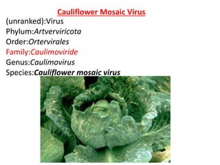
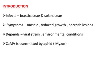
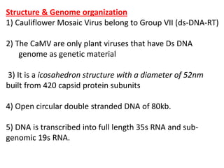
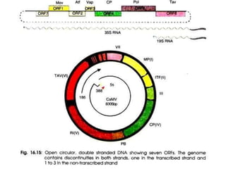
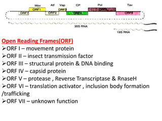
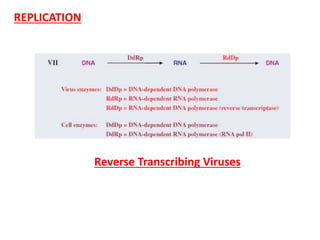
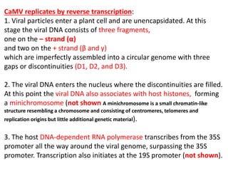
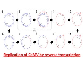
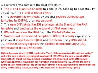
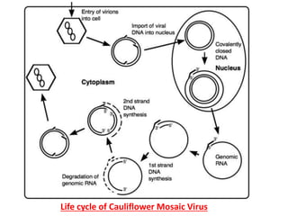
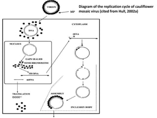
Dr. Pawan Kumar Kanaujia
Assistant Professor
Molecular Virology (Theory)
Cauliflower Mosaic Virus
(Genome organization & their
replication)
Cauliflower Mosaic Virus
(unranked):Virus
Phylum:Artverviricota
Order:Ortervirales
Family:Caulimoviride
Genus:Caulimovirus
Species:Cauliflower mosaic virus
INTRODUCTION
Infects – brassicaceae & solanaceae
 Symptoms – mosaic , reduced growth , necrotic lesions
Depends – viral strain , environmental conditions
CaMV is transmitted by aphid ( Myzus)
Structure & Genome organization
1) Cauliflower Mosaic Virus belong to Group VII (ds-DNA-RT)
2) The CaMV are only plant viruses that have Ds DNA
genome as genetic material
3) It is a icosahedron structure with a diameter of 52nm
built from 420 capsid protein subunits
4) Open circular double stranded DNA of 80kb.
5) DNA is transcribed into full length 35s RNA and sub-
genomic 19s RNA.
Open Reading Frames(ORF)
ORF I – movement protein
ORF II – insect transmission factor
ORF III – structural protein & DNA binding
ORF IV – capsid protein
ORF V – protease , Reverse Transcriptase & RnaseH
ORF VI – translation activator , inclusion body formation
/trafficking
ORF VII – unknown function
REPLICATION
Reverse Transcribing Viruses
CaMV replicates by reverse transcription:
1. Viral particles enter a plant cell and are unencapsidated. At this
stage the viral DNA consists of three fragments,
one on the – strand (α)
and two on the + strand (β and γ)
which are imperfectly assembled into a circular genome with three
gaps or discontinuities (D1, D2, and D3).
2. The viral DNA enters the nucleus where the discontinuities are filled.
At this point the viral DNA also associates with host histones, forming
a minichromosome (not shown A minichromosome is a small chromatin-like
structure resembling a chromosome and consisting of centromeres, telomeres and
replication origins but little additional genetic material).
3. The host DNA-dependent RNA polymerase transcribes from the 35S
promoter all the way around the viral genome, surpassing the 35S
promoter. Transcription also initiates at the 19S promoter (not shown).
Replication of CaMV by reverse transcription
4. The viral RNAs pass into the host cytoplasm.
5. The 3′ end of a tRNA anneals to a site corresponding to discontinuity
1 (D1) near the 5′ end of the 35S RNA.
6. The tRNAprimes synthesis, by the viral reverse transcriptase
(encoded by ORF V), of a new α strand.
7. This new DNA binds the 35S promoter at the 3′ end of the RNA
template and synthesize the α strand of DNA continues.
8. RNase H removes the RNA from the DNA–RNA duplex.
9. Synthesis of the α strand completes. RNase H activity exposes the
position of discontinuity 3 (D3), and synthesize the γ DNA strand.
10. RNase H activity exposes the position of discontinuity 2 (D2),
synthesize of the β DNA strand.
(When the new γ strand of DNA reaches the 5′ end of the new α strand it switches to the 5′
end of the new α strand, recreating discontinuity 1 (D1). When the new γ strand of DNA
reaches the 5′ end of the new β strand, it displaces the primer and some of the newly
synthesized β strand, resulting in the recreation of discontinuity 2 (D2). When the new β
strand of DNA reaches the 5′ end of the new γ strand, it displaces the primer and some of the
newly synthesized γ strand, resulting in the recreation of discontinuity 3 (D3).)
Life cycle of Cauliflower Mosaic Virus
Diagram of the replication cycle of cauliflower
mosaic virus (cited from Hull, 2002a)
Thank you

Cauliflower mosaic virus

  • 1.
    Dr. Pawan KumarKanaujia Assistant Professor Molecular Virology (Theory) Cauliflower Mosaic Virus (Genome organization & their replication)
  • 2.
  • 3.
    INTRODUCTION Infects – brassicaceae& solanaceae  Symptoms – mosaic , reduced growth , necrotic lesions Depends – viral strain , environmental conditions CaMV is transmitted by aphid ( Myzus)
  • 4.
    Structure & Genomeorganization 1) Cauliflower Mosaic Virus belong to Group VII (ds-DNA-RT) 2) The CaMV are only plant viruses that have Ds DNA genome as genetic material 3) It is a icosahedron structure with a diameter of 52nm built from 420 capsid protein subunits 4) Open circular double stranded DNA of 80kb. 5) DNA is transcribed into full length 35s RNA and sub- genomic 19s RNA.
  • 6.
    Open Reading Frames(ORF) ORFI – movement protein ORF II – insect transmission factor ORF III – structural protein & DNA binding ORF IV – capsid protein ORF V – protease , Reverse Transcriptase & RnaseH ORF VI – translation activator , inclusion body formation /trafficking ORF VII – unknown function
  • 7.
  • 8.
    CaMV replicates byreverse transcription: 1. Viral particles enter a plant cell and are unencapsidated. At this stage the viral DNA consists of three fragments, one on the – strand (α) and two on the + strand (β and γ) which are imperfectly assembled into a circular genome with three gaps or discontinuities (D1, D2, and D3). 2. The viral DNA enters the nucleus where the discontinuities are filled. At this point the viral DNA also associates with host histones, forming a minichromosome (not shown A minichromosome is a small chromatin-like structure resembling a chromosome and consisting of centromeres, telomeres and replication origins but little additional genetic material). 3. The host DNA-dependent RNA polymerase transcribes from the 35S promoter all the way around the viral genome, surpassing the 35S promoter. Transcription also initiates at the 19S promoter (not shown).
  • 9.
    Replication of CaMVby reverse transcription
  • 10.
    4. The viralRNAs pass into the host cytoplasm. 5. The 3′ end of a tRNA anneals to a site corresponding to discontinuity 1 (D1) near the 5′ end of the 35S RNA. 6. The tRNAprimes synthesis, by the viral reverse transcriptase (encoded by ORF V), of a new α strand. 7. This new DNA binds the 35S promoter at the 3′ end of the RNA template and synthesize the α strand of DNA continues. 8. RNase H removes the RNA from the DNA–RNA duplex. 9. Synthesis of the α strand completes. RNase H activity exposes the position of discontinuity 3 (D3), and synthesize the γ DNA strand. 10. RNase H activity exposes the position of discontinuity 2 (D2), synthesize of the β DNA strand. (When the new γ strand of DNA reaches the 5′ end of the new α strand it switches to the 5′ end of the new α strand, recreating discontinuity 1 (D1). When the new γ strand of DNA reaches the 5′ end of the new β strand, it displaces the primer and some of the newly synthesized β strand, resulting in the recreation of discontinuity 2 (D2). When the new β strand of DNA reaches the 5′ end of the new γ strand, it displaces the primer and some of the newly synthesized γ strand, resulting in the recreation of discontinuity 3 (D3).)
  • 11.
    Life cycle ofCauliflower Mosaic Virus
  • 12.
    Diagram of thereplication cycle of cauliflower mosaic virus (cited from Hull, 2002a)
  • 13.