Central Nervous System (CNS)Central Nervous System (CNS)
BrainBrain Spinal CordSpinal Cord
Peripheral Nervous System (PNS)Peripheral Nervous System (PNS)
Sensory NeuronsSensory NeuronsMotor NeuronsMotor Neurons
Somatic Nervous System
• voluntary movements via
skeletal muscles
Somatic Nervous System
• voluntary movements via
skeletal muscles
Autonomic Nervous System
• organs, smooth muscles
Autonomic Nervous System
• organs, smooth muscles
Sympathetic
- “Fight-or-Flight” responses
Sympathetic
- “Fight-or-Flight” responses
Parasympathetic
- maintenance
Parasympathetic
- maintenance
The Nervous System
Nervous SystemNervous System
1. CENTRAL
NERVOUS SYSTEM
MR.ASHOK BISHNOIMR.ASHOK BISHNOI
Assist., Professor ,JINRAssist., Professor ,JINR
• Astrocytes:-
• Abundant, star-shaped cells
• Brace neurons
• Form barrier
between capillaries
and neurons
• Control the chemical
environment of
the brain (CNS)
• Microglia
• Spider-like phagocytes
• Dispose of debris
• Ependymal cells
• Line cavities of the
brain and spinal cord
• Circulate
cerebrospinal
fluid
• Oligodendrocytes
Produce myelin
sheath around
nerve fibers in the
CNS
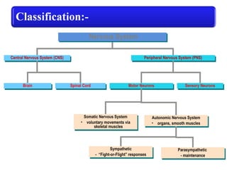
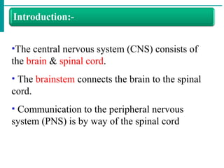
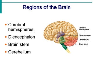
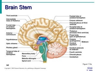
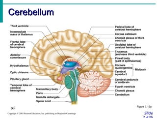
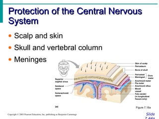
•The central nervous system (CNS) consists of
the brain & spinal cord.
• The brainstem connects the brain to the spinal
cord.
• Communication to the peripheral nervous
system (PNS) is by way of the spinal cord
• The meninges
• Membranes covering brain & spinal cord
• Protect the CNS
Three (3) layers of tissue:-
• Dura mater ( outer layer)
• “Tough mother”
• Venous sinuses
• Arachnoid mater ( middle layer)
• “Spider mother”
• Space contains cerebrospinal
fluid (CSF)
• Pia mater ( inner layer)
• “Little mother”
• Encapsulates blood vessels
Subdural space
Space between dura and arachnoid mater.
Epidural space
Space superior to dura.
Subarachnoid space
Space between arachnoid & pia mater
Filled with CSF
Contains the blood vessels supplying brain.
10
Spinal cord
Spinal cord
Pia mater
Arachnoid mater
Dura mater
Dorsal root
Dorsal root
Spinal nerve
Epidural space
(a) (b)
Ventral root
Dorsal root
ganglion
Thoracic
vertebra
Spinal
nerve
Dorsal root
ganglion
Subarachnoid
space
Dorsal branch
(dorsal ramus)
Ventral branch
(ventral ramus)
Ventral root
Epidural
space
Body of
vertebra
11
• There are four (4) ventricles
• It is interconnected cavities within cerebral
hemispheres and brain stem
• The ventricles are continuous with the
central canal of the spinal cord
• They are filled with CSF
• The four (4) ventricles are:
• Lateral ventricles (2)
• Known as the first and second
ventricles
• Third ventricle (1)
• Fourth ventricle (1)
• Interventricular foramen
• Cerebral aqueduct
Lateral ventricle
Third ventricle
Fourth ventricle
(a)
Interventricular
foramen
Cerebral
aqueduct
To central canal
of spinal cord
Copyright © The McGraw-Hill Companies, Inc. Permission required for reproduction or display.
Third ventricle
(b)
Cerebral
aqueduct
To central canal
of spinal cord
Fourth
ventricle
Lateral
ventricle
Interventricular
foramen
• Secretion of CSF-by the choroid plexus
•About 0.5 ml /mt
•About 20ml/hrs
•About 500-720 ml/day
•Specific gravity is 1.005
•pH of CSF is -7.33
•It is clear ,colorless alkaline fluid present in Subarachnoid space, ventricles of
brain ,Central canal of spinal cord.
•Completely surrounds the brain and spinal cord
Composition of CSF:-
•Water
•Glucose
•Protein
•Nitrogen substance
•Electrolyte eg. Na,K,Cal,Chloride etc.
•Cell (few)
Process of CSF
•CSF secreted by choroid plexus with in the cerebral ventricles (rt & lt) by ultra-
filtration o& active secretion.
•From Rt & Lt lateral ventricle
Foramina
•Third ventricle
Cerebral aqueduct
•Fourth ventricle
Foramina lushka & Foramina magendia
•Sub arachnoid space
•Absorbe in the sinus
Function of CSF:-
1.Support the brain & spinal cord
2.Protect the brain & spinal cord
3.Maintain pressure around structure
4.Keep brain & spinal cord moist
5.Conveys nutrition to brain & spinal cord
6.Remove waste product of brain & spinal cord
Regions of the BrainRegions of the Brain
• Cerebral
hemispheres
• Diencephalon
• Brain stem
• Cerebellum
Cerebral Hemispheres (Cerebrum)Cerebral Hemispheres (Cerebrum)
SlideCopyright © 2003 Pearson Education, Inc. publishing as Benjamin Cummings
• Paired (left
and right)
superior parts
of the brain
• Include more
than half of
the brain
mass
Figure 7.13a
Cerebral Hemispheres (Cerebrum)Cerebral Hemispheres (Cerebrum)
SlideCopyright © 2003 Pearson Education, Inc. publishing as Benjamin Cummings
• The surface
is made of
ridges (gyri)
and grooves
(sulci)
Figure 7.13a
Lobes of the CerebrumLobes of the Cerebrum
SlideCopyright © 2003 Pearson Education, Inc. publishing as Benjamin Cummings
• Fissures (deep grooves) divide the
cerebrum into lobes
• Surface lobes of the cerebrum
•Frontal lobe
•Parietal lobe
•Occipital lobe
•Temporal lobe
Lobes of the CerebrumLobes of the Cerebrum
SlideCopyright © 2003 Pearson Education, Inc. publishing as Benjamin Cummings
Figure 7.15a
Specialized Areas of the CerebrumSpecialized Areas of the Cerebrum
Slide 7.30Copyright © 2003 Pearson Education, Inc. publishing as Benjamin Cummings
• Somatic sensory area – receives
impulses from the body’s sensory
receptors
• Primary motor area – sends impulses to
skeletal muscles
• Broca’s area – involved in our ability to
speak
Sensory and Motor Areas of theSensory and Motor Areas of the
Cerebral CortexCerebral Cortex
Slide 7.31Copyright © 2003 Pearson Education, Inc. publishing as Benjamin Cummings
Figure 7.14
Specialized Area of the CerebrumSpecialized Area of the Cerebrum
SlideCopyright © 2003 Pearson Education, Inc. publishing as Benjamin Cummings
• Cerebral areas involved in special
senses
•Gustatory area (taste)
•Visual area
•Auditory area
•Olfactory area
Specialized Area of the CerebrumSpecialized Area of the Cerebrum
SlideCopyright © 2003 Pearson Education, Inc. publishing as Benjamin Cummings
• Interpretation areas of the cerebrum
•Speech/language region
•Language comprehension region
•General interpretation area
Specialized Area of the CerebrumSpecialized Area of the Cerebrum
SlideCopyright © 2003 Pearson Education, Inc. publishing as Benjamin Cummings
Figure 7.13c
Layers of the CerebrumLayers of the Cerebrum
SlideCopyright © 2003 Pearson Education, Inc. publishing as Benjamin Cummings
• Gray matter
•Outer layer
•Composed
mostly of neuron
cell bodies
Figure 7.13a
Layers of the CerebrumLayers of the Cerebrum
SlideCopyright © 2003 Pearson Education, Inc. publishing as Benjamin Cummings
• White matter
•Fiber tracts
inside the gray
matter
•Example:
corpus callosum
connects
hemispheres
Figure 7.13a
Layers of the CerebrumLayers of the Cerebrum
SlideCopyright © 2003 Pearson Education, Inc. publishing as Benjamin Cummings
• Basal nuclei – internal
islands of gray matter
• Regulates voluntary
motor activities by
modifying info sent to
the motor cortex
• Problems = ie unable
to control muscles,
spastic, jerky
• Involved in
Huntington’s and
Parkinson’s Disease
Figure 7.13a
DiencephalonDiencephalon
SlideCopyright © 2003 Pearson Education, Inc. publishing as Benjamin Cummings
• Sits on top of the brain stem
• Enclosed by the cerebral heispheres
• Made of three parts
•Thalamus
•Hypothalamus
•Epithalamus
DiencephalonDiencephalon
SlideCopyright © 2003 Pearson Education, Inc. publishing as Benjamin Cummings
Figure 7.15
ThalamusThalamus
Slide 7.35Copyright © 2003 Pearson Education, Inc. publishing as Benjamin Cummings
• Surrounds the third ventricle
• The relay station for sensory impulses
• Transfers impulses to the correct part of
the cortex for localization and
interpretation
HypothalamusHypothalamus
SlideCopyright © 2003 Pearson Education, Inc. publishing as Benjamin Cummings
• Under the thalamus
• Important autonomic nervous system
center
•Helps regulate body temperature
•Controls water balance
•Regulates metabolism
HypothalamusHypothalamus
SlideCopyright © 2003 Pearson Education, Inc. publishing as Benjamin Cummings
• An important part of the limbic system
(emotions)
• The pituitary gland is attached to the
hypothalamus
EpithalamusEpithalamus
Slide 7.37Copyright © 2003 Pearson Education, Inc. publishing as Benjamin Cummings
• Forms the roof of the third ventricle
• Houses the pineal body (an endocrine
gland)
• Includes the choroid plexus – forms
cerebrospinal fluid
Brain StemBrain Stem
SlideCopyright © 2003 Pearson Education, Inc. publishing as Benjamin Cummings
• Attaches to the spinal cord
• Parts of the brain stem
•Midbrain
•Pons
•Medulla oblongata
Brain StemBrain Stem
SlideCopyright © 2003 Pearson Education, Inc. publishing as Benjamin Cummings
Figure 7.15a
MidbrainMidbrain
Slide 7.39Copyright © 2003 Pearson Education, Inc. publishing as Benjamin Cummings
• Mostly composed of tracts of nerve
fibers
•Reflex centers for vision and hearing
•Cerebral aquaduct – 3rd
-4th
ventricles
PonsPons
Slide 7.40Copyright © 2003 Pearson Education, Inc. publishing as Benjamin Cummings
• The bulging center part of the brain
stem
• Mostly composed of fiber tracts
• Includes nuclei involved in the control of
breathing
Medulla OblongataMedulla Oblongata
Slide 7.41Copyright © 2003 Pearson Education, Inc. publishing as Benjamin Cummings
• The lowest part of the brain stem
• Merges into the spinal cord
• Includes important fiber tracts
• Contains important control centers
•Heart rate control
•Blood pressure regulation
•Breathing
•Swallowing
•Vomiting
CerebellumCerebellum
SlideCopyright © 2003 Pearson Education, Inc. publishing as Benjamin Cummings
• Two hemispheres with convoluted
surfaces
• Provides involuntary coordination of
body movements
CerebellumCerebellum
SlideCopyright © 2003 Pearson Education, Inc. publishing as Benjamin Cummings
Figure 7.15a
Protection of the Central NervousProtection of the Central Nervous
SystemSystem
SlideCopyright © 2003 Pearson Education, Inc. publishing as Benjamin Cummings
• Scalp and skin
• Skull and vertebral column
• Meninges
Figure 7.16a
Protection of the Central NervousProtection of the Central Nervous
SystemSystem
SlideCopyright © 2003 Pearson Education, Inc. publishing as Benjamin Cummings
• Cerebrospinal fluid
• Blood brain barrier
Figure 7.16a
MeningesMeninges
SlideCopyright © 2003 Pearson Education, Inc. publishing as Benjamin Cummings
• Dura mater
•Double-layered external covering
•Periosteum – attached to surface of the
skull
•Meningeal layer – outer covering of the
brain
•Folds inward in several areas
MeningesMeninges
SlideCopyright © 2003 Pearson Education, Inc. publishing as Benjamin Cummings
• Arachnoid layer
•Middle layer
•Web-like
• Pia mater
•Internal layer
•Clings to the surface of the brain
Cerebrospinal FluidCerebrospinal Fluid
Slide 7.46Copyright © 2003 Pearson Education, Inc. publishing as Benjamin Cummings
• Similar to blood plasma composition
• Formed by the choroid plexus
• Forms a watery cushion to protect the
brain
• Circulated in arachnoid space,
ventricles, and central canal of the
spinal cord
Ventricles and Location of theVentricles and Location of the
Cerebrospinal FluidCerebrospinal Fluid
SlideCopyright © 2003 Pearson Education, Inc. publishing as Benjamin Cummings
Figure 7.17a
Ventricles and Location of theVentricles and Location of the
Cerebrospinal FluidCerebrospinal Fluid
SlideCopyright © 2003 Pearson Education, Inc. publishing as Benjamin Cummings
Figure 7.17b
Blood Brain BarrierBlood Brain Barrier
Slide 7.48Copyright © 2003 Pearson Education, Inc. publishing as Benjamin Cummings
• Includes the least permeable capillaries
of the body
• Excludes many potentially harmful
substances
• Useless against some substances
•Fats and fat soluble molecules
•Respiratory gases
•Alcohol
•Nicotine
•Anesthesia
Traumatic Brain Injuries (TBI)Traumatic Brain Injuries (TBI)
Slide 7.49Copyright © 2003 Pearson Education, Inc. publishing as Benjamin Cummings
• Concussion
•Slight or mild brain injury
•Bleeding & tearing of nerve fibers
happened
•Recovery likely with some memory loss
• Contusion
•A more severe TBI
•Nervous tissue destruction occurs
•Nervous tissue does not regenerate
• Cerebral edema
• Cerebral edema
– Swelling from the inflammatory response
– May compress and kill brain tissue
• Subdural hematoma
– Collection of blood below the dura
• Standards for these conditions were revised
in 2004. Please check out TBIs at
Mayoclinic.com for more current
information on diagnostic terminology.
Cerebrovascular Accident (CVA)Cerebrovascular Accident (CVA)
Slide 7.50Copyright © 2003 Pearson Education, Inc. publishing as Benjamin Cummings
• Commonly called a stroke
• The result of a ruptured blood vessel
supplying a region of the brain
• Brain tissue supplied with oxygen from
that blood source dies
• Loss of some functions or death may
result
Alzheimer’s DiseaseAlzheimer’s Disease
Slide 7.51Copyright © 2003 Pearson Education, Inc. publishing as Benjamin Cummings
• Progressive degenerative brain disease
• Mostly seen in the elderly, but may
begin in middle age
• Structural changes in the brain include
abnormal protein deposits and twisted
fibers within neurons
• Victims experience memory loss,
irritability, confusion and ultimately,
hallucinations and death
Spinal CordSpinal Cord
Slide 7.52Copyright © 2003 Pearson Education, Inc. publishing as Benjamin Cummings
• Extends from the
medulla oblongata to
the region of T12
• Below T12 is the cauda
equina (a collection of
spinal nerves)
• Enlargements occur in
the cervical and lumbar
regions
Figure 7.18
Spinal Cord AnatomySpinal Cord Anatomy
SlideCopyright © 2003 Pearson Education, Inc. publishing as Benjamin Cummings
• Exterior white mater – conduction tracts
Figure 7.19
Spinal Cord AnatomySpinal Cord Anatomy
SlideCopyright © 2003 Pearson Education, Inc. publishing as Benjamin Cummings
• Internal gray matter - mostly cell bodies
•Dorsal (posterior) horns
•Anterior (ventral) horns
Figure 7.19
Spinal Cord AnatomySpinal Cord Anatomy
SlideCopyright © 2003 Pearson Education, Inc. publishing as Benjamin Cummings
• Central canal filled with cerebrospinal
fluid
Figure 7.19
Spinal Cord AnatomySpinal Cord Anatomy
Slide 7.54Copyright © 2003 Pearson Education, Inc. publishing as Benjamin Cummings
• Meninges cover the spinal cord
• Nerves leave at the level of each
vertebrae
•Dorsal root
• Associated with the dorsal root ganglia –
collections of cell bodies outside the central
nervous system
•Ventral root
Peripheral Nervous SystemPeripheral Nervous System
Slide 7.55Copyright © 2003 Pearson Education, Inc. publishing as Benjamin Cummings
• Nerves and ganglia outside the central
nervous system
• Nerve = bundle of neuron fibers
• Neuron fibers are bundled by
connective tissue
Structure of a NerveStructure of a Nerve
Slide 7.56Copyright © 2003 Pearson Education, Inc. publishing as Benjamin Cummings
• Endoneurium
surrounds each fiber
• Groups of fibers are
bound into fascicles
by perineurium
• Fascicles are bound
together by
epineurium
Figure 7.20
Classification of NervesClassification of Nerves
Slide 7.57Copyright © 2003 Pearson Education, Inc. publishing as Benjamin Cummings
• Mixed nerves – both sensory and motor
fibers
• Afferent (sensory) nerves – carry
impulses toward the CNS
• Efferent (motor) nerves – carry impulses
away from the CNS
Spinal NervesSpinal Nerves
Slide 7.63Copyright © 2003 Pearson Education, Inc. publishing as Benjamin Cummings
• There is a pair of spinal nerves at the
level of each vertebrae.
Spinal NervesSpinal Nerves
Slide 7.64Copyright © 2003 Pearson Education, Inc. publishing as Benjamin Cummings
Figure 7.22a
Autonomic Nervous SystemAutonomic Nervous System
Slide 7.67Copyright © 2003 Pearson Education, Inc. publishing as Benjamin Cummings
• The involuntary branch of the nervous
system
• Consists of only motor nerves
• Divided into two divisions
•Sympathetic division
•Parasympathetic division
Comparison of Somatic andComparison of Somatic and
Autonomic Nervous SystemsAutonomic Nervous Systems
Slide 7.69Copyright © 2003 Pearson Education, Inc. publishing as Benjamin Cummings Figure 7.24
Anatomy of the Autonomic NervousAnatomy of the Autonomic Nervous
SystemSystem
Slide 7.73Copyright © 2003 Pearson Education, Inc. publishing as Benjamin Cummings
Figure 7.25
Autonomic FunctioningAutonomic Functioning
SlideCopyright © 2003 Pearson Education, Inc. publishing as Benjamin Cummings
• Sympathetic – “fight-or-flight”
•Response to unusual stimulus
•Takes over to increase activities
•Remember as the “E” division = exercise,
excitement, emergency, and
embarrassment
Autonomic FunctioningAutonomic Functioning
SlideCopyright © 2003 Pearson Education, Inc. publishing as Benjamin Cummings
• Parasympathetic – housekeeping
activites
•Conserves energy
•Maintains daily necessary body functions
•Remember as the “D” division - digestion,
defecation, and diuresis
Development Aspects of theDevelopment Aspects of the
Nervous SystemNervous System
SlideCopyright © 2003 Pearson Education, Inc. publishing as Benjamin Cummings
• The nervous system is formed during
the first month of embryonic
development
• Any maternal infection can have
extremely harmful effects
• The hypothalamus is one of the last
areas of the brain to develop
Development Aspects of theDevelopment Aspects of the
Nervous SystemNervous System
SlideCopyright © 2003 Pearson Education, Inc. publishing as Benjamin Cummings
• No more neurons are formed after birth,
but growth and maturation continues for
several years (new evidence!)
• The brain reaches maximum weight as
a young adult
• However, we can always grow
dendrites!

ppt on CNS

  • 1.
    Central Nervous System(CNS)Central Nervous System (CNS) BrainBrain Spinal CordSpinal Cord Peripheral Nervous System (PNS)Peripheral Nervous System (PNS) Sensory NeuronsSensory NeuronsMotor NeuronsMotor Neurons Somatic Nervous System • voluntary movements via skeletal muscles Somatic Nervous System • voluntary movements via skeletal muscles Autonomic Nervous System • organs, smooth muscles Autonomic Nervous System • organs, smooth muscles Sympathetic - “Fight-or-Flight” responses Sympathetic - “Fight-or-Flight” responses Parasympathetic - maintenance Parasympathetic - maintenance The Nervous System Nervous SystemNervous System
  • 2.
    1. CENTRAL NERVOUS SYSTEM MR.ASHOKBISHNOIMR.ASHOK BISHNOI Assist., Professor ,JINRAssist., Professor ,JINR
  • 3.
    • Astrocytes:- • Abundant,star-shaped cells • Brace neurons • Form barrier between capillaries and neurons • Control the chemical environment of the brain (CNS)
  • 4.
    • Microglia • Spider-likephagocytes • Dispose of debris • Ependymal cells • Line cavities of the brain and spinal cord • Circulate cerebrospinal fluid
  • 5.
    • Oligodendrocytes Produce myelin sheatharound nerve fibers in the CNS
  • 6.
    •The central nervoussystem (CNS) consists of the brain & spinal cord. • The brainstem connects the brain to the spinal cord. • Communication to the peripheral nervous system (PNS) is by way of the spinal cord
  • 7.
    • The meninges •Membranes covering brain & spinal cord • Protect the CNS Three (3) layers of tissue:- • Dura mater ( outer layer) • “Tough mother” • Venous sinuses • Arachnoid mater ( middle layer) • “Spider mother” • Space contains cerebrospinal fluid (CSF) • Pia mater ( inner layer) • “Little mother” • Encapsulates blood vessels
  • 8.
    Subdural space Space betweendura and arachnoid mater. Epidural space Space superior to dura. Subarachnoid space Space between arachnoid & pia mater Filled with CSF Contains the blood vessels supplying brain.
  • 10.
    10 Spinal cord Spinal cord Piamater Arachnoid mater Dura mater Dorsal root Dorsal root Spinal nerve Epidural space (a) (b) Ventral root Dorsal root ganglion Thoracic vertebra Spinal nerve Dorsal root ganglion Subarachnoid space Dorsal branch (dorsal ramus) Ventral branch (ventral ramus) Ventral root Epidural space Body of vertebra
  • 11.
    11 • There arefour (4) ventricles • It is interconnected cavities within cerebral hemispheres and brain stem • The ventricles are continuous with the central canal of the spinal cord • They are filled with CSF • The four (4) ventricles are: • Lateral ventricles (2) • Known as the first and second ventricles • Third ventricle (1) • Fourth ventricle (1) • Interventricular foramen • Cerebral aqueduct Lateral ventricle Third ventricle Fourth ventricle (a) Interventricular foramen Cerebral aqueduct To central canal of spinal cord Copyright © The McGraw-Hill Companies, Inc. Permission required for reproduction or display. Third ventricle (b) Cerebral aqueduct To central canal of spinal cord Fourth ventricle Lateral ventricle Interventricular foramen
  • 15.
    • Secretion ofCSF-by the choroid plexus •About 0.5 ml /mt •About 20ml/hrs •About 500-720 ml/day •Specific gravity is 1.005 •pH of CSF is -7.33 •It is clear ,colorless alkaline fluid present in Subarachnoid space, ventricles of brain ,Central canal of spinal cord. •Completely surrounds the brain and spinal cord
  • 16.
    Composition of CSF:- •Water •Glucose •Protein •Nitrogensubstance •Electrolyte eg. Na,K,Cal,Chloride etc. •Cell (few)
  • 17.
    Process of CSF •CSFsecreted by choroid plexus with in the cerebral ventricles (rt & lt) by ultra- filtration o& active secretion. •From Rt & Lt lateral ventricle Foramina •Third ventricle Cerebral aqueduct •Fourth ventricle Foramina lushka & Foramina magendia •Sub arachnoid space •Absorbe in the sinus
  • 18.
    Function of CSF:- 1.Supportthe brain & spinal cord 2.Protect the brain & spinal cord 3.Maintain pressure around structure 4.Keep brain & spinal cord moist 5.Conveys nutrition to brain & spinal cord 6.Remove waste product of brain & spinal cord
  • 20.
    Regions of theBrainRegions of the Brain • Cerebral hemispheres • Diencephalon • Brain stem • Cerebellum
  • 21.
    Cerebral Hemispheres (Cerebrum)CerebralHemispheres (Cerebrum) SlideCopyright © 2003 Pearson Education, Inc. publishing as Benjamin Cummings • Paired (left and right) superior parts of the brain • Include more than half of the brain mass Figure 7.13a
  • 22.
    Cerebral Hemispheres (Cerebrum)CerebralHemispheres (Cerebrum) SlideCopyright © 2003 Pearson Education, Inc. publishing as Benjamin Cummings • The surface is made of ridges (gyri) and grooves (sulci) Figure 7.13a
  • 23.
    Lobes of theCerebrumLobes of the Cerebrum SlideCopyright © 2003 Pearson Education, Inc. publishing as Benjamin Cummings • Fissures (deep grooves) divide the cerebrum into lobes • Surface lobes of the cerebrum •Frontal lobe •Parietal lobe •Occipital lobe •Temporal lobe
  • 24.
    Lobes of theCerebrumLobes of the Cerebrum SlideCopyright © 2003 Pearson Education, Inc. publishing as Benjamin Cummings Figure 7.15a
  • 25.
    Specialized Areas ofthe CerebrumSpecialized Areas of the Cerebrum Slide 7.30Copyright © 2003 Pearson Education, Inc. publishing as Benjamin Cummings • Somatic sensory area – receives impulses from the body’s sensory receptors • Primary motor area – sends impulses to skeletal muscles • Broca’s area – involved in our ability to speak
  • 27.
    Sensory and MotorAreas of theSensory and Motor Areas of the Cerebral CortexCerebral Cortex Slide 7.31Copyright © 2003 Pearson Education, Inc. publishing as Benjamin Cummings Figure 7.14
  • 28.
    Specialized Area ofthe CerebrumSpecialized Area of the Cerebrum SlideCopyright © 2003 Pearson Education, Inc. publishing as Benjamin Cummings • Cerebral areas involved in special senses •Gustatory area (taste) •Visual area •Auditory area •Olfactory area
  • 29.
    Specialized Area ofthe CerebrumSpecialized Area of the Cerebrum SlideCopyright © 2003 Pearson Education, Inc. publishing as Benjamin Cummings • Interpretation areas of the cerebrum •Speech/language region •Language comprehension region •General interpretation area
  • 30.
    Specialized Area ofthe CerebrumSpecialized Area of the Cerebrum SlideCopyright © 2003 Pearson Education, Inc. publishing as Benjamin Cummings Figure 7.13c
  • 31.
    Layers of theCerebrumLayers of the Cerebrum SlideCopyright © 2003 Pearson Education, Inc. publishing as Benjamin Cummings • Gray matter •Outer layer •Composed mostly of neuron cell bodies Figure 7.13a
  • 32.
    Layers of theCerebrumLayers of the Cerebrum SlideCopyright © 2003 Pearson Education, Inc. publishing as Benjamin Cummings • White matter •Fiber tracts inside the gray matter •Example: corpus callosum connects hemispheres Figure 7.13a
  • 33.
    Layers of theCerebrumLayers of the Cerebrum SlideCopyright © 2003 Pearson Education, Inc. publishing as Benjamin Cummings • Basal nuclei – internal islands of gray matter • Regulates voluntary motor activities by modifying info sent to the motor cortex • Problems = ie unable to control muscles, spastic, jerky • Involved in Huntington’s and Parkinson’s Disease Figure 7.13a
  • 34.
    DiencephalonDiencephalon SlideCopyright © 2003Pearson Education, Inc. publishing as Benjamin Cummings • Sits on top of the brain stem • Enclosed by the cerebral heispheres • Made of three parts •Thalamus •Hypothalamus •Epithalamus
  • 35.
    DiencephalonDiencephalon SlideCopyright © 2003Pearson Education, Inc. publishing as Benjamin Cummings Figure 7.15
  • 36.
    ThalamusThalamus Slide 7.35Copyright ©2003 Pearson Education, Inc. publishing as Benjamin Cummings • Surrounds the third ventricle • The relay station for sensory impulses • Transfers impulses to the correct part of the cortex for localization and interpretation
  • 37.
    HypothalamusHypothalamus SlideCopyright © 2003Pearson Education, Inc. publishing as Benjamin Cummings • Under the thalamus • Important autonomic nervous system center •Helps regulate body temperature •Controls water balance •Regulates metabolism
  • 38.
    HypothalamusHypothalamus SlideCopyright © 2003Pearson Education, Inc. publishing as Benjamin Cummings • An important part of the limbic system (emotions) • The pituitary gland is attached to the hypothalamus
  • 39.
    EpithalamusEpithalamus Slide 7.37Copyright ©2003 Pearson Education, Inc. publishing as Benjamin Cummings • Forms the roof of the third ventricle • Houses the pineal body (an endocrine gland) • Includes the choroid plexus – forms cerebrospinal fluid
  • 40.
    Brain StemBrain Stem SlideCopyright© 2003 Pearson Education, Inc. publishing as Benjamin Cummings • Attaches to the spinal cord • Parts of the brain stem •Midbrain •Pons •Medulla oblongata
  • 41.
    Brain StemBrain Stem SlideCopyright© 2003 Pearson Education, Inc. publishing as Benjamin Cummings Figure 7.15a
  • 42.
    MidbrainMidbrain Slide 7.39Copyright ©2003 Pearson Education, Inc. publishing as Benjamin Cummings • Mostly composed of tracts of nerve fibers •Reflex centers for vision and hearing •Cerebral aquaduct – 3rd -4th ventricles
  • 43.
    PonsPons Slide 7.40Copyright ©2003 Pearson Education, Inc. publishing as Benjamin Cummings • The bulging center part of the brain stem • Mostly composed of fiber tracts • Includes nuclei involved in the control of breathing
  • 44.
    Medulla OblongataMedulla Oblongata Slide7.41Copyright © 2003 Pearson Education, Inc. publishing as Benjamin Cummings • The lowest part of the brain stem • Merges into the spinal cord • Includes important fiber tracts • Contains important control centers •Heart rate control •Blood pressure regulation •Breathing •Swallowing •Vomiting
  • 45.
    CerebellumCerebellum SlideCopyright © 2003Pearson Education, Inc. publishing as Benjamin Cummings • Two hemispheres with convoluted surfaces • Provides involuntary coordination of body movements
  • 46.
    CerebellumCerebellum SlideCopyright © 2003Pearson Education, Inc. publishing as Benjamin Cummings Figure 7.15a
  • 47.
    Protection of theCentral NervousProtection of the Central Nervous SystemSystem SlideCopyright © 2003 Pearson Education, Inc. publishing as Benjamin Cummings • Scalp and skin • Skull and vertebral column • Meninges Figure 7.16a
  • 48.
    Protection of theCentral NervousProtection of the Central Nervous SystemSystem SlideCopyright © 2003 Pearson Education, Inc. publishing as Benjamin Cummings • Cerebrospinal fluid • Blood brain barrier Figure 7.16a
  • 49.
    MeningesMeninges SlideCopyright © 2003Pearson Education, Inc. publishing as Benjamin Cummings • Dura mater •Double-layered external covering •Periosteum – attached to surface of the skull •Meningeal layer – outer covering of the brain •Folds inward in several areas
  • 50.
    MeningesMeninges SlideCopyright © 2003Pearson Education, Inc. publishing as Benjamin Cummings • Arachnoid layer •Middle layer •Web-like • Pia mater •Internal layer •Clings to the surface of the brain
  • 51.
    Cerebrospinal FluidCerebrospinal Fluid Slide7.46Copyright © 2003 Pearson Education, Inc. publishing as Benjamin Cummings • Similar to blood plasma composition • Formed by the choroid plexus • Forms a watery cushion to protect the brain • Circulated in arachnoid space, ventricles, and central canal of the spinal cord
  • 52.
    Ventricles and Locationof theVentricles and Location of the Cerebrospinal FluidCerebrospinal Fluid SlideCopyright © 2003 Pearson Education, Inc. publishing as Benjamin Cummings Figure 7.17a
  • 53.
    Ventricles and Locationof theVentricles and Location of the Cerebrospinal FluidCerebrospinal Fluid SlideCopyright © 2003 Pearson Education, Inc. publishing as Benjamin Cummings Figure 7.17b
  • 54.
    Blood Brain BarrierBloodBrain Barrier Slide 7.48Copyright © 2003 Pearson Education, Inc. publishing as Benjamin Cummings • Includes the least permeable capillaries of the body • Excludes many potentially harmful substances • Useless against some substances •Fats and fat soluble molecules •Respiratory gases •Alcohol •Nicotine •Anesthesia
  • 55.
    Traumatic Brain Injuries(TBI)Traumatic Brain Injuries (TBI) Slide 7.49Copyright © 2003 Pearson Education, Inc. publishing as Benjamin Cummings • Concussion •Slight or mild brain injury •Bleeding & tearing of nerve fibers happened •Recovery likely with some memory loss • Contusion •A more severe TBI •Nervous tissue destruction occurs •Nervous tissue does not regenerate • Cerebral edema
  • 56.
    • Cerebral edema –Swelling from the inflammatory response – May compress and kill brain tissue • Subdural hematoma – Collection of blood below the dura • Standards for these conditions were revised in 2004. Please check out TBIs at Mayoclinic.com for more current information on diagnostic terminology.
  • 57.
    Cerebrovascular Accident (CVA)CerebrovascularAccident (CVA) Slide 7.50Copyright © 2003 Pearson Education, Inc. publishing as Benjamin Cummings • Commonly called a stroke • The result of a ruptured blood vessel supplying a region of the brain • Brain tissue supplied with oxygen from that blood source dies • Loss of some functions or death may result
  • 58.
    Alzheimer’s DiseaseAlzheimer’s Disease Slide7.51Copyright © 2003 Pearson Education, Inc. publishing as Benjamin Cummings • Progressive degenerative brain disease • Mostly seen in the elderly, but may begin in middle age • Structural changes in the brain include abnormal protein deposits and twisted fibers within neurons • Victims experience memory loss, irritability, confusion and ultimately, hallucinations and death
  • 59.
    Spinal CordSpinal Cord Slide7.52Copyright © 2003 Pearson Education, Inc. publishing as Benjamin Cummings • Extends from the medulla oblongata to the region of T12 • Below T12 is the cauda equina (a collection of spinal nerves) • Enlargements occur in the cervical and lumbar regions Figure 7.18
  • 60.
    Spinal Cord AnatomySpinalCord Anatomy SlideCopyright © 2003 Pearson Education, Inc. publishing as Benjamin Cummings • Exterior white mater – conduction tracts Figure 7.19
  • 61.
    Spinal Cord AnatomySpinalCord Anatomy SlideCopyright © 2003 Pearson Education, Inc. publishing as Benjamin Cummings • Internal gray matter - mostly cell bodies •Dorsal (posterior) horns •Anterior (ventral) horns Figure 7.19
  • 62.
    Spinal Cord AnatomySpinalCord Anatomy SlideCopyright © 2003 Pearson Education, Inc. publishing as Benjamin Cummings • Central canal filled with cerebrospinal fluid Figure 7.19
  • 63.
    Spinal Cord AnatomySpinalCord Anatomy Slide 7.54Copyright © 2003 Pearson Education, Inc. publishing as Benjamin Cummings • Meninges cover the spinal cord • Nerves leave at the level of each vertebrae •Dorsal root • Associated with the dorsal root ganglia – collections of cell bodies outside the central nervous system •Ventral root
  • 64.
    Peripheral Nervous SystemPeripheralNervous System Slide 7.55Copyright © 2003 Pearson Education, Inc. publishing as Benjamin Cummings • Nerves and ganglia outside the central nervous system • Nerve = bundle of neuron fibers • Neuron fibers are bundled by connective tissue
  • 65.
    Structure of aNerveStructure of a Nerve Slide 7.56Copyright © 2003 Pearson Education, Inc. publishing as Benjamin Cummings • Endoneurium surrounds each fiber • Groups of fibers are bound into fascicles by perineurium • Fascicles are bound together by epineurium Figure 7.20
  • 66.
    Classification of NervesClassificationof Nerves Slide 7.57Copyright © 2003 Pearson Education, Inc. publishing as Benjamin Cummings • Mixed nerves – both sensory and motor fibers • Afferent (sensory) nerves – carry impulses toward the CNS • Efferent (motor) nerves – carry impulses away from the CNS
  • 67.
    Spinal NervesSpinal Nerves Slide7.63Copyright © 2003 Pearson Education, Inc. publishing as Benjamin Cummings • There is a pair of spinal nerves at the level of each vertebrae.
  • 68.
    Spinal NervesSpinal Nerves Slide7.64Copyright © 2003 Pearson Education, Inc. publishing as Benjamin Cummings Figure 7.22a
  • 69.
    Autonomic Nervous SystemAutonomicNervous System Slide 7.67Copyright © 2003 Pearson Education, Inc. publishing as Benjamin Cummings • The involuntary branch of the nervous system • Consists of only motor nerves • Divided into two divisions •Sympathetic division •Parasympathetic division
  • 70.
    Comparison of SomaticandComparison of Somatic and Autonomic Nervous SystemsAutonomic Nervous Systems Slide 7.69Copyright © 2003 Pearson Education, Inc. publishing as Benjamin Cummings Figure 7.24
  • 71.
    Anatomy of theAutonomic NervousAnatomy of the Autonomic Nervous SystemSystem Slide 7.73Copyright © 2003 Pearson Education, Inc. publishing as Benjamin Cummings Figure 7.25
  • 72.
    Autonomic FunctioningAutonomic Functioning SlideCopyright© 2003 Pearson Education, Inc. publishing as Benjamin Cummings • Sympathetic – “fight-or-flight” •Response to unusual stimulus •Takes over to increase activities •Remember as the “E” division = exercise, excitement, emergency, and embarrassment
  • 73.
    Autonomic FunctioningAutonomic Functioning SlideCopyright© 2003 Pearson Education, Inc. publishing as Benjamin Cummings • Parasympathetic – housekeeping activites •Conserves energy •Maintains daily necessary body functions •Remember as the “D” division - digestion, defecation, and diuresis
  • 74.
    Development Aspects oftheDevelopment Aspects of the Nervous SystemNervous System SlideCopyright © 2003 Pearson Education, Inc. publishing as Benjamin Cummings • The nervous system is formed during the first month of embryonic development • Any maternal infection can have extremely harmful effects • The hypothalamus is one of the last areas of the brain to develop
  • 75.
    Development Aspects oftheDevelopment Aspects of the Nervous SystemNervous System SlideCopyright © 2003 Pearson Education, Inc. publishing as Benjamin Cummings • No more neurons are formed after birth, but growth and maturation continues for several years (new evidence!) • The brain reaches maximum weight as a young adult • However, we can always grow dendrites!