Introductio
n
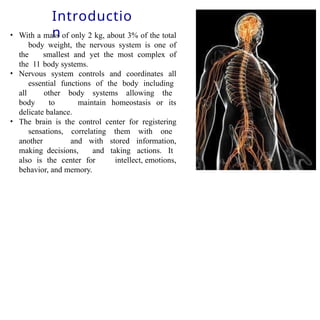
• With amass of only 2 kg, about 3% of the total
body weight, the nervous system is one of
the smallest and yet the most complex of
the 11 body systems.
• Nervous system controls and coordinates all
essential functions of the body including
all other body systems allowing the
body to maintain homeostasis or its
delicate balance.
• The brain is the control center for registering
sensations, correlating them with one
another and with stored information,
making decisions, and taking actions. It
also is the center for intellect, emotions,
behavior, and memory.
3.
Introductio
n
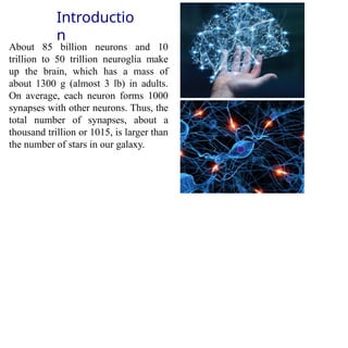
About 85 billionneurons and 10
trillion to 50 trillion neuroglia make
up the brain, which has a mass of
about 1300 g (almost 3 lb) in adults.
On average, each neuron forms 1000
synapses with other neurons. Thus, the
total number of synapses, about a
thousand trillion or 1015, is larger than
the number of stars in our galaxy.
4.
Functions of Nervous
system
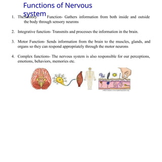
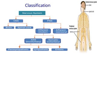
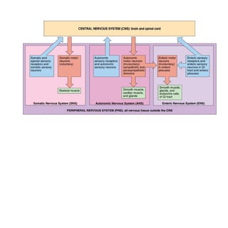
1.TheSensory Function- Gathers information from both inside and outside
the body through sensory neurons
2. Integrative function- Transmits and processes the information in the brain.
3. Motor Function- Sends information from the brain to the muscles, glands, and
organs so they can respond appropriately through the motor neurons
4. Complex functions- The nervous system is also responsible for our perceptions,
emotions, behaviors, memories etc.
•The central nervoussystem (CNS) consists of the brain
& spinal cord.
•The brain is the part of the CNS that is located in the
skull and contains about 85 billion neurons.
•The brain is connected to the spinal cord.
•The spinal cord is connected to the Communication to
the peripheral nervous system (PNS) and transmits the
information from the body to the brain and vice versa.
9.
Nervous
Tissues
There are 2types of nervous tissues
1. Excitatory – Neurons
2. Non-excitatory- Neuroglia
Neurons have the potential the excite in response to specific stimuli
Neuroglia are smaller cells but they greatly outnumber neurons,
perhaps by as much as 25 times
Neuroglia support, nourish, and protect neurons, and maintain the
interstitial fluid that bathes them.
10.
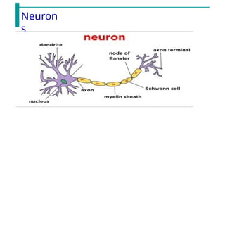
Neurons-
Parts
•Dendrite – receivestimulus and carries its
impulses toward the cell body
• Cell Body with the nucleus
• Axon – fiber which carries impulses away from the
cell body
• Schwann Cells- cells which produce the myelin
sheath
• Myelin sheath – dense lipid layer which insulates the
axon
• Node of Ranvier – gaps or nodes in the myelin
sheath
• Impulses travel from dendrite to cell body to axon
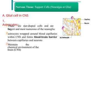
A. Glial cellin CNS
1.
Astrocytes:-
These are star-shaped cells and are
the
largest and most numerous of the neuroglia.
astrocytes wrapped around blood capillaries
within CNS and forms blood-brain barrier
between capillaries and neurons
Maintain the
chemical environment of the
brain (CNS)
13.
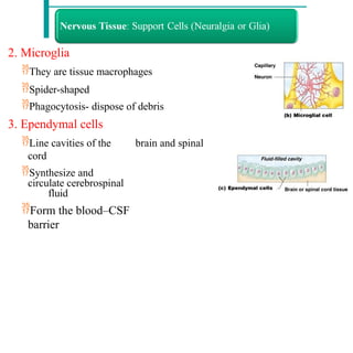
2. Microglia
They aretissue macrophages
Spider-shaped
Phagocytosis- dispose of debris
3. Ependymal cells
Line cavities of the brain and spinal
cord
Synthesize and
circulate cerebrospinal
fluid
Form the blood–CSF
barrier
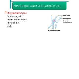
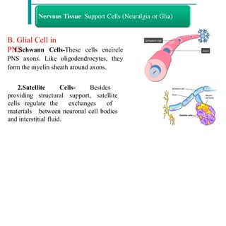
B. Glial Cellin
PNS
1.Schwann Cells-These cells encircle
PNS axons. Like oligodendrocytes, they
form the myelin sheath around axons.
2.Satellite Cells- Besides
providing structural support, satellite
cells regulate the exchanges of
materials between neuronal cell bodies
and interstitial fluid.
16.
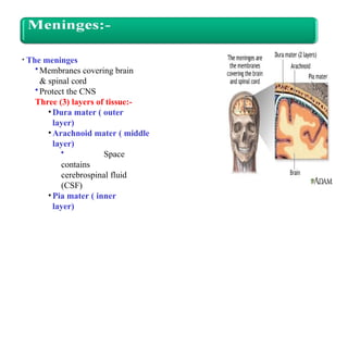
• The meninges
•Membranescovering brain
& spinal cord
•Protect the CNS
Three (3) layers of tissue:-
• Dura mater ( outer
layer)
•Arachnoid mater ( middle
layer)
• Space
contains
cerebrospinal fluid
(CSF)
• Pia mater ( inner
layer)
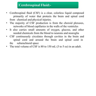
• Cerebrospinal fluid(CSF) is a clear, colorless liquid composed
primarily of water that protects the brain and spinal cord
from chemical and physical injuries.
• The majority of CSF production is from the choroid plexuses,
networks of blood capillaries in the walls of the ventricles
• It also carries small amounts of oxygen, glucose, and other
needed chemicals from the blood to neurons and neuroglia
• CSF continuously circulates through cavities in the brain and
spinal cord and around the brain and spinal cord in
the subarachnoid space
• The total volume of CSF is 80 to 150 mL (3 to 5 oz) in an adult.
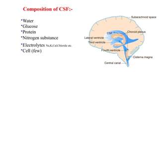
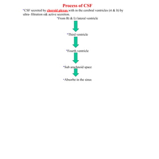
Process of CSF
•CSFsecreted by choroid plexus with in the cerebral ventricles (rt & lt) by
ultra- filtration o& active secretion.
•From Rt & Lt lateral ventricle
•Third ventricle
•Fourth ventricle
•Sub arachnoid space
•Absorbe in the sinus
22.
Function of CSF:-
1.Supportthe brain & spinal cord
2.Protect the brain & spinal cord
3. Maintain pressure around the structure
4. Keep brain & spinal cord moist
5. Conveys nutrition to brain & spinal cord
6. Remove waste product of brain & spinal
cord
23.
Space is superiorto dura matter.
Subdural space
Space between dura and arachnoid
mater.
Subarachnoid space
Space between arachnoid & pia
mater
Filled with CSF
Contains the blood vessels supplying brain.
Epidural space
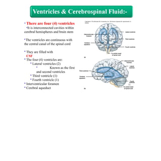
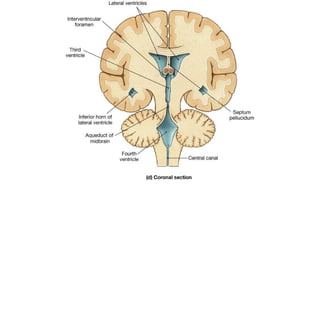
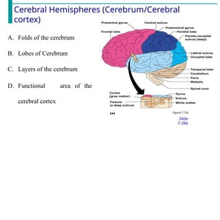
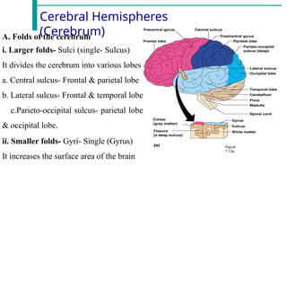
Cerebral Hemispheres
(Cerebrum)
Figure
7.13a
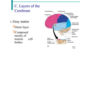
A. Foldsof the cerebrum
i. Larger folds- Sulci (single- Sulcus)
It divides the cerebrum into various lobes
a. Central sulcus- Frontal & parietal lobe
b. Lateral sulcus- Frontal & temporal lobe
c.Parieto-occipital sulcus- parietal lobe
& occipital lobe.
ii. Smaller folds- Gyri- Single (Gyrus)
It increases the surface area of the brain
28.
B. Lobes ofCerebrum
Sulcus divides the cerebrum into
lobes
Surface lobes of the cerebrum
Frontal lobe
Parietal lobe
Occipital lobe
Temporal lobe
29.
C. Layers ofthe
Cerebrum
i. Gray matter
Outer layer
Composed
mostly of
neuron cell
bodies
Figure
7.13a
30.
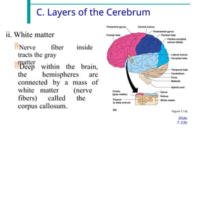
C. Layers ofthe Cerebrum
ii. White matter
Nerve fiber
tracts the gray
matter
inside
Deep within the brain,
the hemispheres are
connected by a mass of
white matter (nerve
fibers) called the
corpus callosum.
Figure 7.13a
Slide
7.33b
31.
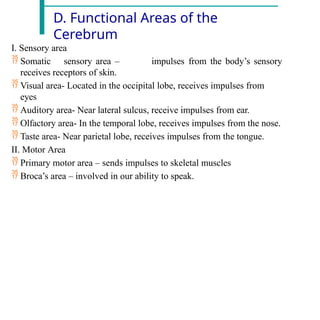
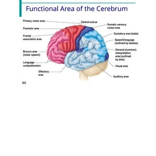
D. Functional Areasof the
Cerebrum
I. Sensory area
Somatic sensory area –
receives receptors of skin.
impulses from the body’s sensory
Visual area- Located in the occipital lobe, receives impulses from
eyes
Auditory area- Near lateral sulcus, receive impulses from ear.
Olfactory area- In the temporal lobe, receives impulses from the nose.
Taste area- Near parietal lobe, receives impulses from the tongue.
II. Motor Area
Primary motor area – sends impulses to skeletal muscles
Broca’s area – involved in our ability to speak.
32.
D. Functional Areasof the
Cerebrum
iii. Interpretation areas of the
cerebrum
Wernicke’s
Area-
Speech/language region- interprets the
meaning of speech by recognizing spoken words
The prefrontal cortex- concerned with the makeup of a person’s
personality, intellect, complex learning abilities, recall of
information, initiative, judgment, foresight, reasoning,
conscience, intuition, mood, planning for the future
General interpretation area- Receive impulses from all the
above areas
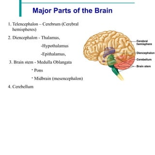
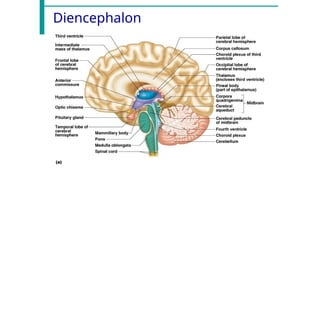
2.
Diencephalon
The diencephalon formsa central core of brain
tissue
completely surrounded by the cerebral hemispheres.
Enclosed by the cerebral
hemispheres
Made of three parts
Thalamus
Hypothalamus
Epithalamus
Thalamus
The thalamus makesup 80% of the diencephalon, consists of
paired oval masses of gray matter.
The relay station for sensory impulses that reaches the sensory
area of the cerebrum.
The thalamus is the major relay station for most sensory
impulses that reach the primary sensory areas of the cerebral
cortex from the spinal cord and brain stem
The thalamus also relays nerve impulses between different
areas of the cerebrum and plays a role in the maintenance of
consciousness
37.
Hypothalamus
small part ofthe diencephalon located inferior to
the thalamus
The hypothalamus controls many body activities and
is one of the major regulators of homeostasis.
38.
Functions of Hypothalamus
Control of the ANS. The hypothalamus controls and integrates
activities of the autonomic nervous system, which regulates the
contraction of smooth muscle and cardiac muscle and glands.
Production of hormones- releasing hormones and inhibiting
hormones that control anterior pituitary hormones and
synthesized oxytocin and antidiuretic hormones produced in
the paraventricular and supraoptic nuclei respectively.
Regulation
participates
of emotional
in expressions of
and
rage,
behavioral patterns-
aggression, pain
and
pleasure, and sexual arousal
39.
Functions of Hypothalamus
Regulationof eating and drinking.- the presence of feeding
center and Thirst center
Control of body temperature- senses body temperature from
the blood flowing through the hypothalamus.
Regulation of circadian rhythms and states of
consciousness- sleep-wake cycle) that occur on a circadian
schedule (cycle of about 24 hours).
40.
Epithalamus
a small regionsuperior and posterior to the
thalamus, consists of the pineal gland
The pineal gland is part of the endocrine system because
it secretes the hormone melatonin.
As more melatonin is liberated during darkness than
in light, this hormone is thought to promote sleepiness.
41.
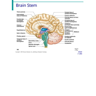
3. Brain Stem
Itis the part of the brain between the spinal cord and
the diencephalon.
Parts of the brain stem
Midbrain
Pons
Medulla oblongata
Midbrain
The midbrain ormesencephalon extends from the diencephalon to the pons.
The midbrain contains 2 imp nuclei,
Substantia nigra- large and darkly pigmented nuclei. Neurons, extending
from the substantia nigra to the basal nuclei release dopamine, which
helps control subconscious muscle activities. Loss of these neurons is
associated with Parkinson’s disease
Red nuclei- look reddish due to their rich blood supply and an iron-
containing pigment in their neuronal cell bodies. Axons from the
cerebellum and cerebral cortex form synapses in the red nuclei, which
help control muscular movements.
44.
Pons
The pons isa bridge that connects parts of the brain such
as the cerebrum, diencephalon, and cerebellum.
Various ascending sensory tracts and descending motor
tracts pass through the pons.
There are nuclei within the pons that act as relay
stations.
pneumotaxic and apnoustic centers (nuclei) in pons
operate in conjunction with the respiratory center in the
medulla oblongata to control respiration
45.
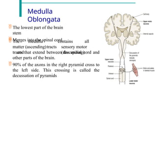
Medulla
Oblongata
The lowestpart of the brain
stem
Merges into the spinal cord
The medulla
matter (ascending)tracts
and
contains all
sensory motor
(descending)
tracts that extend between the spinal cord and
other parts of the brain.
90% of the axons in the right pyramid cross to
the left side. This crossing is called the
decussation of pyramids
46.
Medulla Oblongata-
Functions
The medullaalso contains several nuclei that act as control centers
The cardiovascular center- regulates the rate and force
of the heartbeat and the diameter of the blood vessel.
The medullary respiratory center- adjusts the basic
rhythm of breathing.
The vomiting center of the medulla causes vomiting.
The deglutition center of the medulla promotes deglutition
(swallowing) of a mass of food.
Sneezing and coughing center involves spasmodic contraction of
breathing muscles that forcefully expel air through the nose and
mouth.
47.
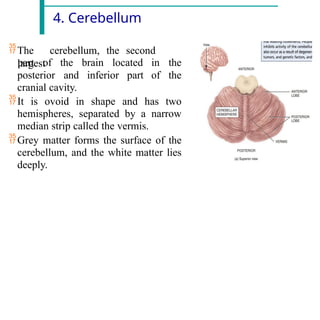
4. Cerebellum
The cerebellum,the second
largest
posterior
part of the brain located
and inferior part
in the
of the
cranial cavity.
It is ovoid in shape and has two
hemispheres, separated by a narrow
median strip called the vermis.
Grey matter forms the surface of the
cerebellum, and the white matter lies
deeply.
Cerebellum- Functions
Thecerebellum smooths and coordinates the complex sequence
of contractions of skeletal muscles that help in learning the
skilled muscular movements.
It coordinates activities associated with the maintenance of
posture, balance and equilibrium. The sensory input for these
functions is derived from the muscles and joints, the eyes and
the ears.
impulses from the eyes and the semicircular canals in the ears
provide information about the position of the head in space.
The cerebellum may also have a role in learning and language
processing.
50.
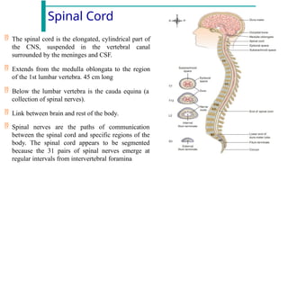
Spinal Cord
Thespinal cord is the elongated, cylindrical part of
the CNS, suspended in the vertebral canal
surrounded by the meninges and CSF.
Extends from the medulla oblongata to the region
of the 1st lumbar vertebra. 45 cm long
Below the lumbar vertebra is the cauda equina (a
collection of spinal nerves).
Link between brain and rest of the body.
Spinal nerves are the paths of communication
between the spinal cord and specific regions of the
body. The spinal cord appears to be segmented
because the 31 pairs of spinal nerves emerge at
regular intervals from intervertebral foramina
51.
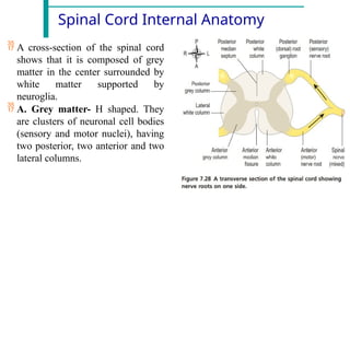
Spinal Cord InternalAnatomy
A cross-section of the spinal cord
shows that it is composed of grey
matter in the center surrounded by
white matter supported by
neuroglia.
A. Grey matter- H shaped. They
are clusters of neuronal cell bodies
(sensory and motor nuclei), having
two posterior, two anterior and two
lateral columns.
52.
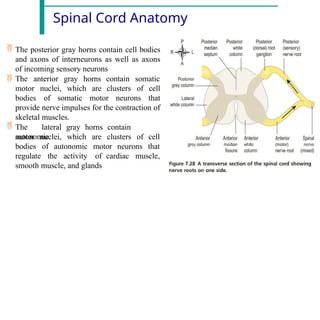
Spinal Cord Anatomy
The posterior gray horns contain cell bodies
and axons of interneurons as well as axons
of incoming sensory neurons
The anterior gray horns somatic
motor nuclei, which are
contain
clusters
bodies of somatic motor neurons
of cell
that
provide nerve impulses for the contraction of
skeletal muscles.
The lateral gray horns contain
autonomic
motor nuclei, which are clusters of
neurons
cell
that
bodies of autonomic motor
regulate the activity of cardiac muscle,
smooth muscle, and glands
53.
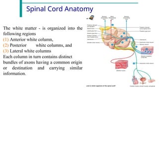
Spinal Cord Anatomy
Thewhite matter - is organized into the
following regions
(1) Anterior white column,
(2) Posterior white columns, and
(3) Lateral white columns
Each column in turn contains distinct
bundles of axons having a common origin
or destination and carrying similar
information.
54.
Spinal Cord- Physiology
Thespinal cord has two principal functions
1. Sensory and Motor Tracts- Nerve impulses from sensory receptors
propagate up the spinal cord to the brain and motor output from the
brain travels down the to body parts
2. Reflexes and Reflex Arcs- A reflex is a fast, involuntary, unplanned
sequence of actions that occurs in response to a particular stimulus.
Some reflexes are inborn, such as pulling your hand away from a hot
surface before you even feel that it is hot. Other reflexes are learned or
acquired.
55.
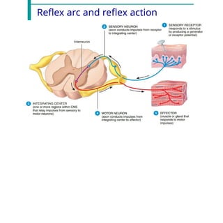
Reflex arc andreflex action
Nerve impulses propagating into, through, and out of the CNS follow specific pathways.
The pathway followed by nerve impulses that produce a reflex is a reflex arc. A reflex
arc includes the following five functional components.
1. Sensory receptor- present in sensory neurons of sense organs, It responds to a
specific stimulus.
2. Sensory neuron- The nerve impulses propagate from the sensory neuron to the
gray matter of the spinal cord.
3. Integrating center- a. Monosynaptic reflex- single synapse between a sensory
neuron and a motor neuron. b. Polysynaptic reflex- the integrating center consists of
one or more interneurons.
4. Motor neuron- Impulses triggered by the integrating center propagate out of the
spinal cord along a motor neuron to the part of the body
5. Effector- The part of the body that responds to the motor nerve impulse, such as a
muscle or gland, is the effector. Its action is called a reflex.
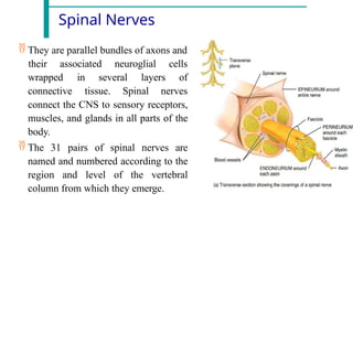
Spinal Nerves
Theyare parallel bundles of axons and
their associated neuroglial cells
of
wrapped in several layers
connective tissue. Spinal nerves
connect the CNS to sensory receptors,
muscles, and glands in all parts of the
body.
The 31 pairs of spinal nerves are
named and numbered according to the
region and level of the vertebral
column from which they emerge.
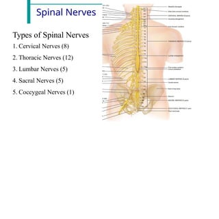
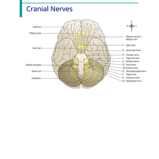
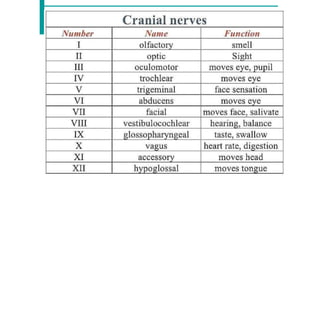
Cranial Nerves
The 12pairs of cranial nerves as they pass through the bones of the cranium
and arise from the brain inside the cranial cavity.
Types of cranial nerves-
1. sensory nerves- Three cranial nerves (I, II, and VIII) carry sensory
neurons and thus are called special sensory nerves.
2. motor nerves- Five cranial nerves (III, IV, VI, XI, and XII) are
classified as motor nerves because they contain only motor neurons as
they leave the brain stem.
3. The remaining four cranial nerves (V, VII, IX, and X) are mixed
nerves—they contain both sensory neurons entering the brain stem and
motor neurons leaving the brain stem.
References
1. Tortora, G.J., & Grabowski, S. R. (2012). 15th edition, Principles of
anatomy and physiology. New York.
2. Ross and Wilson Anatomy and Physiology in Health and
Illness International Edition, 13th Edition.
63.
There is asolution to all your problems/doubts, the
main question is how deep you can go.
View publication
stats