Downloaded 160 times


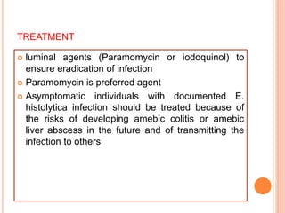


Amebiasis is caused by the intestinal parasite Entamoeba histolytica. It is endemic in areas with poor sanitation. Infection occurs by ingesting E. histolytica cysts in contaminated food or water. Most infections are asymptomatic, but some cause intestinal diseases like dysentery or liver abscesses. Symptoms of intestinal amebiasis include diarrhea and abdominal pain. Liver abscesses appear as round lesions containing anchovy paste-like material. Diagnosis involves detecting the parasite in stool or biopsy. Tinidazole or metronidazole are prescribed to treat intestinal or liver infections, with luminal agents added to clear the infection.


