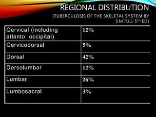
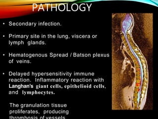
HISTORY
• Documented in5000-year-old mummies
“YAKSHMA”
Yakṣhma (यक्ष्म) in the Rigveda and the Atharvaveda
frequently denotes ‘illness’, in general, perhaps as
rendering the body emaciated
IN 1779, PERCIVAL
POT T
One fifth of TB
population is in India.
3%suffering from
skeletal TB.
Vertebral TB - form of
skeletal TB(50%).
Almost 50% are from
pediatric group.
2nd greatest killer next
to HIV.
In 2017, WHO released a report which revealed as many
as 4.23 lakh deaths from TB in India
19.36 lakh TB cases came into picture in India in 2016.
• Early diagnosis.
• Expeditious medical
treatment.
• Aggressive surgical
approach.
• Prevent deformity.
• Best outcome.
Cervical (including
atlanto occipital)
12%
Cervicodorsal 5%
Dorsal 42%
Dorsolumbar 12%
Lumbar 26%
Lumbosacral 3%
REGIONAL DISTRIBUTION
(TUBERCULOSIS OF THE SKELETAL SYSTEM BY
S.M.TULI 5TH ED)
• Secondary infection.
• Primary site in the lung, viscera or
lymph glands.
• Hematogenous Spread / Batson plexus
of veins.
• Delayed hypersensitivity immune
reaction. Inflammatory reaction with
Langhan’s giant cells, epithelioid cells,
and lymphocytes.
The granulation tissue
proliferates, producing
PATHOLOGY
PATHOLOGY
• Granulomatous
• inflammation leads to erosion
of vertebrae.
• Associated disc degeneration due to
end arteritis, finally complete
destruction.
• Weakening of trabeculae
compression collapse. –
Deformity.
•Formation of cold abscess
•Collect under ant-long-ligament
•Vertebral collapse
•Expression of collection of tuberculous debris
Diverted forward along
different anatomical sites
Slides along VB and invade
the
vertebral canal through
intervertebral foramen.
COLD ABSCESS
•Abscess- collection of liquefied
tissue in the body which is
body’s defense reaction to
foreign material
•NO signs of inflammation
•Collection of dead tubercular
bacilli, serum, leucocytes,
bone debris and caseous
material.
•Can track in to any direction-
along musculo-facial planes or
• Paraspinal regions at the back
• Anterior/ posterior cervical triangles
• Brachial plexus in the axilla
• Intercostal spaces on the chest wall
• Abscesses from dorsolumbar and lumbar
spine- track
down the psoas sheath.
• Palpable in the iliac fossa, lumbar triangle,
upper part of the thigh below inguinal ligament
or even track downwards upto the knee or
sometimes upto the great toe
• Iliac abscess contained
in the sheath of the iliac
muscle.
• The abscess that has
tracked down the psoas
sheath penetrates
through the iliacus
muscle sheath.
• Becomes palpable as a
mass in the iliac fossa
• Abscess that remains
confined to the psoas
sheath may not be
palpable clinically.
TYPES OF VERTEBRAL
INVOLVEMENT
1.PARADISCAL (arterial spread)
2.CENTRAL ( venous spread)
3.ANTERIOR (sub periosteal
tracking of pus)
4.APPENDICIAL
• Most common pattern of spinal
tuberculosis.
• Narrowing of the disc space.
• Destruction of subchondral bone.
• Subsequent herniation of the disc.
1. PARADISCAL LESIONS :
• Subperiosteal lesion under the ALL.
• The periosteal stripping renders the
vertebrae avascular and susceptible
to infection.
• Both pressure and ischemia combine to
produce anterior scalloping. (multiple
vertebrae)
• Collapse of the VB &diminution of the disc
space is minimal
2. ANTERIOR LESIONS :
• Centered on the vertebral body.
• Disc is not involved.
• Infection starts from the center of the
vertebral body.
Batson’s venous
plexus Posterior
vertebral artery
• Concentric collapse producing a
vertebra plana appearance.
3. CENTRAL LESIONS :
• Isolated infection involving
pedicles , laminae (neural
arch), transverse processes, &
spinous process.
• Uncommon lesion (< 5%).
• In conjunction with the
typical paradiscal variant
in 30%.
• Rarely present as synovitis of
facet joints.
4. APPENDICIAL LESIONS :
• Constitutional symptoms
• Pain in the back ( m/c )
• Swelling
• Stiffness
• Neurological symptoms
• Deformity
1. ATTITUDE AND GAIT
• In upper cervical disease – wry neck
• In upper thoracic disease – Military
attitude
• In lumbosacral – Alderman’s Gait
• In lower lumbar – Pronounced lordosis
2. DEFORMITY OF SPINE:
• Kyphosis
1 vertebrae – Knuckle
gibbus 2-3 vertebrae -
Angular gibbus
>3 vertebrae – Round
kyphosis
• Scoliosis
• Lordosis
3. ABSCESS / SINUS FORMATION :
• Dysphagia and dyspnoea – Retropharyngeal abscess
• Hoarseness of voice due to – Abscess in disease of
upper thoracic region.
• Flexion contracture of hip – Psoas abscess
4. MOVEMENT OF SPINE :
• painful due to protective muscular
spasm
5. Paralysis :
• Association - 10-30%
• Type - Incomplete generally
• More common in thoracic
region.
RADIOLOGICAL
DIAGNOSIS:
1. X-RAYS
2. CT SCAN
3. MRI SPINE
4. BONE
SCAN
1. X RAY FINDINGS :
• Early changes :-
haziness and local osteoporosis of end
plates of
two adjacent vertebrae
 narrowing of intervertebral disc space.
• Late changes :-
 paravertebral shadow
 ant wedge compression collapse - deformity
 central or concertina collapse
 destruction of post element
X ray changes appear after 3-5 months.
PARA VERTEBRAL
SHADOW- X RAY
1. Cervical
region
- Shadow in Retropharyngeal
space
- V-shaped
shadow
2. Upper
thoracic
mediastinu
m
- Change in contour of tracheal
shadow
3. Below 4th
thoracic
- Fusiform or bird nest
shadow
4. Below
D10
5.
Aneurysma
l
phenomen
- Bilateral widening of psoas
shadow
-tense thoracic vertebral abscess
showing scalloping effect
2. CT SCAN
3.
MRI
• Increased uptake (60% patients) with active
tuberculosis
• > 5mm lesion size can be detected.
• Avascular segments and abscesses show a cold
spot due to decreased uptake.
• Highly sensitive but nonspecific.
• Aid to localize the site of active disease and to detect
multilevel involvement
4. BONE SCAN (TECHNITIUM
(TC) – 99 M )
• Mantoux / Tuberculin skin test
• ESR may be markedly elevated (neither specific nor
reliable).
• ELISA : for antibody to mycobacterial
antigen-6 , sensitivity 94% and specificity
of 100%
• Polymerase chain reaction PCR : sensitivity 40% only.
LABORATORY TESTS
Biopsy : For definitive diagnosis
• CT or ultrasound guided or open biopsy during a
surgical procedure.
1. Ziehl-Neelsen staining: a quick and inexpensive
method
2. Culture : - results are available only after a few
weeks
- positive only in 60% of cases; most
specific.
3. Histology: demonstration of tubercle, 80% cases.
MICROBIOLOGY STUDIES:
• Most dreaded and crippling
complication.
• Incidence 10-30%.
• age group – first three decades of
life.
• region – thoracic.
1)To eradicate or at least arrest the
disease.
2) To treat major complications
like paraplegia.
3) To prevent or correct deformity.
TULI’S MIDDLE PATH REGIME FOR
TREATMENT OF KOCH’S SPINE
1) Bed rest - with or without traction
2) Drugs – ATTany one regime as preferred
3)Radiograph &ESR – radiologically the kyphosis
and disease activity by ESR is measured 3
monthly.
4)Gradual mobilization with exercise
5)Abscess:
* Repeated aspiration.
* Streptomycin and/or INH instillation.
6) Sinuses :
* Usually heal by 6-12 weeks of ATT.
* Excision of the tract with or without debridement.
7) Neurological complication :
5 indications for surgery (mainly decompression
surgery)
(I) Not showing progressive recovery after 3-6 weeks of
Rx.
(ii) Pt. developing neurological complication during Rx.
(iii) Neurological status becoming worse while
undergoing Rx.
(iv) Recurrence of neurological complication.
(v)In advanced cases with motor, sensory or
sphincter involvement or having severe flexor spasm
8) Operative debridement
- in nonresponsive 3-6m of chemotherapy.
- cases with recurrence of disease.
9) Excisional surgery:
- posterior spinal disease associated with
abscess / sinus formation +/- neural
involvement.
10) Posterior SpinalArthrodesis:
- severe kyphotic deformity
(prevention / correction).
- mechanical instability.
- spine at risk signs.
11)Post –operative:
- hard bed for 2-3 weeks/ neurological
recovery.
- brace for 2 years.
INDICATIONS- SURGICALTREATMENT
•Doubtful diagnosis.
•Failure to respond to conservative Rx after 3-
6 weeks therapy.
•Symptomatic abscess.
•Neurological indications.
•Mechanical instability.
•Deformity.
•Recurrence of disease.
•Posterior spinal disease.
•Spinal tumor syndrome.
TYPES OF SURGERY
•Nursed on a hard bed / POP posterior shell
(children) upto 3 months.
•Careful and assisted turning of the patient is
permitted from the first day.
•At the end of 3-6 months / neurological
recovery pt. mobilized with the help of spinal
brace.
•Spinal brace is discarded after 1- 1 ½ years.
•CERVICAL SPINE :
Four post cervical
brace
Minerva Jacket
SOMI Brace
•UPPER DORSAL SPINE D1-D3:
No simple brace to control the spine
effectively.
Only satisfactory method is to extend the
usual spinal brace upward with the
attachment of a cervical collar.
D4-L2
VERTEBRAE
- acceptable to young girl as
it gets accommodated
according to body
contour.
MILWAUKEE BRACE:
- growing age ; mainly used for
correction of scoliosis.
• Maintain high suspicion not to overlook diagnosis.
• Earlydiagnosis is essential for good results.
• EarlyMRIis anessential tool for diagnosis of Potts
spine.
• Not all patients can be treated bychemotherapy alone
and neither do all patients requiresurgery.
THANK YOU

Tb spine

  • 2.
  • 3.
    “YAKSHMA” Yakṣhma (यक्ष्म) inthe Rigveda and the Atharvaveda frequently denotes ‘illness’, in general, perhaps as rendering the body emaciated
  • 4.
  • 5.
    One fifth ofTB population is in India. 3%suffering from skeletal TB. Vertebral TB - form of skeletal TB(50%). Almost 50% are from pediatric group. 2nd greatest killer next to HIV.
  • 6.
    In 2017, WHOreleased a report which revealed as many as 4.23 lakh deaths from TB in India 19.36 lakh TB cases came into picture in India in 2016.
  • 7.
    • Early diagnosis. •Expeditious medical treatment. • Aggressive surgical approach. • Prevent deformity. • Best outcome.
  • 9.
    Cervical (including atlanto occipital) 12% Cervicodorsal5% Dorsal 42% Dorsolumbar 12% Lumbar 26% Lumbosacral 3% REGIONAL DISTRIBUTION (TUBERCULOSIS OF THE SKELETAL SYSTEM BY S.M.TULI 5TH ED)
  • 10.
    • Secondary infection. •Primary site in the lung, viscera or lymph glands. • Hematogenous Spread / Batson plexus of veins. • Delayed hypersensitivity immune reaction. Inflammatory reaction with Langhan’s giant cells, epithelioid cells, and lymphocytes. The granulation tissue proliferates, producing PATHOLOGY
  • 11.
    PATHOLOGY • Granulomatous • inflammationleads to erosion of vertebrae. • Associated disc degeneration due to end arteritis, finally complete destruction. • Weakening of trabeculae compression collapse. – Deformity.
  • 13.
    •Formation of coldabscess •Collect under ant-long-ligament •Vertebral collapse •Expression of collection of tuberculous debris Diverted forward along different anatomical sites Slides along VB and invade the vertebral canal through intervertebral foramen.
  • 14.
    COLD ABSCESS •Abscess- collectionof liquefied tissue in the body which is body’s defense reaction to foreign material •NO signs of inflammation •Collection of dead tubercular bacilli, serum, leucocytes, bone debris and caseous material. •Can track in to any direction- along musculo-facial planes or
  • 15.
    • Paraspinal regionsat the back • Anterior/ posterior cervical triangles • Brachial plexus in the axilla • Intercostal spaces on the chest wall • Abscesses from dorsolumbar and lumbar spine- track down the psoas sheath. • Palpable in the iliac fossa, lumbar triangle, upper part of the thigh below inguinal ligament or even track downwards upto the knee or sometimes upto the great toe
  • 16.
    • Iliac abscesscontained in the sheath of the iliac muscle. • The abscess that has tracked down the psoas sheath penetrates through the iliacus muscle sheath. • Becomes palpable as a mass in the iliac fossa • Abscess that remains confined to the psoas sheath may not be palpable clinically.
  • 17.
    TYPES OF VERTEBRAL INVOLVEMENT 1.PARADISCAL(arterial spread) 2.CENTRAL ( venous spread) 3.ANTERIOR (sub periosteal tracking of pus) 4.APPENDICIAL
  • 18.
    • Most commonpattern of spinal tuberculosis. • Narrowing of the disc space. • Destruction of subchondral bone. • Subsequent herniation of the disc. 1. PARADISCAL LESIONS :
  • 20.
    • Subperiosteal lesionunder the ALL. • The periosteal stripping renders the vertebrae avascular and susceptible to infection. • Both pressure and ischemia combine to produce anterior scalloping. (multiple vertebrae) • Collapse of the VB &diminution of the disc space is minimal 2. ANTERIOR LESIONS :
  • 22.
    • Centered onthe vertebral body. • Disc is not involved. • Infection starts from the center of the vertebral body. Batson’s venous plexus Posterior vertebral artery • Concentric collapse producing a vertebra plana appearance. 3. CENTRAL LESIONS :
  • 24.
    • Isolated infectioninvolving pedicles , laminae (neural arch), transverse processes, & spinous process. • Uncommon lesion (< 5%). • In conjunction with the typical paradiscal variant in 30%. • Rarely present as synovitis of facet joints. 4. APPENDICIAL LESIONS :
  • 26.
    • Constitutional symptoms •Pain in the back ( m/c ) • Swelling • Stiffness • Neurological symptoms • Deformity
  • 27.
    1. ATTITUDE ANDGAIT • In upper cervical disease – wry neck • In upper thoracic disease – Military attitude • In lumbosacral – Alderman’s Gait • In lower lumbar – Pronounced lordosis
  • 28.
    2. DEFORMITY OFSPINE: • Kyphosis 1 vertebrae – Knuckle gibbus 2-3 vertebrae - Angular gibbus >3 vertebrae – Round kyphosis • Scoliosis • Lordosis
  • 29.
    3. ABSCESS /SINUS FORMATION : • Dysphagia and dyspnoea – Retropharyngeal abscess • Hoarseness of voice due to – Abscess in disease of upper thoracic region. • Flexion contracture of hip – Psoas abscess
  • 30.
    4. MOVEMENT OFSPINE : • painful due to protective muscular spasm 5. Paralysis : • Association - 10-30% • Type - Incomplete generally • More common in thoracic region.
  • 31.
    RADIOLOGICAL DIAGNOSIS: 1. X-RAYS 2. CTSCAN 3. MRI SPINE 4. BONE SCAN
  • 32.
    1. X RAYFINDINGS : • Early changes :- haziness and local osteoporosis of end plates of two adjacent vertebrae  narrowing of intervertebral disc space. • Late changes :-  paravertebral shadow  ant wedge compression collapse - deformity  central or concertina collapse  destruction of post element X ray changes appear after 3-5 months.
  • 33.
    PARA VERTEBRAL SHADOW- XRAY 1. Cervical region - Shadow in Retropharyngeal space - V-shaped shadow 2. Upper thoracic mediastinu m - Change in contour of tracheal shadow 3. Below 4th thoracic - Fusiform or bird nest shadow 4. Below D10 5. Aneurysma l phenomen - Bilateral widening of psoas shadow -tense thoracic vertebral abscess showing scalloping effect
  • 35.
  • 36.
  • 38.
    • Increased uptake(60% patients) with active tuberculosis • > 5mm lesion size can be detected. • Avascular segments and abscesses show a cold spot due to decreased uptake. • Highly sensitive but nonspecific. • Aid to localize the site of active disease and to detect multilevel involvement 4. BONE SCAN (TECHNITIUM (TC) – 99 M )
  • 41.
    • Mantoux /Tuberculin skin test • ESR may be markedly elevated (neither specific nor reliable). • ELISA : for antibody to mycobacterial antigen-6 , sensitivity 94% and specificity of 100% • Polymerase chain reaction PCR : sensitivity 40% only. LABORATORY TESTS
  • 42.
    Biopsy : Fordefinitive diagnosis • CT or ultrasound guided or open biopsy during a surgical procedure. 1. Ziehl-Neelsen staining: a quick and inexpensive method 2. Culture : - results are available only after a few weeks - positive only in 60% of cases; most specific. 3. Histology: demonstration of tubercle, 80% cases. MICROBIOLOGY STUDIES:
  • 43.
    • Most dreadedand crippling complication. • Incidence 10-30%. • age group – first three decades of life. • region – thoracic.
  • 44.
    1)To eradicate orat least arrest the disease. 2) To treat major complications like paraplegia. 3) To prevent or correct deformity.
  • 45.
    TULI’S MIDDLE PATHREGIME FOR TREATMENT OF KOCH’S SPINE 1) Bed rest - with or without traction 2) Drugs – ATTany one regime as preferred 3)Radiograph &ESR – radiologically the kyphosis and disease activity by ESR is measured 3 monthly. 4)Gradual mobilization with exercise 5)Abscess: * Repeated aspiration. * Streptomycin and/or INH instillation.
  • 46.
    6) Sinuses : *Usually heal by 6-12 weeks of ATT. * Excision of the tract with or without debridement. 7) Neurological complication : 5 indications for surgery (mainly decompression surgery) (I) Not showing progressive recovery after 3-6 weeks of Rx. (ii) Pt. developing neurological complication during Rx. (iii) Neurological status becoming worse while undergoing Rx. (iv) Recurrence of neurological complication. (v)In advanced cases with motor, sensory or sphincter involvement or having severe flexor spasm 8) Operative debridement - in nonresponsive 3-6m of chemotherapy. - cases with recurrence of disease.
  • 47.
    9) Excisional surgery: -posterior spinal disease associated with abscess / sinus formation +/- neural involvement. 10) Posterior SpinalArthrodesis: - severe kyphotic deformity (prevention / correction). - mechanical instability. - spine at risk signs. 11)Post –operative: - hard bed for 2-3 weeks/ neurological recovery. - brace for 2 years.
  • 48.
    INDICATIONS- SURGICALTREATMENT •Doubtful diagnosis. •Failureto respond to conservative Rx after 3- 6 weeks therapy. •Symptomatic abscess. •Neurological indications. •Mechanical instability. •Deformity. •Recurrence of disease. •Posterior spinal disease. •Spinal tumor syndrome.
  • 49.
  • 50.
    •Nursed on ahard bed / POP posterior shell (children) upto 3 months. •Careful and assisted turning of the patient is permitted from the first day. •At the end of 3-6 months / neurological recovery pt. mobilized with the help of spinal brace. •Spinal brace is discarded after 1- 1 ½ years.
  • 51.
    •CERVICAL SPINE : Fourpost cervical brace Minerva Jacket SOMI Brace
  • 52.
    •UPPER DORSAL SPINED1-D3: No simple brace to control the spine effectively. Only satisfactory method is to extend the usual spinal brace upward with the attachment of a cervical collar.
  • 53.
    D4-L2 VERTEBRAE - acceptable toyoung girl as it gets accommodated according to body contour. MILWAUKEE BRACE: - growing age ; mainly used for correction of scoliosis.
  • 55.
    • Maintain highsuspicion not to overlook diagnosis. • Earlydiagnosis is essential for good results. • EarlyMRIis anessential tool for diagnosis of Potts spine. • Not all patients can be treated bychemotherapy alone and neither do all patients requiresurgery.
  • 56.