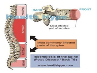
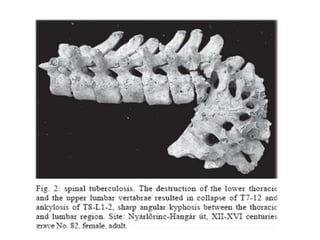
INTERNATIONAL SCHOOL OF
MEDICINE
PROJECT
TUBERCULOSIS OF VERTEBRA AND
BONES
GAURAV LAKHANPAL
3RD GROUP
INTRODUCTION
• The usual sites to be involved are the lower thoracic and
upper lumbar vertebrae.
• The source of infection is usually outside the spine. It is
most often spread from the lungs via the blood.
• There is a combination of osteomyelitis and infective
arthritis.
• Usually more than one vertebra is involved. The area most
affected is the anterior part of the vertebral body adjacent
to the subchondral plate. Tuberculosis may spread from
that area to adjacent intervertebral discs.
• vertebral narrowing and eventually vertebral collapse and
spinal damage. A dry soft tissue mass often forms and
superinfection is rare
SYMPTOMS
• The onset is gradual.
• Back pain is localised.
• Fever, night sweats, anorexia and weight loss.
• Signs may include kyphosis (common) and/or a
paravertebral swelling.
• Affected patients tend to assume a protective
upright, stiff position.
• If there is neural involvement there will be
neurological signs.
A) parasagittal T2 weighted short inversion recovery images
(B) show the inflammatory granulation or abscesses in Th12, L3, L4, and L5
COMPLICATION
– The spinal canal can be narrowed by abscesses, granulation tissue, or
direct dural invasion. This leads to spinal cord compression and
neurological signs (Pott's paralysis).
– Kyphosis occurs because of collapse in the anterior spine and can be
severe.
– Lesions in the thoracic spine have a greater risk of kyphosis than those
in the lumbar spine.
– Neurological problems can be prevented by early diagnosis and
prompt treatment. It can reverse paralysis and minimise disability.
– A combination of conservative management and surgical
decompression gives success in most patients.
– Late-onset paraplegia is best avoided by prevention of the
development of severe kyphosis.
– Patients with tuberculosis of the spine who are likely to have severe
kyphosis should have surgery in the active stage of disease
TEST AND DIAGNOSIS
• Elevated ESR.
• Strongly positive Mantoux skin test.
• Spinal X-ray may be normal in early disease, as 50% of the bone mass
must be lost for changes to be visible on X-ray. Plain X-ray can show
vertebral destruction and narrowed disc space.
• MRI scanning may demonstrate the extent of spinal compression and can
show changes at an early stage. Bone elements visible within the swelling.
• CT scans and nuclear bone scans can also be used but MRI is best to assess
risk to the spinal cord.
• A needle biopsy of bone or synovial tissue is usual. If it shows tubercle
bacilli this is diagnostic but usually culture is required.
SURGERY
• In patients with spinal tuberculosis, anterior spinal fusion should be
considered if there is spinal instability or evidence of spinal cord
compression.
• chemotherapy

shaharukh ahamd

  • 1.
    INTERNATIONAL SCHOOL OF MEDICINE PROJECT TUBERCULOSISOF VERTEBRA AND BONES GAURAV LAKHANPAL 3RD GROUP
  • 2.
    INTRODUCTION • The usualsites to be involved are the lower thoracic and upper lumbar vertebrae. • The source of infection is usually outside the spine. It is most often spread from the lungs via the blood. • There is a combination of osteomyelitis and infective arthritis. • Usually more than one vertebra is involved. The area most affected is the anterior part of the vertebral body adjacent to the subchondral plate. Tuberculosis may spread from that area to adjacent intervertebral discs. • vertebral narrowing and eventually vertebral collapse and spinal damage. A dry soft tissue mass often forms and superinfection is rare
  • 4.
    SYMPTOMS • The onsetis gradual. • Back pain is localised. • Fever, night sweats, anorexia and weight loss. • Signs may include kyphosis (common) and/or a paravertebral swelling. • Affected patients tend to assume a protective upright, stiff position. • If there is neural involvement there will be neurological signs.
  • 7.
    A) parasagittal T2weighted short inversion recovery images (B) show the inflammatory granulation or abscesses in Th12, L3, L4, and L5
  • 8.
    COMPLICATION – The spinalcanal can be narrowed by abscesses, granulation tissue, or direct dural invasion. This leads to spinal cord compression and neurological signs (Pott's paralysis). – Kyphosis occurs because of collapse in the anterior spine and can be severe. – Lesions in the thoracic spine have a greater risk of kyphosis than those in the lumbar spine. – Neurological problems can be prevented by early diagnosis and prompt treatment. It can reverse paralysis and minimise disability. – A combination of conservative management and surgical decompression gives success in most patients. – Late-onset paraplegia is best avoided by prevention of the development of severe kyphosis. – Patients with tuberculosis of the spine who are likely to have severe kyphosis should have surgery in the active stage of disease
  • 9.
    TEST AND DIAGNOSIS •Elevated ESR. • Strongly positive Mantoux skin test. • Spinal X-ray may be normal in early disease, as 50% of the bone mass must be lost for changes to be visible on X-ray. Plain X-ray can show vertebral destruction and narrowed disc space. • MRI scanning may demonstrate the extent of spinal compression and can show changes at an early stage. Bone elements visible within the swelling. • CT scans and nuclear bone scans can also be used but MRI is best to assess risk to the spinal cord. • A needle biopsy of bone or synovial tissue is usual. If it shows tubercle bacilli this is diagnostic but usually culture is required. SURGERY • In patients with spinal tuberculosis, anterior spinal fusion should be considered if there is spinal instability or evidence of spinal cord compression. • chemotherapy