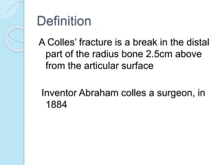
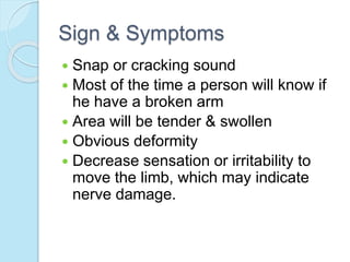
Mahashin akond presented on Colles' fracture, which is a break in the distal part of the radius bone 2.5cm above the articular surface. Colles' fractures most commonly occur in young adults and those over 40 years old from falls or direct blows. Treatment involves reduction, immobilization with a plaster cast or surgery, followed by physiotherapy to regain range of motion, strength, and function. Complications can include malunion, tendon rupture, osteoporosis, and delayed healing, though most fractures heal within 6-8 weeks with proper treatment and rehabilitation.
Colles fracture is the fracture at the distal end of radius, at its
cortico cancellous junction(about 2cm from the distal articular
surface).
It is not just the fracture of distal radius but the fracture
dislocation of the inferior radio-ulnar joint.
ANKLE FRACTURES
Pott’s fracture
A Pott’s fracture is a type of ankle fracture that is characterized by a break in one or more bony prominences on the sides of the ankle known as the malleoli.
Also known as Broken Ankle, Ankle Fracture and malleolar fracture.
Pott’s fracture often occurs in combination with other injuries such as a sprained ankle or other fractures of the foot, ankle or lower leg.
This is a brief presentation of most common fracture around the wrist i.e Colles fracture. This presentation gives a brief information on anatomy, mechanism of injury, causes and treatment methods of colles fracture.
My seminar presentation slides. Please correct me if I've did something wrong in it. You can also leave any message thru my email, nurhanisahzainoren@gmail.com
ELBOW JOINT PATHOLOGY AND REHABILITATION 1.pptxSrishti Mahadik
Elbow joint pathomechanics and rehabilitation in physiotherapy explained in detailed manner.Each and every point from reference books mentiuoned above is included.
In this i have covered the different sports injuries of upper extremities, their causes and their orthotic management.
Helpful for those, who are in the field of P & O.
Hip and Thigh injuries in sports such as- Perthes Disease, Osteitis Pubis, Avascular Necrosis of The Femoral Head, Hip Pointer, Classic Groin Strain, ‘Pull’ Or Adductor Tendinopathy, Slipped Capital Femoral Epiphysis, Trochanteric Bursitis/Gluteus Medius Tendinopathy, Iliopsoas strain, Quadriceps strain, Irritable Hip etc.
Tendonitis details and it's physiotherapy management.
It is define as inflammation of the tendon, tendonitis occur due to overuse and trauma. Depending upon involvement of tendon special test are used. it is treated with PRICE protocol.
SPORTS INJURIES OF ANKLE AND FOOT original.pptxMeghaPrakash9
sports injuries of ankle and foot is a seminar done by Ms. Megha ck on behalf of completing her master of physiotherapy program during the year 2020-2022
4. Definition
A Colles’ fracture is a break in the distal
part of the radius bone 2.5cm above
from the articular surface
Inventor Abraham colles a surgeon, in
1884
5. Incidence
Not more common fracture
But Frequently occur in young adult
Basically those who are over 40 years.
7. Rick Factors
Factors which increase the chance of
fractures are
1.Osteoporosis
2.Poor nutrition
3.Condition that increase risk of falling
Snow ,Ice, Loss of agility or muscle
strength
4.Advancing age
5.Post menopause
6.Decrease muscle mass.
9. Associated injury
Tendon:Rupture of extensor policis
longus and peritendinious adhesion of
both the flexor and extensor
compartments may occur.
Nerves:Nerve injuries include median
nerve contusion ,resulting in the
development of carpal tunnel
syndrom.
10. Types
Open fracture
Close fracture
Intra articular fracture
Extra articular fracture
Comminuted fracture.
11. Sign & Symptoms
Snap or cracking sound
Most of the time a person will know if
he have a broken arm
Area will be tender & swollen
Obvious deformity
Decrease sensation or irritability to
move the limb, which may indicate
nerve damage.
12. Deformity
Volar angulations of the apex of the
fracture
The deformity is called “Silver fork
deformity ”or “Dinner fork deformity”
17. Exercise programme
During reduction plaster cast
-Uninvolved joint active rang of
motion
-Wrist joint passive supination &
pronation
-Isometric hand muscle exercise
18. continue
After removal the cast
-Mobilized the affected wrist
-Start some strengthening
exercise
-Start weight bearing exercise
Later stage Advance exercise ,full
weight bearing exercise
20. Prognosis
Expected time for healing 6th to 8th
weeks until the fracture is stable.
Earlier treatment usually improves the
result
Chronic disease such as osteoporosis
and diabetes may slow the healing
time.