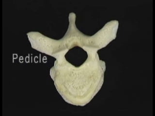
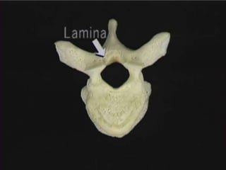
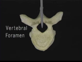
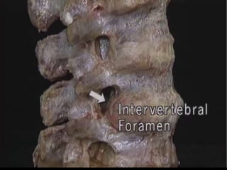
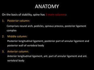
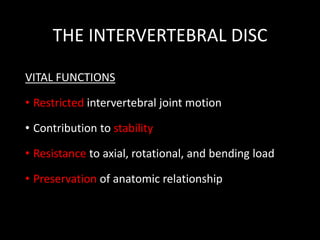
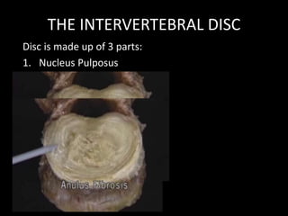
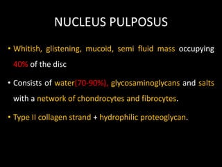
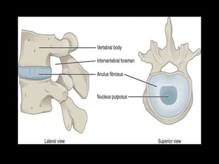
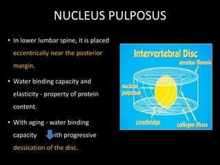
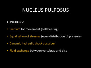
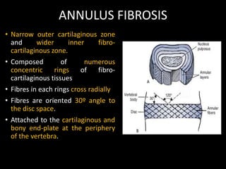
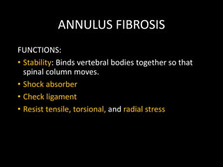
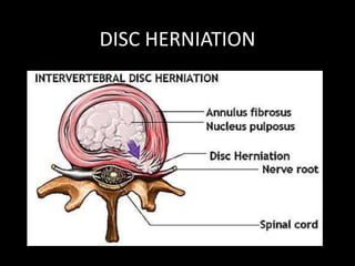
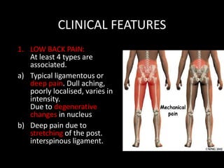
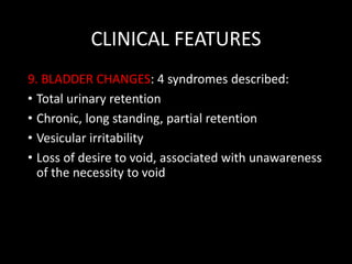
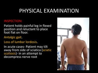
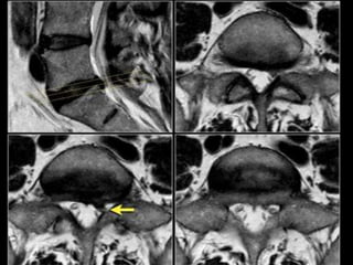
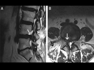
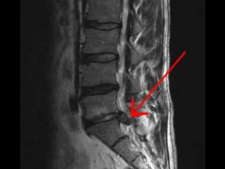
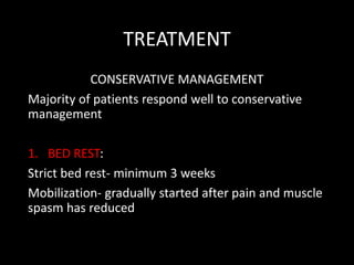
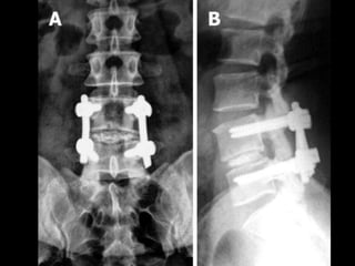
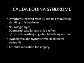
Bicipital tendonitis is inflammation of long head of the biceps tendon under the bicipital groove.
In early stage, tendon becomes red and swollen, as tendonitis develops the tendon sheath can thicken.
In late stage, often become dark red in color due to inflammation.
Lumbar spinal canal stenosis is one of the difficult topic of spine. All the information are taken from Campbell's operative orthopedics Thirteen edition and from internet. I also took help from the lectures of renowned orthopedics professors of Bangladesh.
A fractured neck of femur (NOF) is a serious injury, especially in older people. It is likely to be life changing and for some people life threatening.
Neck of femur fractures (NOF) are common injuries sustained by older patients who are both more likely to have unsteadiness of gait and reduced bone mineral density, predisposing to fracture. Elderly osteoporotic women are at greatest risk.
ANKLE FRACTURES
Pott’s fracture
A Pott’s fracture is a type of ankle fracture that is characterized by a break in one or more bony prominences on the sides of the ankle known as the malleoli.
Also known as Broken Ankle, Ankle Fracture and malleolar fracture.
Pott’s fracture often occurs in combination with other injuries such as a sprained ankle or other fractures of the foot, ankle or lower leg.
As part of a class presentation, we attempted to make this to briefly explain what Torticollis meas, the Types of presentation of Torticollis, and Management strategies for a Physiotherapist for Congenital Torticollis especially.
I hope this helps. :)
The pictures and information had been taken from internet, complied to make a brief presentation for the purpose of class presentation.
I do not own any content.
Introduction:-
Hoffa's pad syndrome also known as Infrapatellar fat syndrome is an intrinsic disease of Hoffa's fat pad and a problem of knee joint which causes the pain at the front of knee joint so this pain is known as anterior knee pain.
Hoffa's fat pad contains pluripotent cells that can differentiate into osteoblasts and chondrocytes.
Hoffa's pad becomes inflamed or Damaged by The crushing of the pad between the femur and tibia during extension, causes inflammation of Hoffa's fat pad.
Nerve supply of fat pad is excellent( It receives branches of the femoral , common peroneal and saphenous nerves) so if it occurs any injury ,causes a sharp anterior knee pain.
The infrapatellar fat pad is a pad of adipose tissue underneath or deep to the patella tendon and the top of the fat pad attaches to the lower part of patella (knee cap).
hoffa's pad is a shock absorber ,when there is a direct force on the patella can result in pinching of the fat pad between femur and tibial plateau. The tibial plateau is the proximal tibial surface on which the femur rests.
HFP is surrounded anteriorly by the patellar tendon and the joint capsule, superiorly by the inferior pole of the patella, inferiorly by the proximal tibia and the deep infrapatellar bursa, and posteriorly by the joint synovium .
The main function of the HFP is to reduce friction between the patella, the patellar tendon, and the deep skeletal structures. In addition, it prevents pinching of the synovial membrane and it facilitates the vascularization of adjacent structures.
Causes:-
cause is usually due to single or repetitive traumatic episodes.
when you extend your knee the fat pad act as a cushion and reduces friction between outer patella facets and quadriceps tendons .
when you flex your knee ,upper part of fat pad becomes tensioned, it moves backwards in the knee.
it develops gradually over time if you repeatedly move your knee.
This is when your knee is forced to move forward from its completely straight normal position.
You may have always been able to over straighten your knee, which is called knee hyperextension or genu recurvatum .
hyperextension sports such as basketball, volleyball or high jumping may also cause inflammation of Hoffa's pad.
Hoffa's disease is more frequent in young women and the symptoms are anterior knee pain when upstairs and downstairs.
Sign and Symptoms:-
Complaints of anterior knee pain occurs when playing hyperextension sports such as basketball ,volleyball or high jumping .effusion and inflammation may be occurs and decreases the ROM of joint , stair negotiation .
Symptoms may worsen if the knee is overly straightened or bent for too long a period. Complications may include an inability to fully straighten the knee.
Diagnosis:-
Hoffa's syndrome completely diagnosed by MRI .we have requirement of an experienced orthopaedics to diagnose it.
primary Assessment have to check the Active and Passive Range Of Motion(AROM/PROM) of Hip joint and Knee joint.
Recomendações da OMS sobre cuidados maternos e neonatais para uma experiência pós-natal positiva.
Em consonância com os ODS – Objetivos do Desenvolvimento Sustentável e a Estratégia Global para a Saúde das Mulheres, Crianças e Adolescentes, e aplicando uma abordagem baseada nos direitos humanos, os esforços de cuidados pós-natais devem expandir-se para além da cobertura e da simples sobrevivência, de modo a incluir cuidados de qualidade.
Estas diretrizes visam melhorar a qualidade dos cuidados pós-natais essenciais e de rotina prestados às mulheres e aos recém-nascidos, com o objetivo final de melhorar a saúde e o bem-estar materno e neonatal.
Uma “experiência pós-natal positiva” é um resultado importante para todas as mulheres que dão à luz e para os seus recém-nascidos, estabelecendo as bases para a melhoria da saúde e do bem-estar a curto e longo prazo. Uma experiência pós-natal positiva é definida como aquela em que as mulheres, pessoas que gestam, os recém-nascidos, os casais, os pais, os cuidadores e as famílias recebem informação consistente, garantia e apoio de profissionais de saúde motivados; e onde um sistema de saúde flexível e com recursos reconheça as necessidades das mulheres e dos bebês e respeite o seu contexto cultural.
Estas diretrizes consolidadas apresentam algumas recomendações novas e já bem fundamentadas sobre cuidados pós-natais de rotina para mulheres e neonatos que recebem cuidados no pós-parto em unidades de saúde ou na comunidade, independentemente dos recursos disponíveis.
É fornecido um conjunto abrangente de recomendações para cuidados durante o período puerperal, com ênfase nos cuidados essenciais que todas as mulheres e recém-nascidos devem receber, e com a devida atenção à qualidade dos cuidados; isto é, a entrega e a experiência do cuidado recebido. Estas diretrizes atualizam e ampliam as recomendações da OMS de 2014 sobre cuidados pós-natais da mãe e do recém-nascido e complementam as atuais diretrizes da OMS sobre a gestão de complicações pós-natais.
O estabelecimento da amamentação e o manejo das principais intercorrências é contemplada.
Recomendamos muito.
Vamos discutir essas recomendações no nosso curso de pós-graduação em Aleitamento no Instituto Ciclos.
Esta publicação só está disponível em inglês até o momento.
Prof. Marcus Renato de Carvalho
www.agostodourado.com
Ozempic: Preoperative Management of Patients on GLP-1 Receptor Agonists Saeid Safari
Preoperative Management of Patients on GLP-1 Receptor Agonists like Ozempic and Semiglutide
ASA GUIDELINE
NYSORA Guideline
2 Case Reports of Gastric Ultrasound
Tom Selleck Health: A Comprehensive Look at the Iconic Actor’s Wellness Journeygreendigital
Tom Selleck, an enduring figure in Hollywood. has captivated audiences for decades with his rugged charm, iconic moustache. and memorable roles in television and film. From his breakout role as Thomas Magnum in Magnum P.I. to his current portrayal of Frank Reagan in Blue Bloods. Selleck's career has spanned over 50 years. But beyond his professional achievements. fans have often been curious about Tom Selleck Health. especially as he has aged in the public eye.
Follow us on: Pinterest
Introduction
Many have been interested in Tom Selleck health. not only because of his enduring presence on screen but also because of the challenges. and lifestyle choices he has faced and made over the years. This article delves into the various aspects of Tom Selleck health. exploring his fitness regimen, diet, mental health. and the challenges he has encountered as he ages. We'll look at how he maintains his well-being. the health issues he has faced, and his approach to ageing .
Early Life and Career
Childhood and Athletic Beginnings
Tom Selleck was born on January 29, 1945, in Detroit, Michigan, and grew up in Sherman Oaks, California. From an early age, he was involved in sports, particularly basketball. which played a significant role in his physical development. His athletic pursuits continued into college. where he attended the University of Southern California (USC) on a basketball scholarship. This early involvement in sports laid a strong foundation for his physical health and disciplined lifestyle.
Transition to Acting
Selleck's transition from an athlete to an actor came with its physical demands. His first significant role in "Magnum P.I." required him to perform various stunts and maintain a fit appearance. This role, which he played from 1980 to 1988. necessitated a rigorous fitness routine to meet the show's demands. setting the stage for his long-term commitment to health and wellness.
Fitness Regimen
Workout Routine
Tom Selleck health and fitness regimen has evolved. adapting to his changing roles and age. During his "Magnum, P.I." days. Selleck's workouts were intense and focused on building and maintaining muscle mass. His routine included weightlifting, cardiovascular exercises. and specific training for the stunts he performed on the show.
Selleck adjusted his fitness routine as he aged to suit his body's needs. Today, his workouts focus on maintaining flexibility, strength, and cardiovascular health. He incorporates low-impact exercises such as swimming, walking, and light weightlifting. This balanced approach helps him stay fit without putting undue strain on his joints and muscles.
Importance of Flexibility and Mobility
In recent years, Selleck has emphasized the importance of flexibility and mobility in his fitness regimen. Understanding the natural decline in muscle mass and joint flexibility with age. he includes stretching and yoga in his routine. These practices help prevent injuries, improve posture, and maintain mobilit
Lung Cancer: Artificial Intelligence, Synergetics, Complex System Analysis, S...Oleg Kshivets
RESULTS: Overall life span (LS) was 2252.1±1742.5 days and cumulative 5-year survival (5YS) reached 73.2%, 10 years – 64.8%, 20 years – 42.5%. 513 LCP lived more than 5 years (LS=3124.6±1525.6 days), 148 LCP – more than 10 years (LS=5054.4±1504.1 days).199 LCP died because of LC (LS=562.7±374.5 days). 5YS of LCP after bi/lobectomies was significantly superior in comparison with LCP after pneumonectomies (78.1% vs.63.7%, P=0.00001 by log-rank test). AT significantly improved 5YS (66.3% vs. 34.8%) (P=0.00000 by log-rank test) only for LCP with N1-2. Cox modeling displayed that 5YS of LCP significantly depended on: phase transition (PT) early-invasive LC in terms of synergetics, PT N0—N12, cell ratio factors (ratio between cancer cells- CC and blood cells subpopulations), G1-3, histology, glucose, AT, blood cell circuit, prothrombin index, heparin tolerance, recalcification time (P=0.000-0.038). Neural networks, genetic algorithm selection and bootstrap simulation revealed relationships between 5YS and PT early-invasive LC (rank=1), PT N0—N12 (rank=2), thrombocytes/CC (3), erythrocytes/CC (4), eosinophils/CC (5), healthy cells/CC (6), lymphocytes/CC (7), segmented neutrophils/CC (8), stick neutrophils/CC (9), monocytes/CC (10); leucocytes/CC (11). Correct prediction of 5YS was 100% by neural networks computing (area under ROC curve=1.0; error=0.0).
CONCLUSIONS: 5YS of LCP after radical procedures significantly depended on: 1) PT early-invasive cancer; 2) PT N0--N12; 3) cell ratio factors; 4) blood cell circuit; 5) biochemical factors; 6) hemostasis system; 7) AT; 8) LC characteristics; 9) LC cell dynamics; 10) surgery type: lobectomy/pneumonectomy; 11) anthropometric data. Optimal diagnosis and treatment strategies for LC are: 1) screening and early detection of LC; 2) availability of experienced thoracic surgeons because of complexity of radical procedures; 3) aggressive en block surgery and adequate lymph node dissection for completeness; 4) precise prediction; 5) adjuvant chemoimmunoradiotherapy for LCP with unfavorable prognosis.
TEST BANK for Operations Management, 14th Edition by William J. Stevenson, Ve...kevinkariuki227
TEST BANK for Operations Management, 14th Edition by William J. Stevenson, Verified Chapters 1 - 19, Complete Newest Version.pdf
Title: Sense of Taste
Presenter: Dr. Faiza, Assistant Professor of Physiology
Qualifications:
MBBS (Best Graduate, AIMC Lahore)
FCPS Physiology
ICMT, CHPE, DHPE (STMU)
MPH (GC University, Faisalabad)
MBA (Virtual University of Pakistan)
Learning Objectives:
Describe the structure and function of taste buds.
Describe the relationship between the taste threshold and taste index of common substances.
Explain the chemical basis and signal transduction of taste perception for each type of primary taste sensation.
Recognize different abnormalities of taste perception and their causes.
Key Topics:
Significance of Taste Sensation:
Differentiation between pleasant and harmful food
Influence on behavior
Selection of food based on metabolic needs
Receptors of Taste:
Taste buds on the tongue
Influence of sense of smell, texture of food, and pain stimulation (e.g., by pepper)
Primary and Secondary Taste Sensations:
Primary taste sensations: Sweet, Sour, Salty, Bitter, Umami
Chemical basis and signal transduction mechanisms for each taste
Taste Threshold and Index:
Taste threshold values for Sweet (sucrose), Salty (NaCl), Sour (HCl), and Bitter (Quinine)
Taste index relationship: Inversely proportional to taste threshold
Taste Blindness:
Inability to taste certain substances, particularly thiourea compounds
Example: Phenylthiocarbamide
Structure and Function of Taste Buds:
Composition: Epithelial cells, Sustentacular/Supporting cells, Taste cells, Basal cells
Features: Taste pores, Taste hairs/microvilli, and Taste nerve fibers
Location of Taste Buds:
Found in papillae of the tongue (Fungiform, Circumvallate, Foliate)
Also present on the palate, tonsillar pillars, epiglottis, and proximal esophagus
Mechanism of Taste Stimulation:
Interaction of taste substances with receptors on microvilli
Signal transduction pathways for Umami, Sweet, Bitter, Sour, and Salty tastes
Taste Sensitivity and Adaptation:
Decrease in sensitivity with age
Rapid adaptation of taste sensation
Role of Saliva in Taste:
Dissolution of tastants to reach receptors
Washing away the stimulus
Taste Preferences and Aversions:
Mechanisms behind taste preference and aversion
Influence of receptors and neural pathways
Impact of Sensory Nerve Damage:
Degeneration of taste buds if the sensory nerve fiber is cut
Abnormalities of Taste Detection:
Conditions: Ageusia, Hypogeusia, Dysgeusia (parageusia)
Causes: Nerve damage, neurological disorders, infections, poor oral hygiene, adverse drug effects, deficiencies, aging, tobacco use, altered neurotransmitter levels
Neurotransmitters and Taste Threshold:
Effects of serotonin (5-HT) and norepinephrine (NE) on taste sensitivity
Supertasters:
25% of the population with heightened sensitivity to taste, especially bitterness
Increased number of fungiform papillae
- Video recording of this lecture in English language: https://youtu.be/lK81BzxMqdo
- Video recording of this lecture in Arabic language: https://youtu.be/Ve4P0COk9OI
- Link to download the book free: https://nephrotube.blogspot.com/p/nephrotube-nephrology-books.html
- Link to NephroTube website: www.NephroTube.com
- Link to NephroTube social media accounts: https://nephrotube.blogspot.com/p/join-nephrotube-on-social-media.html
Title: Sense of Smell
Presenter: Dr. Faiza, Assistant Professor of Physiology
Qualifications:
MBBS (Best Graduate, AIMC Lahore)
FCPS Physiology
ICMT, CHPE, DHPE (STMU)
MPH (GC University, Faisalabad)
MBA (Virtual University of Pakistan)
Learning Objectives:
Describe the primary categories of smells and the concept of odor blindness.
Explain the structure and location of the olfactory membrane and mucosa, including the types and roles of cells involved in olfaction.
Describe the pathway and mechanisms of olfactory signal transmission from the olfactory receptors to the brain.
Illustrate the biochemical cascade triggered by odorant binding to olfactory receptors, including the role of G-proteins and second messengers in generating an action potential.
Identify different types of olfactory disorders such as anosmia, hyposmia, hyperosmia, and dysosmia, including their potential causes.
Key Topics:
Olfactory Genes:
3% of the human genome accounts for olfactory genes.
400 genes for odorant receptors.
Olfactory Membrane:
Located in the superior part of the nasal cavity.
Medially: Folds downward along the superior septum.
Laterally: Folds over the superior turbinate and upper surface of the middle turbinate.
Total surface area: 5-10 square centimeters.
Olfactory Mucosa:
Olfactory Cells: Bipolar nerve cells derived from the CNS (100 million), with 4-25 olfactory cilia per cell.
Sustentacular Cells: Produce mucus and maintain ionic and molecular environment.
Basal Cells: Replace worn-out olfactory cells with an average lifespan of 1-2 months.
Bowman’s Gland: Secretes mucus.
Stimulation of Olfactory Cells:
Odorant dissolves in mucus and attaches to receptors on olfactory cilia.
Involves a cascade effect through G-proteins and second messengers, leading to depolarization and action potential generation in the olfactory nerve.
Quality of a Good Odorant:
Small (3-20 Carbon atoms), volatile, water-soluble, and lipid-soluble.
Facilitated by odorant-binding proteins in mucus.
Membrane Potential and Action Potential:
Resting membrane potential: -55mV.
Action potential frequency in the olfactory nerve increases with odorant strength.
Adaptation Towards the Sense of Smell:
Rapid adaptation within the first second, with further slow adaptation.
Psychological adaptation greater than receptor adaptation, involving feedback inhibition from the central nervous system.
Primary Sensations of Smell:
Camphoraceous, Musky, Floral, Pepperminty, Ethereal, Pungent, Putrid.
Odor Detection Threshold:
Examples: Hydrogen sulfide (0.0005 ppm), Methyl-mercaptan (0.002 ppm).
Some toxic substances are odorless at lethal concentrations.
Characteristics of Smell:
Odor blindness for single substances due to lack of appropriate receptor protein.
Behavioral and emotional influences of smell.
Transmission of Olfactory Signals:
From olfactory cells to glomeruli in the olfactory bulb, involving lateral inhibition.
Primitive, less old, and new olfactory systems with different path
micro teaching on communication m.sc nursing.pdfAnurag Sharma
Microteaching is a unique model of practice teaching. It is a viable instrument for the. desired change in the teaching behavior or the behavior potential which, in specified types of real. classroom situations, tends to facilitate the achievement of specified types of objectives.
Acute scrotum is a general term referring to an emergency condition affecting the contents or the wall of the scrotum.
There are a number of conditions that present acutely, predominantly with pain and/or swelling
A careful and detailed history and examination, and in some cases, investigations allow differentiation between these diagnoses. A prompt diagnosis is essential as the patient may require urgent surgical intervention
Testicular torsion refers to twisting of the spermatic cord, causing ischaemia of the testicle.
Testicular torsion results from inadequate fixation of the testis to the tunica vaginalis producing ischemia from reduced arterial inflow and venous outflow obstruction.
The prevalence of testicular torsion in adult patients hospitalized with acute scrotal pain is approximately 25 to 50 percent
NVBDCP.pptx Nation vector borne disease control programSapna Thakur
NVBDCP was launched in 2003-2004 . Vector-Borne Disease: Disease that results from an infection transmitted to humans and other animals by blood-feeding arthropods, such as mosquitoes, ticks, and fleas. Examples of vector-borne diseases include Dengue fever, West Nile Virus, Lyme disease, and malaria.
1. PROLPASED INTER-
VERTEBRAL DISC (PIVD)
Presenter: Dr. Souradeep Mitra
Junior Resident
Dept. Of Orthopaedics
SDUMC, Kolar
Moderator: Dr. Arun H.S.
Professor and HOD
Dept. Of Orthopaedics
SDUMC, Kolar
2. DEFINITION
• Herniation OR Prolapse - Rupture or protrusion of all or part
of an organ through a tear or abnormal opening in the wall of
the containing cavity
• Outpouching of the disc nucleus pulposus through an annulus
tear - “prolapsed”, “ruptured”, “herniated” or “slipped” disc
3. HISTORY
• AURELIAMIS (5th century) - Described symptoms of sciatica
• ANDREAS VESALIUS (1543) - Described intervertebral disc
• VIRCHOW (1857) - Described acute traumatic rupture of intervertebral
disc.
• FORST (1811) - Described Lasegue sign
• OPPENHEIM (1909) - Performed 1st successful surgical excision of
herniated disc
• KAMBIN AND GELLMAN (1983) - Percutaneous approach for lumbar
discectomy
4. ANATOMY
Central axis of human skeleton is formed by the
vertebral column. This has
Vertebrae
Intervertebral disc
Spinal cord in its cavity
15. ANATOMY
On the basis of stability, spine has 3 main columns:
1. Posterior column:
Comprises neural arch, pedicles, spinous process, posterior ligament
complex
2. Middle column:
Posterior longitudinal ligament, posterior part of annular ligament and
posterior wall of vertebral body
3. Anterior column:
Anterior longitudinal ligament, ant. part of annular ligament and ant.
vertebral body
16.
17. THE INTERVERTEBRAL DISC
• 23 discs exist in spine- absent at atalanto-axial
articulation
• Thinnest- Thoracic region
Thickest- Lumbar region
• Interposed between bodies of vertebrae
• Body- Covered superiorly and inferiorly by an end
plate.
• Ant. longitudinal ligament- Present anteriorly and
laterally and intimately blends with the disc
18.
19. THE INTERVERTEBRAL DISC
• Makes up 33% of lumbar vertebral height. (24% of
cervical and 20% of thoracic)
• Thicker anteriorly in the cervical and lumbar
regions - contributing to anterior convexity.
20. THE INTERVERTEBRAL DISC
VITAL FUNCTIONS
• Restricted intervertebral joint motion
• Contribution to stability
• Resistance to axial, rotational, and bending load
• Preservation of anatomic relationship
22. NUCLEUS PULPOSUS
• Whitish, glistening, mucoid, semi fluid mass occupying
40% of the disc
• Consists of water(70-90%), glycosaminoglycans and salts
with a network of chondrocytes and fibrocytes.
• Type II collagen strand + hydrophilic proteoglycan.
23.
24. NUCLEUS PULPOSUS
• In lower lumbar spine, it is placed
eccentrically near the posterior
margin.
• Water binding capacity and
elasticity - property of protein
content.
• With aging - water binding
capacity with progressive
dessication of the disc.
25. NUCLEUS PULPOSUS
FUNCTIONS:
• Fulcrum for movement (ball bearing)
• Equalization of stresses (even distribution of pressure)
• Dynamic hydraulic shock absorber
• Fluid exchange between vertebrae and disc
26. ANNULUS FIBROSIS
• Narrow outer cartilaginous zone
and wider inner fibro-
cartilaginous zone.
• Composed of numerous
concentric rings of fibro-
cartilaginous tissues
• Fibres in each rings cross radially
• Fibres are oriented 30º angle to
the disc space.
• Attached to the cartilaginous and
bony end-plate at the periphery
of the vertebra.
27. ANNULUS FIBROSIS
FUNCTIONS:
• Stability: Binds vertebral bodies together so that
spinal column moves.
• Shock absorber
• Check ligament
• Resist tensile, torsional, and radial stress
28. BLOOD SUPPLY
• In adults, it is AVASCULAR.
• Sustain by diffusion of nutrients into disc through
porous central concavity of end plate.
• Diffusion is maintained by- Motion and weight
bearing.
29. NERVE SUPPLY
• Sinu-vertebral nerve
(recurrent branch of
vertebral nerve)
• Supply periosteum, post.
longitudinal ligament and
OUTERMOST layers of
annulus fibrosus.
• Nucleus pulposus and
innermost layers of
annulus fibrosus have NO
nerve supply
30. DISTRIBUTION OF LOAD
A. In the normal, healthy
disc, the nucleus
distributes the load
equally throughout the
annulus.
B. As the disc undergoes
degeneration, the nucleus
loses some of its
cushioning ability and
transmits the load
unequally to the annulus.
31. DISTRIBUTION OF LOAD
C. In the severely
degenerated disc, the
nucleus has lost all
of its ability to
cushion the load,
which can lead to disc
herniation
32. DISC HERNIATION
• Due to trauma, lifting injuries or idiopathic, a tear
in the outer, fibrous ring (annulus fibrosus) of an
intervertebral disc allows the soft, central portion
(nucleus pulposus) to bulge out beyond the
damaged outer rings.
• Tear in the disc ring may result in the release of
inflammatory chemical mediators which may
directly cause severe pain, even in the absence of
nerve root compression
33. BIOMECHANICS
• Supine patient weighing 70kg has a load of 20kg on
his L3 spine.
• This increases to
100kg on standing with 20kg in his hand and to
270kg when sitting and leaning forward with 20kg
weight in his hands.
• Young adults- IVD are strong and it’s impossible to
damage a healthy disc, except by forcible flexion.
34. BIOMECHANICS
• After 2nd decade- degenerative changes may result
in:
Necrosis, sequestration of nucleus pulposus.
Softening and weakening of the annulus fibrosus.
• Comparatively minor strains may cause either
internal derangement with eccentric displacement
of nucleus OR external derangement
35. BIOMECHANICS
• In internal derangement- Unequal tension in the
joint causes muscle spasm and sudden violent pain.
• In external derangement- Nucleus pulposus
herniates through the annulus fibrosus (usually
posterolaterally) into the verterbral canal, where it
produces pressure on the nerve root.
• MOST COMMON- L4-L5, L5-S1, L3-L4
36. BIOMECHANICS
• A large central rupture may produce pressure on
cauda equina
• Intra-discal pressures, myoelectric activity and
intra-abdominal pressure measurements have
shown that distance between the weight and body
influences stress on the back.
• Disc pressures and myoelectric activity are highest
in anterior unsupported sitting and lowest when
sitting straight
37.
38. NATURAL HISTORY OF DISC
DISEASE
• Recurrent strains produce small, circumferential
tears in the annulus fibrosus.
• Later, they enlarge and coalesce to form radial tears
that run from annulus to nucleus pulposus.
• Still later, these tears increase further in size until
disc is completely disrupted internally.
39. NATURAL HISTORY OF DISC
DISEASE
• The normal disc height is
reduced because of loss of
proteoglycans and water from
nucleus.
• Annulus become lax and bulges
around circumference of disc.
• This bulge must be
distinguished from disc
herniation, which is a “local
protrusion”
• With further loss of disc
content, disc space is
represented by a “thin slit”
filled with fibrous tissue.
40. NATURAL HISTORY OF DISC
DISEASE
• Vertebral body on either side
of the disc is dense and
sclerotic- called ‘disc
resorption’
• Finally disc is anchored by
peripheral osteophytes that
pass around its
circumference.
• End result is bony ankylosis
41. STAGES OF DISC HERNIATION
1. DISC DEGENERATION:
Chemical changes
associated with aging
causes discs to weaken,
without herniation.
2. PROTRUSION:
When nuclear material
causes bulging of
outermost annular fibres
(prolapsed disc)
42. STAGES OF DISC HERNIATION
3. EXTRUSION:
IVD rupture occurs when
nuclear material escapes
through annular fibres but
still remains connected
within the disc.
4. SEQUESTRATION:
Nucleus pulposus breaks
through the annulus
fibrosus and lies outside
the disc in the spinal canal.
43. VARIETIES OF DISC HERNIATION
1. POSTEROLATERAL DISC
HERNIATION – Protrusion is
usually posterolateral into
vertebral canal and compress
the roots of a spinal nerve.
Protruded disc usually
compresses the lower nerve
as that nerve crosses level of
disc in its path to its foramen.
(eg. protrusion of L5 disc
usually affects S1 instead)
44.
45. VARIETIES OF DISC HERNIATION
2. CENTRAL (POSTERIOR) HERNIATION:
When nuclear material is extruded through
central portion of annulus but contained by post.
longitudinal ligament.
Complete rupture- Rare
May rupture when spine is subjected to violent
flexion forces.
Nerve roots not commonly involved, but post.
ligament may be stretched causing severe back
pain without radicular pain
46. VARIETIES OF DISC HERNIATION
3. LATERAL DISC HERNIATION:
May compress the nerve root
above the level of the
herniation
L4 nerve root is most often
involved & patients typically
have intense radicular pain.
47. FREQUENCY
TWO MOST COMMON FORMS
• Lumbar disc herniation
• Cervical disc herniation
Most disc herniations occur in the age group of 30-40
years (when nucleus is still gelatin like)
With age nucleus dries out and risk of herniation is
greatly reduced.
At 50-60, spondylosis or spinal stenosis are more likely
causes of low back pain/leg pain
48. FREQUENCY
• Lumbar disc herniation:
15 times more common than cervical disc herniation.
LEVEL PERCENTAGE
L5-S1 40
L4-L5 49
L3-L4 7.5
L2-L3 3
L1-L2 0.5
49. FREQUENCY
• Cervical disc herniation:
Most often between C5-C6 and C6-C7.
Symptoms can affect the back of the skull, neck,
shoulder girdle, scapula, shoulder, arm and hand
50. CAUSES
• Repetitive mechanical activities:
Frequent bending, twisting, lifting, and other similar
activities without breaks and proper stretching can leave
the discs damaged.
• Living a sedentary lifestyle:
Individuals who rarely if ever engage in physical activity
are more prone to herniated discs because the muscles
that support the back and neck weaken, which increases
strain on the spine.
• Traumatic injury to lumbar discs commonly occurs when
lifting while bent at the waist, rather than lifting with the
legs while the back is straight.
51. CAUSES
• Obesity:
Spinal degeneration can be quickened as a result of the
burden of supporting excess body fat.
• Practicing poor posture:
Improper spinal alignment while sitting, standing, or
lying down strains the back and neck.
• Tobacco abuse:
The chemicals commonly found in cigarettes can
interfere with the disc’s ability to absorb nutrients,
which results in the weakening of the disc.
52. CLINICAL FEATURES
1. LOW BACK PAIN:
At least 4 types are
associated.
a) Typical ligamentous or
deep pain. Dull aching,
poorly localised, varies in
intensity.
Due to degenerative
changes in nucleus
b) Deep pain due to
stretching of the post.
interspinous ligament.
53. CLINICAL FEATURES
c) Direct irritation of nerve root by posterior
protrusion produces pain referred to
cutaneous distribution of affected root.
Patient is suddenly seized with an acute and
agonising pain in lumbar region due to locking of
joint by a nuclear sequesterum.
Relief of symptoms- when there is shrinkage
of sequesterum
d) Last stages of disc lesion, is when affected
joint is undergoing arthritic changes, it
becomes a source of pain associated with degenerative
arthritis
54. CLINICAL FEATURES
• REFERRED PAIN:
Begins in lower back and is referred to SI region and
buttocks or posterior thigh.
Can be elicited from many areas of spine- Facet
joints, longitudinal ligaments and periosteum
These are mesodermal structures which when
irritated give rise to referred pain.
55. CLINICAL FEATURES
• RADICULAR PAIN:
Extends below knee and follows
dermatome of involved nerve root.
Pressure on root produces pain and
motor and/or sensory signs.
• SCIATICA:
Pathognomic of disc herniation.
Pain- Gradual or sudden in onset.
Gnawing or burning- continuously
present or occurs paroxysmally.
Worse at night and while sitting.
Pain starts in back and spreads
downwards.
Generally worse at he back of thigh and
hip.
56. CLINICAL FEATURES
2. MUSCLE SPASM:
Reflex spasm of the erector spinae muscle- to protect
and immobilize affected joint.
3. ABNORMALITIES OF POSTURE:
Flattening of lumbar lordosis- due to joint
derangement.
57. CLINICAL FEATURES
4. DISORDERS OF MOVEMENT:
IVDP produces limitation and pain on movement of
lumbar spine. Forward flexion and extension- more
affected.
5. LOCAL TENDERNESS:
Deep tenderness present- about 2 inches from
midline at level of lesion.
58. CLINICAL FEATURES
6. MOTOR CHANGES:
Muscle weakness and wasting occurs with prolonged and
severe root irritation by increased tension.
7. REFLEX CHANGES:
There is associated diminution or complete loss of reflex
tendon jerks in the affected root.
Alteration of these reflexes is an early and reliable sign of
root involvement.
59. CLINICAL FEATURES
REFLEX CHANGES (contd)
Once reflex has been lost- recovery is slow even if
nerve root is completely freed by operating.
Classically, ankle jerk- absent, knee jerk- present with
herniation of lower lumbar discs.
60. CLINICAL FEATURES
8. SENSORY CHANGES:
Involvement of nerve root associated with sensory
changes in skin area which it supplies.
Tingling/ pins and needles/ blunting/ loss of
sensation.
Due to overlap of dermatomes- difficult to identify
specific root involved.
Sensory finding below ankle- more reliable.
Loss of proprioception
61.
62.
63. CLINICAL FEATURES
9. BLADDER CHANGES: 4 syndromes described:
• Total urinary retention
• Chronic, long standing, partial retention
• Vesicular irritability
• Loss of desire to void, associated with unawareness
of the necessity to void
64. PHYSICAL EXAMINATION
INSPECTION:
Patient holds painful leg in flexed
position and reluctant to place
foot flat on floor.
Antalgic gait.
Loss of lumbar lordosis.
In acute cases- Patient may tilt
away from side of sciatica (sciatic
scoliosis)- in an attempt to
decompress nerve root
65. PHYSICAL EXAMINATION
PALPATION:
Tenderness on palpation- at level of symptomatic
degenerative disc.
Paraspinal muscle spasm +
Patients with radiculopathy have tender motor
points corresponding to probable segmental level of
nerve root.
67. PHYSICAL EXAMINATION
SCIATIC TENSION SIGNS:
1. Valsalva manoeuvre:
Coughing, sneezing and
straining may produce sudden
increase in intra discal
pressure and thus- stretch
pain
68. PHYSICAL EXAMINATION
2. Straight leg raising test:
Positive in 90% cases
Patient lies supine with head flat or on a pillow
Examiner stabilizes pelvis and with other hand, slowly
elevates leg by the heel with knee straight.
Patient is questioned if this produces leg pain.
Leg pain or radicular symptoms produced- positive
69. PHYSICAL EXAMINATION
• If pain produced below 40˚-impingement of
protruding disc on a nerve root.
• If pain produced above 40˚-tension on nerve root
(not necessarily from disc prolapse)
• At the angle when patient experiences pain,
dorsiflex the ankle- causes aggravation of pain due
to sciatic nerve stretching- LASSEUGE SIGN
70. PHYSICAL EXAMINATION
3. Contralateral SLRT
Performed in same manner as SLRT
Except- non painful leg is raised
If sciatica produced in opposite limb- positive
Suggestive of herniated disc.
71. PHYSICAL EXAMINATION
4. Bowstring sign
Most reliable test of root tension- MACNAB
SLRT is performed as usual till pain is elicited.
At this point, knee is flexed- symptoms will reduce.
Finger pressure applied in popliteal space over
terminal end of sciatic nerve- Pain reappears.
72. PHYSICAL EXAMINATION
5. Figure of ‘4’ test (FABER):
Patient is supine.
Flex, abduct and externally rotate the lower limb on
affected side
Flex the knee and rest the limb on the opposite
thigh.
Give jerky pressure over medial aspect of knee.
Pain occurs at sciatic notch and along sciatic nerve.
73. PHYSICAL EXAMINATION
6. Femoral nerve stretch test:
Seen in cases of disc prolapse at higher levels. (when
roots of femoral nerve involved)
Reverse SLRT
Patient placed prone and knee is flexed and hip
extended.
Pain produced over anterior thigh.
74. PHYSICAL EXAMINATION
7. Cross-over test:
Determinant of compression of lumbosacral roots.
Raise the affected leg and this produces symptoms
down asymptomatic contralateral extremity- positive
Indicates- large central disc protrusion.
75. INVESTIGATIONS
PLAIN RADIOGRAPHY
1. AP VIEW (LS Spine)
• Vertical alignment of spinous process
• Intervertebral disc space uniformity
2. LATERAL VIEW (LS spine)
• Vertebral body
• Facet joint
• Lordotic curvature
• Intervertebral foramen
• Disc space height shortening
• Spndylolisthesis
77. INVESTIGATIONS
MYELOGRAPHY
Currently outdated
Water soluble contrast compounds- 3 to 5ml of solution
injected into sub-arachnoid space followed by Xray.
Typical appearance:
1. Lateral indentation and deformation of contrast
column by posterolateral disc
2. Hourglass deformity from midline herniation
3. Root pouch filling defects
4. Complete or partial blocks at the level of disc
78. INVESTIGATIONS
CT SCAN
Lumbar disc herniation found to be- Focal, asymmetric and
dorsolateral
ADVANTAGE:
Ability to see beyond dural sac and root sleeves and size of
spinal canal
LIMITATION:
Cannot differentiate between-
• Scar tissue and disc herniation
• Annulus and nucleus
79. INVESTIGATIONS
MRI
• Clearly superior in the detection of disc
degeneration.
• Allow evaluation of complete spinal group
• Shows:
• Intervertebral disc protrusion.
• Compression of nerve root
80.
81.
82.
83.
84. TREATMENT
CONSERVATIVE MANAGEMENT
Majority of patients respond well to conservative
management
1. BED REST:
Strict bed rest- minimum 3 weeks
Mobilization- gradually started after pain and muscle
spasm has reduced
85. TREATMENT
Lying in SEMI-FOWLER position
OR
On the side with both hips and knee flexed with a
pillow between the legs- relieves pressure on disc
Pelvic or skin traction may also be used
86. TREATMENT
2. DRUG THERAPY:
Bed rest supplemented by- NSAIDS, Muscle relaxants,
night sedation
3. PHYSIOTHERAPY:
• Patients with acute back pain eased by passive
extension of spine can benefit with extension exercises.
• Should not be forced in extreme pain.
• Education should be given regarding posture and
biomechanics.
87. TREATMENT
• Some patients respond to-
TENS
Skin/pelvic traction
Back braces/corsets
Ultrasound and diathermy
88. TREATMENT
4. EPIDURAL STEROID:
Provides long pain relief without excessive narcotic
intake.
Usually used is-
Methylprednisolone (80-120mg)/ Triamcinolone (40mg)
WITH
2% Xylocaine mixed with NS to make 10cc
Acts by reducing inflammation around affected nerve
root.
89. TREATMENT
INDICATIONS:
• Painful SLRT
• Patients with neurological deficit
• Patient with acute on chronic symptoms, with different level of disc
pathology
CONTRAINDICATIONS:
• Infection
• Haemorrhagic and bleeding diasthases
• Neurological diseases
COMPLICATIONS:
• Bacterial meningitis
• Transient hypotension
• Headache
90. TREATMENT
5. CHEMONUCLEOSIS
Enzymatic dissolution of disc.
Various substances used are- chymopapain,
collagenase, apoprotein, chondroitinase.
Limited to lumbar disc preferably at one level.
Confirmed by MRI/CT
91. TREATMENT
SURGICAL MANAGEMENT
INDICATIONS:
• Paraplegia or acute bladder paralysis due to cauda
equina- an absolute indication.
• Neurological impairment- which is progressing
• Failure of conservative management
• Recurrent sciatica
92. TREATMENT
Options available are:
1. Posterior approach
-Standard laminectomy and discectomy
-Fenestration operation
-Microsurgical laminotomy with disc
fragment excision.
2. Anterior approach with or interbody fusion
3. Percutaneous approach- suction, laser or
arthroscopic discectomy
95. TREATMENT
SPINAL FUSION
INDICATIONS:
• Young patients with signs of spondylolisthesis
• Post laminectomy who show instability
• Advanced intervertebral arthritis
• Need to return back to heavy manual work.
101. TREATMENT
4. PERCUTANEOUS LASER
DISECTOMY:
A thin cannula is inserted
through the back
Laser catheter is then inserted
Pulses of laser light will shrink
the disc wall.
Because no muscles or bone is
cut, recovery is faster
102. TREATMENT
5. LUMBAR ARTIFICIAL DISC
REPLACEMENT
These replace entire disc unit
(annulus and nucleus).
Less invasive- Only nucleus
pulposus is replaced
Implant is designed to bear
load through spine and
prevent collapse.
103. TREATMENT
6. INTRADISCAL
ELECTROTHERMIC THERAPY
(IDET)
Advanced procedure by
electrothermal catheters that
allow for careful and accurate
temperature control.
The procedure works by
cauterizing the nerve endings
within the disc wall to help block
the pain signals
Minimally invasive outpatient
surgical procedure
105. CAUDA EQUINA SYNDROME
• Seen post-operatively after lumbar
disc excision OR in a large
midline/higher disc herniation.
CLINICAL FEATURES:
• Difficulty in walking for a specified
distance associated with pins and
needles.
• Difficulty with urination, frequency
or overflow incontinence
• Paraesthesia in gluteal region,
perineum and urethra
• Loss of anal reflex
106. CAUDA EQUINA SYNDROME
• Symptoms relieved after 30 sec to 3 minutes by
standing or lying down.
• Neurologic signs:
Depressed patellar and ankle reflex.
B/L muscle wasting in glutei, hamstring and calf.
• Hypoalgesia and hypoesthesia in all sacral
segments.
• Absolute indication for surgery.