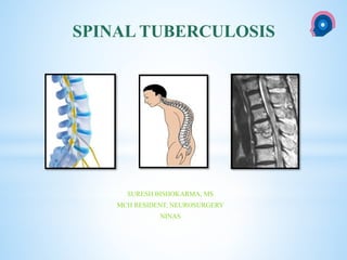
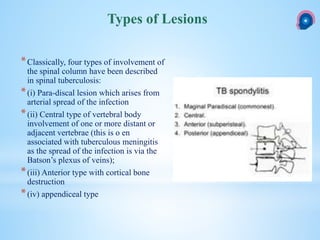
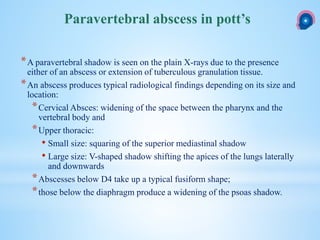
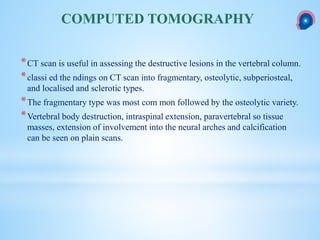
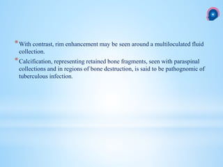
This document provides information on spinal tuberculosis, including its history, types of lesions, clinical presentation, imaging findings, treatment, and indications for surgery. It discusses how spinal tuberculosis is usually secondary to a primary infection elsewhere in the body that spreads hematogenously to the spine. The most common type of spinal lesion is a paradiscal lesion that begins in the vertebral body. Clinical presentation varies from asymptomatic to paraplegia. Imaging like CT and MRI are useful to identify bone destruction and abscesses. Treatment involves anti-tuberculosis medications for 18 months along with rest. Surgery is indicated for neurological deterioration, advanced disease, or diagnostic uncertainty.
Colles fracture is the fracture at the distal end of radius, at its
cortico cancellous junction(about 2cm from the distal articular
surface).
It is not just the fracture of distal radius but the fracture
dislocation of the inferior radio-ulnar joint.
Colles fracture is the fracture at the distal end of radius, at its
cortico cancellous junction(about 2cm from the distal articular
surface).
It is not just the fracture of distal radius but the fracture
dislocation of the inferior radio-ulnar joint.
Cervical radiculopathy is the clinical description of when a nerve root in the cervical spine becomes inflamed or damaged, resulting in a change in neurological function. Neurological deficits, such as numbness, altered reflexes, or weakness, may radiate anywhere from the neck into the shoulder, arm, hand, or fingers. Pins-and-needles tingling and/or pain, which can range from achy to shock-like or burning, may also radiate down into the arm and/or hand.
Water dynamic of UBE Unilateral Biportal Endoscopy.pptxsuresh Bishokarma
Unilateral Biportal Endoscopy (UBE) is a fluid medium surgery. Continuous saline output is critical
Hydrostatic pressure. Managing the fluid is the key to successful surgery. It use the principle of Bernauli’s and Pascal law. Explore the water dynamic of UBE surgery.
Posterior lumbar fusion vs Lumbar interbody fusion Evidence based.pptxsuresh Bishokarma
Lumbar degenerative disc diseases (LDDD): irreversible process in lumbar disk architecture.
Sparse literature to choose proper technique to address these pathology with or without fusion surgery.
A clear benefit of lumbar fusion surgery: lowered pain and disability scores.
Lumbar surgery rates have increased steadily over time, and hence related complications.
Evidence of the superiority of one technique over the other is sparse.
Surgery offers greater improvement compared with non-operative treatment in LDDD.
Surgery in disc herniation resulted in faster recovery, However no added benefit of fusion surgery.
There was no obvious disadvantage of posterolateral fusion without internal fixation in patient with spondylosis.
Among patients with lumbar spinal stenosis without spondylolisthesis, decompression plus fusion surgery may not result in better clinical outcomes.
In patient with spondylolisthesis with or without stenosis, fusion is more effective than laminectomy in achieving a satisfactory outcome. Decompression only had the least satisfactory outcome.
Patients who underwent interbody fusion may have significantly higher fusion rates compared to posterior lumbar fusion only.
TLIF has advantages over PLIF in the complication rate, blood loss, and operation duration. The clinical outcome is similar, with a slightly lower postoperative ODI score for TLIF.
In the end, The choice of technique is still greatly based on the surgeons’ preference and experience.
Brain abscess may have hematogenous spread: Pneumococcus common or via Contiguous spread. Risk factors includes pulmonary abscess or AV fistulas, congenital cyanotic heart disease, immunocompromised, chronic sinusitis/otitis, dental procedures. Intraventricular rupture of abscess is life threatening. Timely diagnosis and treatment is the goal.
Pituitary tumor accounts for ~10% ICT. They are common in 3-4 decade and shows association with MEN I.
About 5% of PT are invasive usually with giant tumor (>4cm). Tumor can be classified as functional (hormone secreting) or non functional. This slides details the algorithmic approach in management of pituitary tumors.
Medulloblastoma- A primitive neuroectodermal tumors (PNETs) is the most common malignant brain tumor of childhood (WHO IV)
arising from the vermis in the inferior medullary velum.
It comprises up to 18% of all pediatric brain tumors.
WNT and Shh pathway plays major role in its pathogenesis.
c-erbB-2 (HER2/neu) oncogene expression has prognostic value. Norcantharidin, Vismodegib, Sonidegib are the future in medulloblastoma.
Pineal gland is essentially an extra axial midline structure lying at the roof of dienchephalon rostral to the quadrigeminal cistern surrounded by important neurovascular structure, occurring in the geometric center of brain with same depth of trajectory had made the surgery in this region a formidable challenge to neurosurgeons, however radical resection must be the goal in selected pathologies, if not pure germ cell tumor.
Before embarking on an approach, the surgeon should be familiar with both the ventricular anatomy and the options for optimally Accessing lesions in third ventricle is a surgical challenge because of its difficult corridor as well as deeper location, need of neural incision, preservation of vascular, thalamus and hypothalamus and likely risk of fornix injury.
Brain arteriovenous malformations (bAVM) are abnormal connections of arteries and veins in the brain, forming a tangled web of vessels instead of a normal capillary network treated with multimodalities including, SRS, embolisation and Microneurosurgery.
This slides updates the management of AVM highlighting the importance of SM grading, Pollock radiation grading etc.
Brain abscess is a common neurosurgical emergencies, of which periventricular warrants urgent attention either medically or surgically. This algorithmic approach may help understand the very essentials of Brain abscess.
Angulation, trajectory and depth of screw placement in spine is not everyone's cup of tea unless you have a very clear idea of its ergonomics and dynamics.
Radiosurgery is a discipline that utilizes externally generated ionizing radiation in certain cases to inactivate or eradicate a defined target(s) in the head or spine without the need to make an incision. Its uses in Neurosurgery is immense.
Gliomas are the commonest tumor of brain arising from the supportive cells of the brain with diverse form and presentation the treatment of which is surgical and demands adjuvant therapy for most of circumstances.
Foramen magnum meningiomas are challenging tumors, requiring special considerations because of the vicinity of the medulla oblongata, the lower cranial nerves, and the vertebral artery. It accounts for 1-3% of all intracranial Meningioma.
Brain metastasis is an advance diseases with poor overall prognosis management of which is full of controversies. This slide aims to make metastasis simplified.
Dandy–Walker malformation (DWM) encompasses cystic dilatation of the fourth ventricle, complete or partial agenesis of cerebella vermis and enlarged posterior fossa while Dandy–Walker variant (DWV) comprises cystic posterior mass with variable hypoplasia of the cerebella vermis and no enlargement of the posterior fossa.
The caroticocavernous fistula is a specific type of dural arteriovenousfistula characterized by abnormal arteriovenous shunting within the cavernous sinus.
A caroticocavernous fistula results in high-pressure arterial blood entering the low-pressure venous cavernous sinus.
This interferes with normal venous drainage patterns and compromises blood flow within the cavernous sinus and the orbit.
Vascular crowding in the ventricle of brain is the chorioid plexus, the primary function of which is to secrete CSF has immensely diverse function which is still the huge scope in neuroscience exploration.
CRISPR-Cas9, a revolutionary gene-editing tool, holds immense potential to reshape medicine, agriculture, and our understanding of life. But like any powerful tool, it comes with ethical considerations.
Unveiling CRISPR: This naturally occurring bacterial defense system (crRNA & Cas9 protein) fights viruses. Scientists repurposed it for precise gene editing (correction, deletion, insertion) by targeting specific DNA sequences.
The Promise: CRISPR offers exciting possibilities:
Gene Therapy: Correcting genetic diseases like cystic fibrosis.
Agriculture: Engineering crops resistant to pests and harsh environments.
Research: Studying gene function to unlock new knowledge.
The Peril: Ethical concerns demand attention:
Off-target Effects: Unintended DNA edits can have unforeseen consequences.
Eugenics: Misusing CRISPR for designer babies raises social and ethical questions.
Equity: High costs could limit access to this potentially life-saving technology.
The Path Forward: Responsible development is crucial:
International Collaboration: Clear guidelines are needed for research and human trials.
Public Education: Open discussions ensure informed decisions about CRISPR.
Prioritize Safety and Ethics: Safety and ethical principles must be paramount.
CRISPR offers a powerful tool for a better future, but responsible development and addressing ethical concerns are essential. By prioritizing safety, fostering open dialogue, and ensuring equitable access, we can harness CRISPR's power for the benefit of all. (2998 characters)
India Clinical Trials Market: Industry Size and Growth Trends [2030] Analyzed...Kumar Satyam
According to TechSci Research report, "India Clinical Trials Market- By Region, Competition, Forecast & Opportunities, 2030F," the India Clinical Trials Market was valued at USD 2.05 billion in 2024 and is projected to grow at a compound annual growth rate (CAGR) of 8.64% through 2030. The market is driven by a variety of factors, making India an attractive destination for pharmaceutical companies and researchers. India's vast and diverse patient population, cost-effective operational environment, and a large pool of skilled medical professionals contribute significantly to the market's growth. Additionally, increasing government support in streamlining regulations and the growing prevalence of lifestyle diseases further propel the clinical trials market.
Growing Prevalence of Lifestyle Diseases
The rising incidence of lifestyle diseases such as diabetes, cardiovascular diseases, and cancer is a major trend driving the clinical trials market in India. These conditions necessitate the development and testing of new treatment methods, creating a robust demand for clinical trials. The increasing burden of these diseases highlights the need for innovative therapies and underscores the importance of India as a key player in global clinical research.
Navigating Challenges: Mental Health, Legislation, and the Prison System in B...Guillermo Rivera
This conference will delve into the intricate intersections between mental health, legal frameworks, and the prison system in Bolivia. It aims to provide a comprehensive overview of the current challenges faced by mental health professionals working within the legislative and correctional landscapes. Topics of discussion will include the prevalence and impact of mental health issues among the incarcerated population, the effectiveness of existing mental health policies and legislation, and potential reforms to enhance the mental health support system within prisons.
Global launch of the Healthy Ageing and Prevention Index 2nd wave – alongside...ILC- UK
The Healthy Ageing and Prevention Index is an online tool created by ILC that ranks countries on six metrics including, life span, health span, work span, income, environmental performance, and happiness. The Index helps us understand how well countries have adapted to longevity and inform decision makers on what must be done to maximise the economic benefits that comes with living well for longer.
Alongside the 77th World Health Assembly in Geneva on 28 May 2024, we launched the second version of our Index, allowing us to track progress and give new insights into what needs to be done to keep populations healthier for longer.
The speakers included:
Professor Orazio Schillaci, Minister of Health, Italy
Dr Hans Groth, Chairman of the Board, World Demographic & Ageing Forum
Professor Ilona Kickbusch, Founder and Chair, Global Health Centre, Geneva Graduate Institute and co-chair, World Health Summit Council
Dr Natasha Azzopardi Muscat, Director, Country Health Policies and Systems Division, World Health Organisation EURO
Dr Marta Lomazzi, Executive Manager, World Federation of Public Health Associations
Dr Shyam Bishen, Head, Centre for Health and Healthcare and Member of the Executive Committee, World Economic Forum
Dr Karin Tegmark Wisell, Director General, Public Health Agency of Sweden
One of the most developed cities of India, the city of Chennai is the capital of Tamilnadu and many people from different parts of India come here to earn their bread and butter. Being a metropolitan, the city is filled with towering building and beaches but the sad part as with almost every Indian city
Navigating the Health Insurance Market_ Understanding Trends and Options.pdfEnterprise Wired
From navigating policy options to staying informed about industry trends, this comprehensive guide explores everything you need to know about the health insurance market.
Struggling with intense fears that disrupt your life? At Renew Life Hypnosis, we offer specialized hypnosis to overcome fear. Phobias are exaggerated fears, often stemming from past traumas or learned behaviors. Hypnotherapy addresses these deep-seated fears by accessing the subconscious mind, helping you change your reactions to phobic triggers. Our expert therapists guide you into a state of deep relaxation, allowing you to transform your responses and reduce anxiety. Experience increased confidence and freedom from phobias with our personalized approach. Ready to live a fear-free life? Visit us at Renew Life Hypnosis..
Telehealth Psychology Building Trust with Clients.pptxThe Harvest Clinic
Telehealth psychology is a digital approach that offers psychological services and mental health care to clients remotely, using technologies like video conferencing, phone calls, text messaging, and mobile apps for communication.
2. *The classical paper by Pott entitled, “Remarks on that kind of Palsy of the
lower limbs which is frequently found to accompany a curvature of the
spine, and is supposed to be caused by it, together with the Method of Cure”,
has led to paraplegia of tuberculous origin being named Pott’s paraplegia.
HISTORY
3. *The spinal disease is always secondary to a primary lesion, usually from a
visceral focus, and occurs due to haematogenous spread.
*Involvement of different parts of the spine and the presence of associated
visceral lesions imply a bacteremia. Infection may reach the spine due to a
bacillaemia or through the Batson’s plexus of veins.
*The primary focus may be active or quiescent and may be in the lungs,
mediastinal lymph nodes, kidneys or other viscera.
*Simultaneous involvement of the paradiscal part of two contiguous vertebrae
suggests spread of infection via the common blood supply to the region
Route of infection
4.
5. *Classically, four types of involvement of
the spinal column have been described
in spinal tuberculosis:
*(i) Para-discal lesion which arises from
arterial spread of the infection
*(ii) Central type of vertebral body
involvement of one or more distant or
adjacent vertebrae (this is o en
associated with tuberculous meningitis
as the spread of the infection is via the
Batson’s plexus of veins);
*(iii) Anterior type with cortical bone
destruction
*(iv) appendiceal type
Types of Lesions
6. *The paradiscal lesion begins in the vertebral
metaphysis, erodes the cartilage plate and destroys
the disc.
*The cartilaginous end plate acts as a barrier, but
once invaded, destruction of the disc progresses
rapidly due to its relative avascularity, and the
infection goes on to involve the adjacent vertebrae.
*The early resorption of the disc leads to narrowing
of the disc space, although with progressive
involvement of the body and accumulation of
debris, the space may sometimes be widened.
PARADISCAL LESION
7. *In the central type of lesion the infection begins in the midsection of the
body instead of the metaphysis.
*It extends centrifugally to involve the whole body.
*Following the infection, marked hyperaemia and osteoporosis occur.
*The body, which is thus softened, easily yields under gravity and muscle
action, leading to compression, collapse and bony deformation.
CENTRAL TYPE
8. *Anterior lesions lead to cortical bone
destruction beneath the anterior
longitudinal ligament.
*Spread of the infection in the subperiosteal
and subligamentous planes, allows
extension of the infection to adjacent
bodies without involvement of the
intervening disc space.
*Stripping of the periosteum results in loss
of the periosteal blood supply to the body.
*This, along with thromboembolic
phenomena, periarteritis and endarteritis
can lead to ischemic reactions of the bone
contributing to the vertebral collapse.
ANTERIOR LESIONS
9. *In the appendiceal type the pedicle, the lamina, the articular process or the
spinous process is affected primarily
APPENDICEAL TYPE
13. *Infection and infarction lead to the formation of bone and cartilage
sequestrae.
*The granulomatous debris and infective material may be compressed
between normal bony and ligamentous structures, and get pushed along
tissue planes, resulting in lateral extension, retropulsion into the spinal canal
or propulsion anteriorly.
Patho-anatomical consequences
15. The cause of paraplegia in most cases is compression of the spinal cord
due to one of the following mechanisms:
Compressive paraplegia
Mechanism
In healed disease:
(a) Transverse ridge or bone anterior
to the spinal cord;
(b) Stretching or attrition of the cord
due to spinal deformity;
(c) Fibrosis of the dura
In active disease:
(a) Abscess (fluid or caseous);
(b) Granulation tissue;
(c) Sequestrated bone and disc;
(d) Pathological subluxation or
dislocation of vertebrae.
16. *Infective thrombosis of the cord, and the spinal tumour syndrome.
*Non-compressive pathologies may rarely lead to neurological deficits.
*There is no proof that the paraplegia can be caused by toxic or vascular
reactions in the cord secondary to the presence of active tuberculosis in close
relationship to the dura as was proposed by Butler.
17. *The extradural mass, caused by an abscess, sequestrae or granulation tissue,
fills the epidural space and spreads around the dural sac, thus compressing
the spinal cord.
*It may get adherent to the dura, which becomes thickened by the formation
of new fibrous tissue on its outer surface.
*The granulation tissue causes loss of function by not only direct
compression of the cord, but also by impeding venous drainage, thus causing
cord oedema.
*As a rule the dura constitutes a very good barrier against the spread of
infection.
*Rarely, the barrier is broken and the infection spreads intradurally to result in
focal or di use meningitis, arachnoiditis or a granuloma.
Pathology
18. * The usual pathological lesion in the cord, in the early stages, is a vacuolar type of myelin
degenera- tion seen in the lateral or anterolateral columns.
* This is more diffuse than seen in ischaemic lesions and may be secondary to venous
obstruction.
* Wallerian degeneration above or below the lesion is uncommon in spite of considerable
myelin damage at that level.
* Autopsy findings in chronic cases show vacuolisation and myelin degeneration.
* Acute severe lesions of the cord may be produced where there is rapid collapse of a
vertebral body with the cord stretched over a relatively intact intervertebral disc or when
sequestrae are pushed backwards into the spinal canal by a sudden angulation.
* Infarction of the spinal cord may occur due to endarteritis, periarteritis or thrombosis of
the arterial supply of the cord. An important radicular supply to the cord may be
compromised occasionally at the intervertebral foramen before it enters the dura.
* Angulation of the spine on healing may lead to the formation of a bony ridge or spur
called ‘an internal gibbus’ on the anterior wall of the spinal canal, resulting in a slow and
progressive paraparesis.
19. *Stage 1 :Patient unaware of neural deficit, physician detects plantar extensor
and/or ankle clonus.
*Stage II : Patient aware of deficit but manages to walk with support,
clumsiness of gait.
*Stage III : Paralysis in extension, sensory deficit less than 50%
*Stage IV : III + flexor spasm/ paralysis in flexion/ flaccid/ sensory deficit
more than 50%/ sphincters involved.
Neurological assessment staging :
Tuli’s classification
20. *Jain and Sinha, on evaluating the ASIA scoring system and Tuli’s
classication for assessment of the neurological status of patients with
tuberculosis of the spine, found both systems wanting.
*In their study they found the Tuli system insensitive to early detection of
improvement or deterioration, while the ASIA system failed to score all
types of neurological involvement.
*They have suggested a new staging system for Pott’s spine.
Critics of classification system
Jain AK, Sinha S. Evaluation of systems of grading of neurological deficit in
tuberculosis of the spine. Spinal Cord. 2005;43:375−80.
21. *The incidence of associated visceral lesions (of the lung, kidney or lymph
nodes) varies between 40% and 50% in different series.
*Outside the nervous system in 33% (Bahemuka and Murungi)
*Pulmonary tuberculosis in 25% (Chan et al.)
*Simultaneous involvement of other bones and joints : 12% and 15%
Associated Lesions
22. *Weakness of the legs (69%), gibbus (46%), pain (21%) and a palpa- ble
mass (10%) were the most common clinical features seen in 694 cases
reported by Turgut
Clinical presentaion
23. *While usually the onset of symptoms is slow and progressive, in a small
percentage of cases the paraplegia may be of sudden onset and nearly
complete from the beginning.
*Tandon and Pathak have divided the clinical picture of Pott’s disease into
four groups:
(1) Paraplegia arising in a known case of spinal tuberculosis;
(2) Paraplegia as the presenting symptom of spinal tuberculosis;
(3) Spinal tumour syndrome and
(4) Paraplegia due to tuberculosis of the posterior neural arch.
Neurological deficit
Tandon PN, Pathak SN. Tuberculosis of the central nervous system. In: Spillane JD (Ed). Tropical
Neurology. London: Oxford University Press;1973. pp. 37−62.
24. *Lesions of the posterior neural arch, including the lamina and pedicle, while
reported to be rare by orthopaedic surgeons are more frequently seen in
neuro- surgical practice.
*This is probably due to increasing use of the CT scan and the magnetic
resonance (MR) in neurologically compromised patitents.
Posterior element involvement in Pott’s
spine
25. *The paradiscal lesion is the commonest lesion seen.
*Narrowing of the disc space is the earliest radiological finding on plain X-
rays, and when associated with a loss of definition of the paradiscal margins
of the vertebra, the diagnosis of tuberculosis is obvious.
*Lytic areas in the metaphyseal regions of the body may not be seen early, as
foci less than 1.5 cm in diameter are not demonstrable on a conventional
radiograph.
*At least 30−40% of calcium should be lost before a radiolucent area is
visible on a plain X-ray.
*Sclerosis may be seen in up to 50% of patients at presentation.
Imaging
27. Significant pre- and paravertebral cold abscess along with compression of the
thecal sac and the exiting nerve roots
28. *The central type of disease arises from the centre of the vertebral body,
which loses the normal bony trabeculae and may show as areas of
destruction.
*Occasionally, and especially in children, the body may be ballooned out as a
result of the accumulation of inflammatory debris which expands the
weakened cortical bone.
*Towards the later stages, the diseased vertebral body may show a
“concertina” collapse and may resemble a collapse due to secondary
deposits.
29. *In the anterior type of lesion, the infection begins beneath the anterior
longitudinal ligament.
*This results in erosion of the peripheral parts of the vertebral body (in front
and on the sides), which is seen well in the lateral or oblique views as
shallow excavations.
*Destruction of the anterior surface of the vertebral body due to a tense
paravertebral abscess may simulate scalloping due to an aneurysm of the
abdominal aorta
* The tendency for erosion is greater when the aorta is in close proximity to
the paravertebral abscess as the transmitted pulsations compound the
pressure caused by the abscess.
*Stripping of the periosteum deprives the bone of its blood supply, making
the bone more liable to the destructive and scalloping effects of the lesion
30. *Lesions of the pedicle, transverse process and the spinous process appear as
erosion of the region involved and may be missed in the plain X-rays unless
speci cally looked for.
31. *A paravertebral shadow is seen on the plain X-rays due to the presence
either of an abscess or extension of tuberculous granulation tissue.
*An abscess produces typical radiological findings depending on its size and
location:
*Cervical Absces: widening of the space between the pharynx and the
vertebral body and
*Upper thoracic:
• Small size: squaring of the superior mediastinal shadow
• Large size: V-shaped shadow shifting the apices of the lungs laterally
and downwards
*Abscesses below D4 take up a typical fusiform shape;
*those below the diaphragm produce a widening of the psoas shadow.
Paravertebral abscess in pott’s
32. *CT scan is useful in assessing the destructive lesions in the vertebral column.
*classi ed the ndings on CT scan into fragmentary, osteolytic, subperiosteal,
and localised and sclerotic types.
*The fragmentary type was most com mon followed by the osteolytic variety.
*Vertebral body destruction, intraspinal extension, paravertebral so tissue
masses, extension of involvement into the neural arches and calcification
can be seen on plain scans.
COMPUTED TOMOGRAPHY
33. *With contrast, rim enhancement may be seen around a multiloculated fluid
collection.
*Calcification, representing retained bone fragments, seen with paraspinal
collections and in regions of bone destruction, is said to be pathognomic of
tuberculous infection.
35. *MR is also helpful in revealing associated lesions like intraosseous
abscesses, paraspinal cold abscess, vertebral body and disc collapse, spinal
deformity, skip lesions, epidural and intraspinal extension, involvement and
compression of the cord or nerve roots and appen- dicular lesions
36. *Response to therapy may be seen as an increase in the signal intensity on
T1-weighted images from the affected vertebra.
*This has been found to correlate well with clinical signs and symptoms.
*However, radiologically evidenced progression of bone destruction may be
seen up to 14 months after the initiation of anti-tubercular therapy.
*This should not be taken as an indication of treatment failure.
MRI to follow up
37. *Distinguishing spinal tuberculosis from other spinal diseases, especially
pyogenic and fungal spondylitis and also metastatic disease of the spine is
important. Features that are indicative of a tuberculous infection are:
(1) calcification within a paravertebral abscess,
(2) presence of a large paravertebral abscess,
(3) involvement of more than two vertebral levels,
(4) involvement of the posterior elements,
(5) multicentric disease, (6) subligamentous spread and
(7) heteroge- neous MR signal.
*MRI can distinguish tuberculous from pyogenic infection in most instances
D/D
38. *While bone scans are useful in delineating osteomyelitis
*In spinal caries, Technetium scans may be negative in one-third of cases and
gallium scans negative in two- thirds.
* Their diagnostic value is limited and of historic interest only.
Radioisotope Bone Scans
39. *The orthodox conservative treatment in modern times was entirely
constitutional and included recumbency and immobilisation by body casts,
plaster beds and braces, but the results were often unsatisfactory.
*This led surgeons to attempt direct surgical procedures. Unfortunately, these
had disastrous results.
*SO THAT CALOT IN 1930 REMARKED,
* “the surgeon who, so far as tuberculosis is concerned, swears to remove the evil from the very
root, will nd only one result awaiting him—the death of his patient”.
Treatment
40. *Our policy is to advise our patients on chemotherapy to have bed rest
without immobilisation for 4−6 weeks till the pain and spasm disappear and
the general health improves. They are then allowed to get up, but wear
braces which can be discarded after a period of 6−8 weeks.
*The chemotherapy is continued for 18 months.
*The family members and contacts are checked for the presence of
tuberculous infection at the beginning of the treatment
41. 1. Neurological signs not improving or worsening within 4 weeks of
adequate conservative and drug therapy
2. Development of neurological signs with progression while on adequate
therapy.
3. Recurrence of neurological signs after improvement.
4. Rapid onset paraplegia.
5. Paraplegia with flexor or painful spasms.
6. Posterior spinal disease with spinal tumour syndrome.
7. Pre vertebral cervical abscess with difficulty in deglutition.
8. When the diagnosis is in doubt.
9. Late onset paraplegia.
10.Correction of kyphosis.
Indications of Surgical Therapy
42. *Surgery to decompress the spinal cord is indicated in patients:
(i) Failure to respond to conservative treatment;
(ii) Develop paraplegia while on appropriate chemotherapy;
(iii) Patients in an advanced stage of disease when delay in decompression
is risky;
(iv) If the diagnosis is in doubt : Diseases affecting the posterior element.
(v) Patients with posterior neural arch disease;
(vi) Those with “spinal tumour syndrome” and
Surgical indication for paraplegia
43. *Relief of pressure on the spinal cord by the most appropriate technique is the
aim of treatment when parapare-sis starts to appear. Bed rest and anti-
tubercular therapy alone have been found su cient in mild paraparesis.
*However, if there is no obvious improvement within wo or three weeks,
surgical decompression becomes necessary. Simple drainage of the cold
abscess would
*Be sufficient in those cases where the tension inside the abscess is the cause
of cord compression. In cases where the cord is compressed due to debris,
sequestra or granulation tissue, a direct approach becomes necessary.
*Laminectomy is an unsatisfactory procedure except in a few cases when the
compressing element is posterior, as in tuberculous disease of the neural
arch, or in spinal tumour syndrome.
44. 1. Pathological fractures,
2. Involvement of the anterior and posterior spinal elements,
3. Translation or dislocation of the destroyed vertebrae, and
4. Long segment disease with kyphosis
Sign of instability in pott’s spine
45. *Delaying surgical decompression for too long in the presence of paraplegia
may lead to problems, like extradural fibrosis, which may be difficult to
eradicate.
Risk of delay in surgery decompression in
spinal paraplegia
46. *Urgent early surgical intervention in the treatment of Pott’s paraplegia may
have been overstated. (Pattison).
*a series of 89 consecutive cases of Pott’s disease, out of which 85 were
treated conservatively.
*Around 83% of these returned to normal life and full activity.
*Of 41 patients, who deteriorated neurologically in the rest 3 months, 37
recovered completely. (Pattison).
Conservative treatment
Pattison recommend that the conservative regime be used confidently, especially in less privileged countries
where adequate facilities for hospital treatment may not be available
47. *The aims of treatment are to confirm diagnosis, achieve bacteriological cure,
prevent and/or treat deformity and neural compression
*With technological advancements in the field of spinal imaging, early
diagnosis of spinal tuberculosis is possible. With better chemotherapeutic
drugs available that reach pus, granulation, caseous material and bone, non-
operative management has become more effective, especially in cases
diagnosed early.
RECENT TRENDS IN THE
MANAGEMENT OF SPINAL
TUBERCULOSIS
48. *6 and 9 months courses of isoniazid plus rifampicin was excellent and
similar to that of the 18 months course of ethambutol plus PAS.
*They, however, found the 9 months ethambutol plus PAS regime inferior to
the others.
The fourteenth report of the MRC working party on tuberculosis of the spine
49. *Radical resection has been de ned as excision of the disease focus to uncover
the dura mater as completely as possible until healthy bleeding bone is
reached.
*Debridement, on the other hand, has been defined as removal of pus,
granulation tissue, caseous tissue and loose sequestrated bone from lesions
and also part of the viable bone needed to decompress the spinal cord
leaving a relatively stable spine.
*It is now recommended that radical excision is not necessary for infective
spinal lesions, like tuberculosis, where effective anti-tubercular
chemotherapy is available.
Terminology
50. *In cases of tuberculous infection of the lumbosacral region, surgery is
indicated in the group who are younger than 10 years old, as they have a
tendency to a greater kyphosis. (Rajasekharan et al.)
*Instable pott’s spine
Indication of surgery
51. *Lesions affecting the vertebral body should be decompressed anteriorly.
*Performing a posterior decompression like a laminectomy is not recommended
as it removes the healthy segment of the spinal column and renders the spine
unstable
*In the cervical spine the anterior approach is presently established as the
approach of choice.
*In the case of thoracic tuberculosis, a transthoracic transpleural approach gives
an excellent exposure of the diseased segments. However, in the presence of
compromised pulmonary function, systemic morbidity factors and extensive
disease, the risk of surgery increases and one may opt for an extrapleural
anterolateral exposure.
*Jain et al. reported that the extrapleural anterolateral approach provides the
surgeon the exposure to perform anterior decompression as well as a posterior
instrumentation at the same sitting with decreased morbidity.
*The lumbar and lumbosacral spine may be approached retroperitonially.
APPROACH
52. *Single level disease with no major substance loss, anterior debridement and
grafting alone was adequate.
(Klockner and Valencia,retrospective)
*Instrumentation is recommended in the presence of multi-level disease and
extensive kyphotic deformity
Instrumentation
53. *Years afer the primary disease of the spine, paraplegia may develop due to
recrudescence of active disease at the original site or as a result of chronic
compression and ischaemia of the cord due to pronounced kyphosis.
*Anterior decompression is helpful for these patients
Late onset Paraplegia
54. *Craniovertebral tuberculosis is rare, forming less than 1% of spinal tuberculosis
*Karapurkardescribed five types of clinical presentations:
1. The commonest was that of progressive pain in the neck, restriction of
movement and torticollis. The pain radiates to the occipital region.
Neurological deficit is usually progressive, but may be stuttering or
sudden. Some patients in this group had no neurological deficit.
2. Low grade fever, progressive neck pain and stiffness without any
neurological deficit.
3. Fever, neck pain, difficulty in swallowing and an orpharyngeal bulge.
4. Neck pain and stiffness associated with dysphagia and involvement of the
lower cranial nerves.
5. Fever, neck pain and progressive quadriparesis starting in one leg,
successively followed by the other and then the upper limbs.
Craniovertebral tuberculosis
55. *Tuberculosis of the craniovertebral junction may
*cause atlantoaxial dislocation, upward translocation of the dens,
cervicomedullary compression by tubercu- lar abscess or direct invasion by
tuberculous disease
56. *Traction and anti-tubercular drugs are the principal modes of therapy and
stability may be achieved within 3−6 months.
*The neck may be immobilised by a halo vest or a four post-cervical collar
and the patient is allowed normal activities.
*Surgical decompression is indicated in patients who do not stabilise
spontaneously or if there are progressive neurological signs.
*Following such a transoral decompression, halo traction is used till
radiological union (8−17 weeks) becomes apparent, or else a posterior
fusion and internal stabilisation may be done as second stage surgery.
Treatment
Sagi al MRI scan showing apendicular dorsal caries at D3 with dorsal epidural collection causing cord compression.
Axial MRI T1-weighted and T2-weighted images showing the extent of involvement of the posterior elements
It represents the lack of the necessary proteolytic enzymes in Mycobacterium tuberculosis that are necessary to lyse the bone