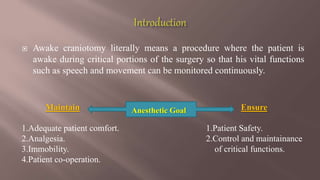
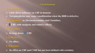
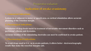
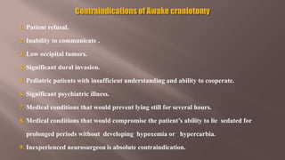
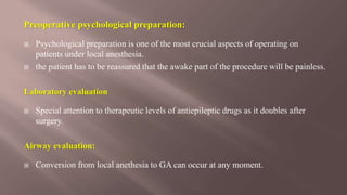
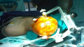
1. Awake craniotomy allows monitoring of brain functions like speech and movement during brain surgery to minimize damage to critical areas. Scalp blocks and sedation are used to keep the patient comfortable but awake.
2. Advantages include mapping of motor and language areas by directly stimulating the brain and observing responses. Risks include seizures, neurological deficits, and other surgical complications if the procedure is not performed carefully.
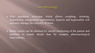
3. Proper patient selection, psychological preparation, monitoring, anesthesia technique and mapping are essential for successful awake craniotomy outcomes. Careful attention to details improves results more than complex interventions.
![ Cerebral perfusion pressure [CPP] is defined as the difference between
mean arterial and intracranial pressures.
CPP = MAP – ICP
Normal cerebral perfusion pressure is 80 mmHg, but when reduced to
less than 50 mmHg there is metabolic evidence of ischemia and
reduced electrical activity.](https://image.slidesharecdn.com/anestheticsafetyinawakecraniotomy-150113105932-conversion-gate02/85/Awake-Craniotomy-8-320.jpg)
![1. Monitored Anesthesia care [MAC]:
MAC is a specific anesthetic protocol that includes careful monitoring and support
of vital functions.
Patients receives fentanyl, with or without either droperidol or midazolam,
followed by a propofol infusion.
The craniotomy proceeds with the patient breathing spontaneously.](https://image.slidesharecdn.com/anestheticsafetyinawakecraniotomy-150113105932-conversion-gate02/85/Awake-Craniotomy-25-320.jpg)
![2. Asleep Awake Asleep [AAA] Technique:
A. LMA with Spontaneous Ventilation Technique:
B. LMA with Controlled Ventilation Technique:
Artificial ventilation allows better control of PaCO2, providing good operative
conditions.
A background infusion of remifentanyl is used to provide additional analgesia
during the awake period.](https://image.slidesharecdn.com/anestheticsafetyinawakecraniotomy-150113105932-conversion-gate02/85/Awake-Craniotomy-26-320.jpg)