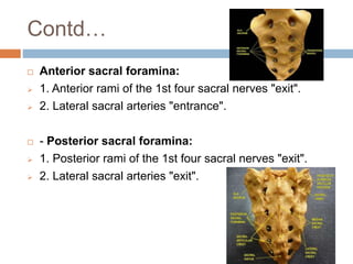
2. History
October 1941 (Robert Andrew Hingson) developed the
technique of continuous caudal anesthesia.
The first use of continuous caudal anesthesia in a
laboring woman was on January 6, 1942.
3. Introduction
Caudal epidural anesthesia is one of the most
commonly used regional techniques in pediatric patients
and for anorectal surgery in adults.
The caudal space is the sacral portion of the epidural
space.
Involves needle or catheter penetration of the
sacrococcygeal ligament covering the sacral hiatus that
is created by the unfused S4 and S5 laminae
5. Embryology
The somites that give rise to the VC begin to develop
from head to tail along the length of the notochord.
6. Anatomy of sacrum
Prior to the adoption of sacrum,
the bone was also called holy bone in English
Posterior bone of the pelvic cavity,
formed of five sacral vertebrae fused together.
Shape: triangular or wedge
with the base above and
its apex below.
It is slightly concave anteriorly.
7. CONTD…
The anterior upper border of the body of first sacral (S1)
vertebra projects inwards. It is called sacral promontory
The lateral mass on each side is a fan-shaped,
called ala of sacrum.
The superior articular process of
S1 vertebra carries articular facet
directed posteriorly.
8. CONTD…
The sacral foramina form together the sacral canal.
The lower opening of sacral canal is called sacral hiatus.
It is surrounded on either sides by sacral cornua, that
are of great importance for identification of sacral hiatus
on the body surface.
9. CONTD…
The anterior surface is smooth and concave
It has four pair of anterior sacral foramina.
The posterior surface is irregular and convex.
It has four pair of posterior sacral foramina.
10. CONTD…
The lateral surface shows articular surface,
called auricular surface for articulation with the
hip bone at sacro-iliac joint.
11. Sex differences
MALE SACRUM FEMALE
SACRUM
Length and width It is longer and narrower It is shorter and wider
Curvature of bone It shows a gentle and
uniform curvature
It descends nearly straight
in its upper part, while the
lower part turns forwards.
Auricular surface Longer Shorter
Upper surface Body is wider than the ala Body is narrower or equal
to ala.
12. Contents of the Sacral canal
Dural sac which ends at the border of the 2ND sacral
vertebra on a line joining the posterior iliac spine.
The pia mater is continued as the filum terminale.
Sacral nerves and the coccygeal nerve
Venous plexus formed by the lower end of the internal
vertebral plexus.
Areolar and fatty tissue.
13. Contd…
Sacral hiatus:
1. One pair of sacral (5th ) nerves.
2. One pair of coccygeal nerves.
3. Filum terminale. It ends through blending with
periosteum at the back of periosteum.
14. Contd…
Anterior sacral foramina:
1. Anterior rami of the 1st four sacral nerves "exit".
2. Lateral sacral arteries "entrance".
- Posterior sacral foramina:
1. Posterior rami of the 1st four sacral nerves "exit".
2. Lateral sacral arteries "exit".
15. Differences
At birth the dura mater ends at the level of the 3rd or 4th
sacral vertebra and the cord (conus medullaris) at the L3
or L4 level.
Sacral hiatus is relatively wider in children.
Palpating sacral hiatus in children is easier.
16. Contd…
In infants –the sacral canal is filled with fluid
fat and loose areolar connective tissue which
allows easy spread of anesthethetic solutions
up to age of 6 or 7 years, also this fluidity of
epidural fat allows catheter insertion easier.
17. Main Anatomic and Physiologic Factors in the Pediatric Period
That Can Influence the Selection or Performance of a Regional
Block Procedure
Pediatric Factors
(Infants Mainly)
Resulting Danger Implications for
Regional Anesthesia
Lower termination of
spinal cord
Increased risk of direct
trauma to the spinal
cord
Avoid epidural
approaches above L3
whenever possible.
Lower projection of
dural sac
Increased risk of
inadvertent
penetration of the dura
mater
Check for
cerebrospinal fluid
reflux, including
during caudal
approaches.
Favor low approaches
to the epidural space
Delayed myelinization
of nerve fibers
Easier intraneural
penetration of local
anesthetics
Onset time is
shortened, and diluted
local anesthetic is as
effective as more
concentrated
18. Contd….
Pediatric Factors
(Infants Mainly)
Resulting Danger Implications for
Regional Anesthesia
Lack of fusion of
sacral vertebrae
Persistence of sacral
intervertebral spaces
Intervertebral sacral
epidural approaches
can be performed
throughout childhood
Changing axis of
coccyx and absence of
growth of sacral hiatus
Sacral hiatus
comparatively smaller
with increasing age
Identification of sacral
hiatus becomes more
difficult above 6-8
years (increased
failure rate of caudal
anesthesia).
Increased fluidity of
epidural fat
Increased diffusion of
local anesthetic up to
6-7 years of age
Excellent blockade
after caudal anesthesia
can be achieved up to
6-7 years of age
19. T.Y
The atlas of Human body
Millers anesthesia 7th edition
Editor's Notes
Wy not in ypotension??? Wy preloadin is not reqired????
The human vertebral column usually consists of 33 vertebrae; the upper 24 are articulating vertebrae, separated by intervertebral discs and the lower nine are fused, five fused in the sacrum and four in the coccyx.
At day 20 of embryogenesis the first four pairs of somites appear in the future occipital bone region. Developing at the rate of three or four a day, the next eight pairs form in the cervical region to develop into the cervical vertebrae; the next twelve pairs will form the thoracic vertebrae; the next five pairs the lumbar vertebrae and by about day 29 the sacral somites will appear to develop into the sacral vertebrae; finally on day 30 the last three pairs will form the coccyx
Usually, it begins as five unfused vertebrae which begin to fuse between the ages of 16–18 years and have usually completely fused into a single bone by the age of 34 years.
Identification of the promontory is important in gynecological examination of the pelvis and at laparotomy
The laminae of the fifth and sometimes of the fourth sacral vertebrae fail to fuse in the midline, the deficiency thus formed is known as the sacral hiatus
These vessels are more numerous anteriorly than posteriorly and so the needle point should be kept as far posteriorly as possible
It is only at the end of the first year of life that adult level is attained, namely, L1 for the conus medullaris and S2 for the dural sac.