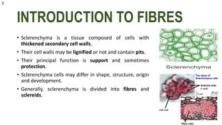
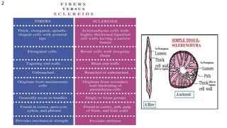
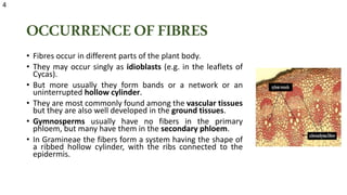
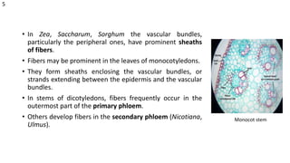
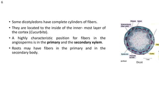
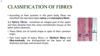
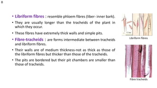
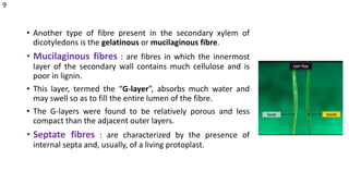
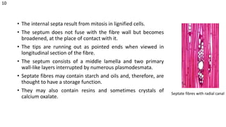
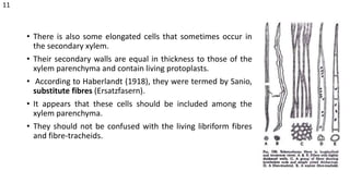
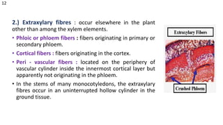
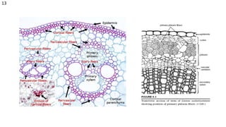
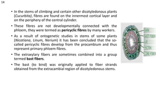
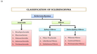
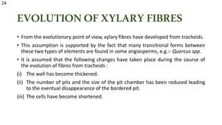
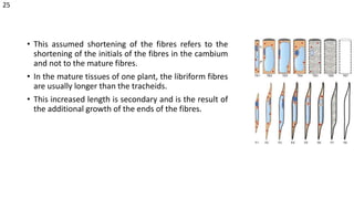
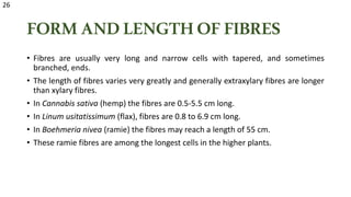
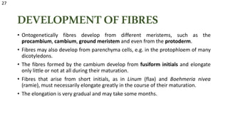
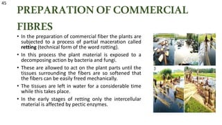
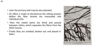
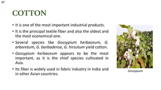
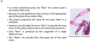
This document summarizes the different types of plant tissues. It describes two main categories of plant tissues - meristematic and permanent tissues. Permanent tissues are further divided into simple and complex tissues. Simple tissues include epidermis, parenchyma, collenchyma, sclerenchyma and cork. Complex tissues include xylem and phloem. Each tissue type is then defined and their structure and functions are explained.
This document summarizes plant tissue systems. It describes the three main types of tissues - meristematic, permanent and secretory tissues. It focuses on mature or permanent tissues, which are divided into simple tissues like parenchyma, collenchyma and sclerenchyma, and complex tissues like xylem and phloem. Parenchyma, the most common simple tissue, includes storage, aerenchyma and transfer cells. Xylem conducts water and minerals, containing tracheids, vessels, fibers and parenchyma. Phloem transports organic compounds using sieve elements, companion cells, fibers and parenchyma. Secretory tissues produce and secrete substances through internal and external structures.
This document summarizes plant tissue systems. It describes the three main types of tissues - meristematic, permanent and secretory. Permanent tissues are further divided into simple tissues like parenchyma, collenchyma and sclerenchyma, and complex tissues like xylem and phloem. Parenchyma is the most common simple tissue and performs many functions. Xylem conducts water and minerals throughout the plant and consists of tracheids, vessels, fibers and parenchyma. Phloem transports organic compounds and consists of sieve elements, companion cells, parenchyma and fibers. The document also discusses secretory tissues and their structures.
The document discusses the structure and composition of plant cell walls. It describes the primary components of plant cell walls including cellulose, hemicellulose, pectin, and lignin. It discusses the basic unit of secondary cell walls known as microfibrils. It also describes different types of secondary cell wall thickenings and pit structures found in cell walls.
This document discusses the three main types of simple permanent plant tissues: parenchyma, collenchyma, and sclerenchyma. Parenchyma tissue is made of living cells found in many plant parts that perform functions like photosynthesis, storage, and structural support. Collenchyma cells provide mechanical support to stems and leaves and have thickened but non-lignified cell walls. Sclerenchyma cells have thick, lignified secondary walls and occur as fibers or sclereids; they provide structure and protection but are dead at maturity.
There are three main tissue systems in plants: epidermal, fundamental (ground), and vascular. The epidermal tissue system includes the outer layer of cells and performs protective and regulatory functions. The fundamental tissue system constitutes the basic cellular framework and includes cells like cortex and pith. The vascular tissue system transports water and nutrients throughout the plant and is composed of xylem and phloem arranged in vascular bundles. Vascular bundles can be arranged collaterally, bicollaterally, or radially depending on the position of the xylem and phloem.
Unit1 part 1 (1).pptx dicot anatomy in which it will show the anatomical stru...prekshanalwaya
1. The document describes the structure and function of different parts of the root, stem, and leaf in plants.
2. The root has key layers including the piliferous layer, cortex, endodermis, pericycle, and vascular bundles. The stem contains an epidermis, cortex with hypodermis, ground tissue, and vascular bundles.
3. Leaves have an upper and lower epidermis, palisade and spongy mesophyll tissue, and vascular bundles. These structures allow the plant to absorb water and nutrients, provide support, transport materials, and perform photosynthesis.
The document summarizes the structure and composition of eukaryotic cell walls. It discusses the key components of plant cell walls including cellulose, hemicellulose, pectin, and lignin. It describes the layers of the plant cell wall - the middle lamella, primary wall, and secondary wall. The secondary wall may have three layers and is thicker than the primary wall. It provides structure and protection to the plant cell.
Connective tissue consists of cells separated by extracellular matrix. The main cell types are fibroblasts that produce collagen, elastic and reticular fibers embedded in ground substance. Bone contains osteoblasts, osteocytes and osteoclasts. It has a hard osseous tissue and ground substance including hydroxyapatite. Cartilage contains chondroblasts and chondrocytes in a matrix with collagen fibers and glycosaminoglycans. There are three types of cartilage - hyaline, elastic and fibrous.
This document discusses different types of permanent tissue in plants. It begins by defining permanent tissue as mature meristematic tissue that has lost the ability to divide. Permanent tissue is classified as simple tissue, complex tissue, or secretory tissue. Simple permanent tissue includes parenchyma, collenchyma, and sclerenchyma. Parenchyma is living tissue that can be photosynthetic, structural, or storage-related. Collenchyma provides support and is located beneath the epidermis. Sclerenchyma includes dead fibers for support and sclereids for hardness. The document outlines characteristics and functions of each tissue type.
This document summarizes plant cell envelopes and cell walls. It describes that plant cells have cell walls that provide structure, protection, and prevent water movement into cells. The cell wall is made of cellulose, hemicellulose, pectin and lignin. It differentiates into a primary wall, secondary wall and sometimes a tertiary wall. The cell wall provides shape, structure and acts as a protective barrier for the cell.
This document describes the different types of plant tissues - parenchyma, collenchyma, sclerenchyma, xylem and phloem. It provides details on the characteristics, structure and functions of each type of tissue. Parenchyma is a simple living tissue made of thin-walled cells involved in plant metabolism. Collenchyma has elongated cells with thick non-lignified primary walls that provide support to growing organs. Sclerenchyma includes thick-walled and often lignified cells that provide mechanical strength. Xylem and phloem are complex tissues - xylem transports water and minerals throughout the plant while phloem transports food.
This document summarizes the different types of plant tissues, including meristematic and permanent tissues. It discusses meristem tissue in plants, which are young, dividing cells capable of growth. There are four types of meristem tissue classified by plane of cell division, development, function, or position. The document also summarizes the three main types of permanent tissues - parenchyma, collenchyma, and sclerenchyma - including their characteristics, distribution in plants, and functions in providing structure and support.
Cells in plants gradually differentiate from meristems into permanent tissues with specialized functions. Permanent tissues can be simple, consisting of a single cell type, or complex. Simple permanent tissues include parenchyma, collenchyma, and sclerenchyma. Parenchyma cells are living cells involved in storage and transport. Collenchyma provides support and flexibility. Sclerenchyma cells, including fibers and sclereids, provide structure and protection through thick lignified cell walls. These mature tissues perform crucial roles in plant structure and function.
The document discusses the internal structure of dicot and monocot roots. It describes the key layers and tissues found in roots, including the epiblema, cortex, endodermis, pericycle, vascular bundles, xylem, phloem and pith. In dicot roots, the vascular bundles vary from 2-6 and are radial, while in monocot roots they vary from 8-20 and are also radial. The pith is poorly developed or absent in dicots but highly developed in monocots.
The secondary growth in dicot stems involves the activity of vascular cambium and cork cambium.
1) The vascular cambium forms a complete ring under the stem and produces secondary xylem inward and secondary phloem outward, increasing the stem's diameter. More xylem is produced, pushing the primary tissues toward the center.
2) With increased growth, the epidermis ruptures and is replaced by cork produced by the cork cambium, forming the periderm and bark tissues. Lenticels in the bark allow for gas exchange.
The document summarizes the three main tissue types in plants: dermal, ground, and vascular tissues. Dermal tissue covers the outer surface and is composed of epidermal cells that secrete a waxy cuticle. There are two types of plant tissues: meristematic tissues which are undifferentiated and can divide, as well as permanent tissues. Meristematic tissue includes apical and lateral meristem while permanent tissues include various ground tissues like parenchyma, collenchyma, and sclerenchyma as well as vascular tissues like xylem and phloem. Xylem contains tracheids and vessels that transport water and minerals upward while phloem contains sieve tubes and companion cells that
1. The internal structure of a dicot stem features an epidermis, hypodermis, cortex, endodermis, pericycle, vascular bundles arranged in rings, pith, and medullary rays.
2. Vascular bundles are conjoint, collateral or bicollateral, and open type, containing xylem, phloem, and cambium.
3. The internal structure of a monocot stem lacks a differentiated cortex and pith, and has scattered vascular bundles surrounded by bundle sheath, lacking medullary rays.
This document discusses different types of plant and animal tissues. It describes in detail the various types of plant tissues including meristematic tissues, permanent tissues (simple and complex), and their functions. The simple permanent tissues discussed are parenchyma, collenchyma, sclerenchyma. The complex permanent tissues discussed are xylem and phloem. It also describes the various types of animal tissues like epithelial tissue, connective tissue, muscular tissue and nervous tissue. Epithelial tissue is described in more detail covering the different types like squamous, cuboidal and columnar epithelium.
The document discusses several key organelles found within eukaryotic cells, including the cell wall, endomembrane system, endoplasmic reticulum, Golgi apparatus, lysosomes, vacuoles, mitochondria, and plastids. The cell wall provides shape, strength, and protection to the cell. The endomembrane system consists of organelles that work together, including the endoplasmic reticulum, Golgi apparatus, and plasma membrane. The endoplasmic reticulum functions in protein transport and processing. The Golgi apparatus packages materials and forms glycoproteins. Lysosomes contain digestive enzymes and vacuoles provide storage space in the cell. Mitochondria perform aerobic respiration and ATP synthesis
This document discusses cell inclusions and sclerenchyma cells. It describes how cell inclusions are non-living substances present in cells that can be organic or inorganic reserves, excretions, or minerals. Common reserves include starch, glycogen, fat droplets, and aleurone grains. Sclerenchyma cells have thick, lignified secondary cell walls that provide structure and support. There are two types: fibers which are long and needle-shaped, and sclereids which are irregularly shaped and found in fruits and seeds. Both fiber and sclereid cell walls strengthen plant tissues.
BIOLOGY STD 11
SANJAY SIDDHAPURA
HELPFUL FOR NEET/ GSET/NET EXAMINATION PREPARATION
TYPES OF PLANT TISSUE, ANATOMY OF ROOT, STEM AND KEAVES OF MONOCOTS AND DICOTS PLANTS, SECONDARY GROWTH, CAMBIUM
The basement membrane is a thin sheet-like structure composed of two layers - the basal lamina and reticular lamina. The basal lamina contains type IV collagen, laminin and other glycoproteins, while the reticular lamina contains collagen fibers and glycosaminoglycans. It is located at the interface between epithelial or other cells and the underlying connective tissue, serving to anchor and support cells as well as acting as a selective barrier.
Similar to Plant fibres.........................pptx (20)
Immersive Learning That Works: Research Grounding and Paths ForwardLeonel Morgado
We will metaverse into the essence of immersive learning, into its three dimensions and conceptual models. This approach encompasses elements from teaching methodologies to social involvement, through organizational concerns and technologies. Challenging the perception of learning as knowledge transfer, we introduce a 'Uses, Practices & Strategies' model operationalized by the 'Immersive Learning Brain' and ‘Immersion Cube’ frameworks. This approach offers a comprehensive guide through the intricacies of immersive educational experiences and spotlighting research frontiers, along the immersion dimensions of system, narrative, and agency. Our discourse extends to stakeholders beyond the academic sphere, addressing the interests of technologists, instructional designers, and policymakers. We span various contexts, from formal education to organizational transformation to the new horizon of an AI-pervasive society. This keynote aims to unite the iLRN community in a collaborative journey towards a future where immersive learning research and practice coalesce, paving the way for innovative educational research and practice landscapes.
Signatures of wave erosion in Titan’s coastsSérgio Sacani
The shorelines of Titan’s hydrocarbon seas trace flooded erosional landforms such as river valleys; however, it isunclear whether coastal erosion has subsequently altered these shorelines. Spacecraft observations and theo-retical models suggest that wind may cause waves to form on Titan’s seas, potentially driving coastal erosion,but the observational evidence of waves is indirect, and the processes affecting shoreline evolution on Titanremain unknown. No widely accepted framework exists for using shoreline morphology to quantitatively dis-cern coastal erosion mechanisms, even on Earth, where the dominant mechanisms are known. We combinelandscape evolution models with measurements of shoreline shape on Earth to characterize how differentcoastal erosion mechanisms affect shoreline morphology. Applying this framework to Titan, we find that theshorelines of Titan’s seas are most consistent with flooded landscapes that subsequently have been eroded bywaves, rather than a uniform erosional process or no coastal erosion, particularly if wave growth saturates atfetch lengths of tens of kilometers.
BIRDS DIVERSITY OF SOOTEA BISWANATH ASSAM.ppt.pptxgoluk9330
Ahota Beel, nestled in Sootea Biswanath Assam , is celebrated for its extraordinary diversity of bird species. This wetland sanctuary supports a myriad of avian residents and migrants alike. Visitors can admire the elegant flights of migratory species such as the Northern Pintail and Eurasian Wigeon, alongside resident birds including the Asian Openbill and Pheasant-tailed Jacana. With its tranquil scenery and varied habitats, Ahota Beel offers a perfect haven for birdwatchers to appreciate and study the vibrant birdlife that thrives in this natural refuge.
Sexuality - Issues, Attitude and Behaviour - Applied Social Psychology - Psyc...PsychoTech Services
A proprietary approach developed by bringing together the best of learning theories from Psychology, design principles from the world of visualization, and pedagogical methods from over a decade of training experience, that enables you to: Learn better, faster!
PPT on Sustainable Land Management presented at the three-day 'Training and Validation Workshop on Modules of Climate Smart Agriculture (CSA) Technologies in South Asia' workshop on April 22, 2024.
(June 12, 2024) Webinar: Development of PET theranostics targeting the molecu...Scintica Instrumentation
Targeting Hsp90 and its pathogen Orthologs with Tethered Inhibitors as a Diagnostic and Therapeutic Strategy for cancer and infectious diseases with Dr. Timothy Haystead.
CLASS 12th CHEMISTRY SOLID STATE ppt (Animated)eitps1506
Description:
Dive into the fascinating realm of solid-state physics with our meticulously crafted online PowerPoint presentation. This immersive educational resource offers a comprehensive exploration of the fundamental concepts, theories, and applications within the realm of solid-state physics.
From crystalline structures to semiconductor devices, this presentation delves into the intricate principles governing the behavior of solids, providing clear explanations and illustrative examples to enhance understanding. Whether you're a student delving into the subject for the first time or a seasoned researcher seeking to deepen your knowledge, our presentation offers valuable insights and in-depth analyses to cater to various levels of expertise.
Key topics covered include:
Crystal Structures: Unravel the mysteries of crystalline arrangements and their significance in determining material properties.
Band Theory: Explore the electronic band structure of solids and understand how it influences their conductive properties.
Semiconductor Physics: Delve into the behavior of semiconductors, including doping, carrier transport, and device applications.
Magnetic Properties: Investigate the magnetic behavior of solids, including ferromagnetism, antiferromagnetism, and ferrimagnetism.
Optical Properties: Examine the interaction of light with solids, including absorption, reflection, and transmission phenomena.
With visually engaging slides, informative content, and interactive elements, our online PowerPoint presentation serves as a valuable resource for students, educators, and enthusiasts alike, facilitating a deeper understanding of the captivating world of solid-state physics. Explore the intricacies of solid-state materials and unlock the secrets behind their remarkable properties with our comprehensive presentation.
SDSS1335+0728: The awakening of a ∼ 106M⊙ black hole⋆Sérgio Sacani
Context. The early-type galaxy SDSS J133519.91+072807.4 (hereafter SDSS1335+0728), which had exhibited no prior optical variations during the preceding two decades, began showing significant nuclear variability in the Zwicky Transient Facility (ZTF) alert stream from December 2019 (as ZTF19acnskyy). This variability behaviour, coupled with the host-galaxy properties, suggests that SDSS1335+0728 hosts a ∼ 106M⊙ black hole (BH) that is currently in the process of ‘turning on’. Aims. We present a multi-wavelength photometric analysis and spectroscopic follow-up performed with the aim of better understanding the origin of the nuclear variations detected in SDSS1335+0728. Methods. We used archival photometry (from WISE, 2MASS, SDSS, GALEX, eROSITA) and spectroscopic data (from SDSS and LAMOST) to study the state of SDSS1335+0728 prior to December 2019, and new observations from Swift, SOAR/Goodman, VLT/X-shooter, and Keck/LRIS taken after its turn-on to characterise its current state. We analysed the variability of SDSS1335+0728 in the X-ray/UV/optical/mid-infrared range, modelled its spectral energy distribution prior to and after December 2019, and studied the evolution of its UV/optical spectra. Results. From our multi-wavelength photometric analysis, we find that: (a) since 2021, the UV flux (from Swift/UVOT observations) is four times brighter than the flux reported by GALEX in 2004; (b) since June 2022, the mid-infrared flux has risen more than two times, and the W1−W2 WISE colour has become redder; and (c) since February 2024, the source has begun showing X-ray emission. From our spectroscopic follow-up, we see that (i) the narrow emission line ratios are now consistent with a more energetic ionising continuum; (ii) broad emission lines are not detected; and (iii) the [OIII] line increased its flux ∼ 3.6 years after the first ZTF alert, which implies a relatively compact narrow-line-emitting region. Conclusions. We conclude that the variations observed in SDSS1335+0728 could be either explained by a ∼ 106M⊙ AGN that is just turning on or by an exotic tidal disruption event (TDE). If the former is true, SDSS1335+0728 is one of the strongest cases of an AGNobserved in the process of activating. If the latter were found to be the case, it would correspond to the longest and faintest TDE ever observed (or another class of still unknown nuclear transient). Future observations of SDSS1335+0728 are crucial to further understand its behaviour. Key words. galaxies: active– accretion, accretion discs– galaxies: individual: SDSS J133519.91+072807.4
Describing and Interpreting an Immersive Learning Case with the Immersion Cub...Leonel Morgado
Current descriptions of immersive learning cases are often difficult or impossible to compare. This is due to a myriad of different options on what details to include, which aspects are relevant, and on the descriptive approaches employed. Also, these aspects often combine very specific details with more general guidelines or indicate intents and rationales without clarifying their implementation. In this paper we provide a method to describe immersive learning cases that is structured to enable comparisons, yet flexible enough to allow researchers and practitioners to decide which aspects to include. This method leverages a taxonomy that classifies educational aspects at three levels (uses, practices, and strategies) and then utilizes two frameworks, the Immersive Learning Brain and the Immersion Cube, to enable a structured description and interpretation of immersive learning cases. The method is then demonstrated on a published immersive learning case on training for wind turbine maintenance using virtual reality. Applying the method results in a structured artifact, the Immersive Learning Case Sheet, that tags the case with its proximal uses, practices, and strategies, and refines the free text case description to ensure that matching details are included. This contribution is thus a case description method in support of future comparative research of immersive learning cases. We then discuss how the resulting description and interpretation can be leveraged to change immersion learning cases, by enriching them (considering low-effort changes or additions) or innovating (exploring more challenging avenues of transformation). The method holds significant promise to support better-grounded research in immersive learning.
1. PLANT FIBERS
Submitted to,
Dr. Chandini V. K
Asst. Professor
ST. Teresa’s College, Ekm
Submitted by,
A.T Milin Sera
Roll no : 1
1st M.sc. Botany
ST. Teresa's College, Ekm
2. INTRODUCTION TO FIBRES
• Sclerenchyma is a tissue composed of cells with
thickened secondary cell walls.
• Their cell walls may be lignified or not and contain pits.
• Their principal function is support and sometimes
protection.
• Sclerenchyma cells may differ in shape, structure, origin
and development.
• Generally, sclerenchyma is divided into fibres and
sclereids.
1
4. FIBRES
• Fibres are elongated cells often with pointed ends, thick
secondary wall and reduced pits.
• Certain other types of fibres possess blunt or even branched
ends.
• As the fibres get old, the protoplasmic contents become
multinucleate and finally disappear.
• But in certain cases, fibres retain protoplast even upto the
permanent stage.
• Some fibre walls contain lignin while certain fibres like those
occurring in flux (Linum usitatissimum) the wall is made up of
pure cellulose.
3
5. OCCURRENCEOFFIBRES
• Fibres occur in different parts of the plant body.
• They may occur singly as idioblasts (e.g. in the leaflets of
Cycas).
• But more usually they form bands or a network or an
uninterrupted hollow cylinder.
• They are most commonly found among the vascular tissues
but they are also well developed in the ground tissues.
• Gymnosperms usually have no fibers in the primary
phloem, but many have them in the secondary phloem.
• In Gramineae the fibers form a system having the shape of
a ribbed hollow cylinder, with the ribs connected to the
epidermis.
4
6. • In Zea, Saccharum, Sorghum the vascular bundles,
particularly the peripheral ones, have prominent sheaths
of fibers.
• Fibers may be prominent in the leaves of monocotyledons.
• They form sheaths enclosing the vascular bundles, or
strands extending between the epidermis and the vascular
bundles.
• In stems of dicotyledons, fibers frequently occur in the
outermost part of the primary phloem.
• Others develop fibers in the secondary phloem (Nicotiana,
Ulmus).
Monocot stem
5
7. • Some dicotyledons have complete cylinders of fibers.
• They are located to the inside of the inner- most layer of
the cortex (Cucurbita).
• A highly characteristic position for fibers in the
angiosperms is in the primary and the secondary xylem.
• Roots may have fibers in the primary and in the
secondary body.
Dicot
6
8. CLASSIFICATIONOFFIBRES
• According to their position in the plant body, fibres are
classified into two basic types xylary and extraxylary fibres.
1.) Xylary fibres : constitute an integral part of the xylem
and they develop from the same meristematic tissues as the
other xylem elements.
• These fibres are of varied shape in spite of their common
origin.
• Two main types of xylary fibres, i.e. libriform fibres and
fibre-tracheids, are distinguished on the basis of wall
thickness and type and amount of pits.
7
9. • Libriform fibres : resemble phloem fibres (liber- inner bark).
• They are usually longer than the tracheids of the plant in
which they occur.
• These fibres have extremely thick walls and simple pits.
• Fibre-tracheids : are forms intermediate between tracheids
and libriform fibres.
• Their walls are of medium thickness-not as thick as those of
the libriform fibres but thicker than those of the tracheids.
• The pits are bordered but their pit chambers are smaller than
those of tracheids.
Libriform fibres
Fibre tracheids
8
10. • Another type of fibre present in the secondary xylem of
dicotyledons is the gelatinous or mucilaginous fibre.
• Mucilaginous fibres : are fibres in which the innermost
layer of the secondary wall contains much cellulose and is
poor in lignin.
• This layer, termed the “G-layer”, absorbs much water and
may swell so as to fill the entire lumen of the fibre.
• The G-layers were found to be relatively porous and less
compact than the adjacent outer layers.
• Septate fibres : are characterized by the presence of
internal septa and, usually, of a living protoplast.
9
11. • The internal septa result from mitosis in lignified cells.
• The septum does not fuse with the fibre wall but becomes
broadened, at the place of contact with it.
• The tips are running out as pointed ends when viewed in
longitudinal section of the fibre.
• The septum consists of a middle lamella and two primary
wall-like layers interrupted by numerous plasmodesmata.
• Septate fibres may contain starch and oils and, therefore, are
thought to have a storage function.
• They may also contain resins and sometimes crystals of
calcium oxalate.
Septate fibres with radial canal
10
12. • There is also some elongated cells that sometimes occur in
the secondary xylem.
• Their secondary walls are equal in thickness to those of the
xylem parenchyma and contain living protoplasts.
• According to Haberlandt (1918), they were termed by Sanio,
substitute fibres (Ersatzfasern).
• It appears that these cells should be included among the
xylem parenchyma.
• They should not be confused with the living libriform fibres
and fibre-tracheids.
11
13. 2.) Extraxylary fibres : occur elsewhere in the plant
other than among the xylem elements.
• Phloic or phloem fibers : fibers originating in primary or
secondary phloem.
• Cortical fibers : fibers originating in the cortex.
• Peri - vascular fibers : located on the periphery of
vascular cylinder inside the innermost cortical layer but
apparently not originating in the phloem.
• In the stems of many monocotyledons, the extraxylary
fibres occur in an uninterrupted hollow cylinder in the
ground tissue.
12
15. • In the stems of climbing and certain other dicotyledonous plants
(Cucurbita), fibres are found on the innermost cortical layer and
on the periphery of the central cylinder.
• These fibres are not developmentally connected with the
phloem, they were termed as pericyclic fibres by many workers.
• As a result of ontogenetic studies in stems of some plants
(Nicotiana, Linum, Nerium) it has been concluded that the so-
called pericyclic fibres develop from the procambium and thus
represent primary phloem fibres.
• The extraxylary fibers are sometimes combined into a group
termed bast fibers.
• The bast (to bind) was originally applied to fiber strands
obtained from the extracambial region of dicotyledonous stems.
14
17. ORIGINOFFIBRES
• Fibers arise from various meristems.
• Fibers of the xylem and the phloem are derived from procambium or cambium.
• In the cambium, the fibers arise from the fusiform initials.
• Extraxylary fibers other than those of the phloem arise from the ground
meristem.
• In some Gramineae and Cyperaceae, fibers originate in the protoderm and
become elements of the epidermis.
• In plants having fibrous bundle sheaths, part of the fibers may be derived from
the procambium and part from the ground meristem.
• The fibrous bundles of monocotyledons appear to be connected with vascular
bundles and they are considered as originating from the procambium.
16
18. STRUCTUREOFFIBRES
(I) Extraxylary Fibers : A long spindle-like shape is considered
typical of extraxylary fibers (fibers in general).
• These elements may vary in length, and their ends are
sometimes blunt, rather than tapering, and may be branched.
• Generally, primary extraxylary fibers are longer than the
secondary.
• The cell walls of the extraxylary fibers are frequently very
thick.
• The pits are simple or slightly bordered.
• Some extraxylary fibers have lignified walls, others non-
lignified.
17
19. • The flax fiber (Linum usitatissimum) is typically not lignified,
its secondary wall consists of almost pure cellulose.
• Concentric lamellations may be observed in extraxylary
fibers with or without treatment with swelling reagents.
• This lamellation appears to result from an alternation of
cellulosic and noncellulosic layers.
• In cotton fiber, the lamellation is a reflection of varying
densities of the cellulosic matrix in the successive lamellae.
18
20. (II) Xylem Fibers : Wood fibers typically have lignified
secondary walls.
• They vary in size, shape, thickness of wall, and type and
abundance of pitting.
• With reference to the possible evolution of the xylem
fibers, different categories of these fibres are understood.
• Phylogenetically, these fibers are believed to be derived
from an imperforate xylem cell combining the functions of
water conduction and support, that is, a tracheid.
19
21. • A good indication that fibers and tracheids are phylogenetically related is the
occurrence of almost imperceptible gradations between these two cell types in
certain angiosperms such as the oak.
• The gradations suggest the following principal changes during the evolution of
the fiber from a tracheid:
(i) increase in wall thickness, (ii) decrease in length, and (iii) reduction in the size of
bordered pits.
• In the extreme condition the pit appears simple or nearly so.
• Wall thickness and the nature of pitting are used to differentiate between the
two main categories of wood fibers, fiber-tracheids and libriform fibers.
• Fiber-tracheids are cells with pits whose borders are reduced as compared with
those in the tracheids.
20
22. • Finally the cells with simple pits are classified as libriform fibers.
• Commonly, the thickness of wall increases in the sequence of tracheid, fiber-
tracheid, libriform fiber.
• The increase in wall thickness results in an increase in the length of the pit canal.
• In the fiber-tracheid these canals lead into small but evident pit chambers.
• In them the inner apertures are lenticular to slit-like and usually extended beyond
the outlines of the border.
• The libriform fibers also have long slit-like canals, but their pit chambers are much
reduced or absent.
• The inner apertures of the pit-pairs in the fiber-tracheids and libriform fibers are
often crossed with each other.
21
23. • The tracheids are usually shorter, the fibers longer, with
the libriform fibers attaining the greatest length.
• The fibers become longer than the associated tracheids
because they undergo a more extensive apical
elongation during tissue differentiation.
• Fiber-tracheids and libriform fibers may both be septate.
• The septa are true walls, but they are formed after the
deposition of the secondary layers on the longitudinal
walls of the element.
22
24. • Parts of the secondary wall of fiber-tracheids and libriform fibers may have a
great capacity for absorption of water.
• In the presence of water such walls swell. Upon drying, these walls shrink again.
• Fibers possessing hygroscopic walls are sometimes termed mucilaginous or
gelatinous.
• The gelatinous wall layers do not contain excessive amounts of pectinaceous,
gummy, or mucilaginous substances.
• They show a peculiar physical condition of the cellulose which possibly is
responsible for their gelatinous nature.
• Many woods contain gelatinous fibers. Oak (Quercus) and black locust (Robinia)
are noteworthy examples.
23
25. EVOLUTIONOFXYLARYFIBRES
• From the evolutionary point of view, xylary fibres have developed from tracheids.
• This assumption is supported by the fact that many transitional forms between
these two types of elements are found in some angiosperms, e.g.:- Quercus spp.
• It is assumed that the following changes have taken place during the course of
the evolution of fibres from tracheids :
(i) The wall has become thickened.
(ii) The number of pits and the size of the pit chamber has been reduced leading
to the eventual disappearance of the bordered pit.
(iii) The cells have become shortened.
24
26. • This assumed shortening of the fibres refers to the
shortening of the initials of the fibres in the cambium
and not to the mature fibres.
• In the mature tissues of one plant, the libriform fibres
are usually longer than the tracheids.
• This increased length is secondary and is the result of
the additional growth of the ends of the fibres.
25
27. FORMANDLENGTHOFFIBRES
• Fibres are usually very long and narrow cells with tapered, and sometimes
branched, ends.
• The length of fibres varies very greatly and generally extraxylary fibres are longer
than xylary fibres.
• In Cannabis sativa (hemp) the fibres are 0.5-5.5 cm long.
• In Linum usitatissimum (flax), fibres are 0.8 to 6.9 cm long.
• In Boehmeria nivea (ramie) the fibres may reach a length of 55 cm.
• These ramie fibres are among the longest cells in the higher plants.
26
28. DEVELOPMENT OFFIBRES
• Ontogenetically fibres develop from different meristems, such as the
procambium, cambium, ground meristem and even from the protoderm.
• Fibres may also develop from parenchyma cells, e.g. in the protophloem of many
dicotyledons.
• The fibres formed by the cambium develop from fusiform initials and elongate
only little or not at all during their maturation.
• Fibres that arise from short initials, as in Linum (flax) and Boehmeria nivea
(ramie), must necessarily elongate greatly in the course of their maturation.
• The elongation is very gradual and may take some months.
27
29. • This gradual elongation of primary phloem fibres involves a very complicated
development of the secondary wall.
• While the fibre still grows symplastically, the wall remains thin.
• Later, when the ends begin to grow by intrusive growth, only the cell walls of
the ends remain thin.
• Secondary wall formation commences from the middle of the fibre in those
parts of wall which have ceased to elongate.
• In Linum and ramie it has been found that this process is gradual.
• So the new lamellae of the secondary wall are added centripetally in the form
of cylinders which are open at both ends.
28
30. • At the same time the first-formed lamellae continue to
elongate towards the fibre ends which they reach only
when the fibre ceases to elongate.
• According to Kundu and Sen (1960) the upper ends of ramie
fibres continue to grow for a longer period than the basal
ends.
• Sometimes not all the lamellae reach the actual fibre end.
• In some fibres, chambers may be formed in the terminal
portions by the ingrowth, towards the cell lumen, of these
lamellae.
• The lamellae of the primary phloem fibres, or at least of the
immature fibres, are often not strongly attached to one
another.
29
31. • In short fibres, such as those found in Agave, Sansevieria, and
Musa textilis, all portions of the cell wall grow at the same
rate.
• Differences exist in the manner of growth of the fibres in the
primary body and of those in the secondary body.
• The initials of the primary fibres appear early, before the
organ in which they occur has elongated.
• So they may grow in length symplastically together with the
neighboring cells which continue to divide.
• The symplastic growth is augmented by intrusive and gliding
growth of the ends which thus penetrate between the
surrounding cells.
• The initials of the secondary fibres develop in organs that
have ceased to elongate.
30
32. • Therefore the growth of secondary fibres can be intrusive only.
• This is apparently the reason why the primary fibres are usually longer than the
secondary fibres of the same plant.
• Electron microscopic studies of wood fibres have shown that during secondary
wall formation the cytoplasm was in general conspicuously vacuolated, although
the vacuolation did not extend to the cell tips.
• The nucleus was large and the organelles which were generally few in number
appeared more crowded near the tips.
• The ER frequently appeared to lie parallel to the wall surface.
• In the process of wall formation, lamellar apposition of cellulose and vesicular
secretion of matrix takes place.
31
33. FIBREPROTOPLAST
• During the development of primary phloem fibres of Nicotiana and Linum, the
protoplast was multinucleate.
• The protoplast in developing secondary fibres usually has a single nucleus.
• Mature libriform fibres and fibre-tracheids were usually regarded as being dead
supporting structures.
• In mature fibres the presence of a living protoplast and nucleus had been
described only in phloem fibres and in septate fibres.
• According to Bailey (1953), libriform fibres sometimes retain their living contents
subsequent to the formation of the thick, lignified secondary wall, thus enabling
these cells to assume a storage function in addition to that of support.
32
34. • Recently, however, living protoplasts and nuclei have been identified in libriform
fibres of many species, and even in fibre-tracheids.
• Such living fibres were found to occur in the wood of Tamarix spp., and in many
other woody species.
• Living protoplasts have also been found in many monocotyledonous fibres.
• In fibres with long, narrow lumina the nuclei are usually elongated.
• During the elongation of the primary phloem fibers in tobacco, the protoplasts
become multinucleate as a result of repeated mitotic divisions.
• Cell plates do not form at the ends of the successive nuclear divisions and the
spindle fibers are less persistent than in ordinary division.
33
35. • At the final stages in fiber ontogeny, usually after secondary walls have
developed, the nuclei appear to fuse or clump.
• In nearly mature fibers the nuclear material frequently occurs as one large
degenerating mass.
• The bast fibers of Linum also exhibit a multinucleate phase during differentiation.
• Haberlandt (1914) observed a multinucleate protoplast in the bast fibers of Linum
and certain members of the Leguminosae.
• He suggested that, the presence of several nuclei appears advantageous when
the length of many bast-cells and their active growth in length and thickness are
taken into account.
34
36. • In certain types of fibers, however, mitosis is
followed by cytokinesis, resulting in a
chambered or septate fibre.
• In Hypericum androsaemum the fiber-
tracheid retains its protoplast after the thick
secondary wall has been laid down.
• Mitosis may then occur in such a cell.
• Cell plate formation then occurs in the
normal manner.
• A thin transverse septum is formed across
the lumen, intersecting the inner edge of the
secondary wall of the “mother cell.”
35
37. STRUCTURE&USEOFCOMMERCIAL
FIBRES
• The term fibre, as used in industry, does not generally have the same meaning as
that defined by botanists.
• For instance, the commercial fibres of Linum, Boehmeria, and Corchorus are, in
reality, a bundle of fibres.
• Those from monocotyledonous leaves, such as from Agave, Musa textilis, and
others, are usually the vascular bundles with the surrounding sheaths of fibres.
• From some plants the commercial fibres comprise the vascular system of the
root, e.g. Muhlenbergia, or of the entire plant, e.g. Tillandsia.
• The commercial fibres of Gossypium (cotton) and kapok are the epidermal hairs
of the seeds.
36
39. • Commercial fibres are divided into two types - hard fibres and soft fibres.
• Hard fibres : are those which have a lignin content in the walls, and are of a stiff
texture.
• They are obtained from monocotyledons.
• Soft fibres : may or may not contain lignin, they are flexible and elastic.
• They are of dicotyledonous origin.
• The best- known plants from which hard fibres are produced are different species
of Agave, especially sisalana (sisal), Tillandsia usneoides (Spanish moss), Musa
textilis (abaca), Fureraea gigantea (Mauritius hemp).
• Soft fibres are mainly produced from Linum usitatissimum (flax), Cannabis sativa
(hemp), Boehmeria nivea (ramie), Corchorus capsularis (jute), Hibiscus
cannabinus (kenaf).
38
41. • The fibres of cotton, which are produced from the indumentum of seeds,
represent the most important commercial fibres in use today.
• Fibres are also classified according to their use :
(a) Textile fibres :- which are used in the manufacture of fabrics.
(b) Cordage fibres :- used for making cordages.
(c) brush fibres :- which are used in the manufacture of brushes and brooms.
(d) filling fibres :- such as those used for stuffing upholstery, mattresses and life-
belts, caulking (barrels, plumbing), and reinforcing (wall plates, plastics).
• In the textile industry the principal fibre used is cotton and, in smaller amounts,
flax, ramie, and hemp.
• For coarser fabrics, such as sacking and bagging, jute is principally used, and
cotton, flax, hemp, and a few other hard fibres are used to a lesser extent.
40
43. • For the manufacture of twine ; jute, cotton, hemp and, to a lesser extent, flax
and several hard fibres are used.
• Ropes and binder twines are manufactured from hard fibres, such as those of
Musa textilis (abaca) and Agave spp. (sisal), and to a small extent from cotton
and other soft fibres.
• Brushes and brooms are made from Agave fibres, fibres from the stems and
leaves of the Palmae and the inflorescences of Sorghum vulgare, etc.
• As filling fibres, the fibres of Ceiba pentandra (kapok), cotton, jute, the fibres of
Tillandsia usneoides, several hard fibres, and others are used.
• Fibres are also used in the paper industry, depending on their physical and
chemical properties different types of paper can be made.
42
45. • From a technological point of view, the shape of the fibre cell, its length, and
wall structure are of importance in the fibre industry.
• Special attention is paid to the length of the fibre, the extent to which
neighboring fibres overlap, and how they are joined to one another.
• The orientation of the cellulose units in the wall has an important effect on the
physical properties of wood and commercial fibres.
• Elasticity and heat conductivity increase as the degree of orientation parallel to
the length of the fibre increases.
Cannabis
44
46. PREPARATIONOFCOMMERCIAL
FIBRES
• In the preparation of commercial fiber the plants are
subjected to a process of partial maceration called
retting (technical form of the word rotting).
• In this process the plant material is exposed to a
decomposing action by bacteria and fungi.
• These are allowed to act on the plant parts until the
tissues surrounding the fibers are so softened that
the fibers can be easily freed mechanically.
• The tissues are left in water for a considerable time
while this takes place.
• In the early stages of retting only the intercellular
material is affected by pectic enzymes.
45
47. • Later the primary wall may be also attacked.
• An effort is made to discontinue the retting process
before the fiber strands are macerated into
individual cells.
• Then the retted stems are dried and passed
between rollers, which separates the fibres from the
other tissues.
• Finally they are combed, beaten out and placed in
bales.
46
48. COTTON
• It is one of the most important industrial products.
• It is the principal textile fiber and also the oldest and
the most economical one.
• Several species like Gossypium herbaceum, G.
arboretum, G. barbadense, G. hirsutum yield cotton.
• Gossypium herbaceum appears to be the most
important, as it is the chief species cultivated in
Asia.
• Its fiber is widely used in fabric industry in India and
in other Asian countries.
Gossypium
47
49. • In a strict anatomical sense, the “fiber” of a cotton seed is
not really a fiber at all.
• Because it is not lignified nor does it occur in the appropriate
part of the plant to be called a fiber.
• The proper anatomical cell name for the seed “fiber” is a
trichome.
• However, we will adopt the term “fiber” to describe the long
hairs on the cotton seed as that is the commonly used term.
• Each “fiber” is produced by the outgrowth of a single
epidermal cell.
• The “fibers” are actually hairs that grow out of the seed
coats.
Processed fibres of Gossypium
48
50. • The seed “Fiber” is composed of :
(i) cuticle: mixture of cutinous, pectinous substance.
(ii) outer cellulose layer: mostly original cell wall.
(iii) layer of secondary deposits: almost all cellulose.
(iv) walls of lumen: spiral structure surrounding the central cavity, most dense part
of the “fiber”.
(v) lumen: filled with structure less substance, nitrogenous nature.
49
51. • Some epidermal cells make long lint hairs.
• Other cells stop growing before they become too long; we call these fuzz hairs.
• The fuzz hairs have a bigger diameter and a thinner cell wall.
• As the boll grows, the “fibers” are maturing within.
• In about four months times the boll begins to slit open and the “fibers” are
exposed.
• Each seed has about 10,000 to 20,000 “fibers”.
• The boll matures and the “fibers” lose their water and die.
• Each “fiber” collapses and appears as a flattened twisted tube.
50
52. • The degree of twist is a result of the type of environment the plant is grown in,
the specific species or cultivar, and the ripeness of the “fibers”.
• The twists cause the “fibers” to cling to each other and improve their spinning
qualities.
51
53. JUTE
• Jute is the bast fiber obtained from the secondary
phloem of plants called Corchorus capsularis and
Corchorus olitorius.
• Their fiber is cheaper than cotton and flax.
• The plant is tall slender annual one with shrubby
nature.
• The fibre surrounds the woody central part of the
stem.
• In fact, it is located in wedge-shaped bundles outside
the xylem.
52
54. • Fibres are grouped in concentric rings alternating with the thin walled tissue.
• This thin walled phloem tissue breaks up through microbiological decomposition
during retting.
• At maturity, these fibres lose their protoplasm and become dead.
Corchorus olitorius Corchorus capsularis
53
55. • They are elongated, unbranched, sclerenchymatous cells
with very thick cell walls.
• These fibres are longer than a meter in comparison to the
fibres in fruits or seeds.
• The stem is retted in water and the long fibers are separated
from the phloem part.
• The pale yellow fibers are obtained during this process.
• Jute is used to produce rough wearing, bags, sacs and
wrapping materials for textile industry.
• The fibers are twisted to produce twine, coarse cloth, and
curtains, manufacture of paper etc.
• India is the best producer of jute.
54
56. COIR
• Coir is a hard, versatile, natural fibre.
• The coir is obtained from the mesocarp of the fruit.
• Coir fibre is generally not very long (0.15—0.28 m).
• The coir fibre is multicellular (the fibre contains 30
to 300 or more cells in its total cross-section) and its
cross-section is polygonal or round.
• The transverse and longitudinal section of coir fibre,
shows the number of cells and distribution of cells
around the central pore called ‘lacuna’.
55
57. • The walls are thin to fairly thick with lense-shaped silicified stegmata on their
surface.
• These are delicate thickenings with a few of them being circular or spiral in shape.
• There is a central cavity in each cell called lumen, which is medium to large in size
(polygonal, round or rounded to elliptic in shape).
56
58. • The surface of the individual cell is smooth or rough with
certain defects like cross-markings, while the surface of
the fibre is coated with a waxy material called cuticle.
• The chemical constituents of pure coir fibre have been
found to be cellulose, lignin, hemicellulose, pectin and
water solubles.
• The ageing of the fibres is found to render the fibres
stiffer and tougher, and leads to an increase in the lignin
content
• Coconut husk is retted and the fibers are entangled by
beating.
• Coir fiber has various uses like making mats, carpets,
cordage, brushes etc.
57
59. REFERENCE
• Cutter, E. (1978). Plant Anatomy Part I. Reading, Mass.: Addison-Wesley Pub. Co.
• Esau, K. (1965). Plant anatomy. Plant Anatomy., (2nd Edition).
• Fahn, A. (1977). Plant Anatomy. Oxford: Pergamon Press.
• Foster, A. (1949). Practical Plant Anatomy. New York: D. Van Nostrand Co.
• Pandey, B. (1996). Plant Anatomy. New Delhi: S Chand & Comp.
• Pandey, S. N., & Chadha, A. (2009). Plant anatomy and embryology. Vikas
Publishing House.
• Satyanarayana, K. G., Kulkarni, A. G., & Rohatgi, P. K. (1981). Structure and
properties of coir fibres. Proceedings of the Indian Academy of Sciences Section
C: Engineering Sciences, 4, 419-436.
• https://labs.plb.ucdavis.edu/rost/cotton/reproduction/frfiber.html
58