Heart (Its Structure & Function).pptx
•Download as PPTX, PDF•
16 likes•29,032 views

THIS CONTENT WILL BE BENEFICIAL FOR B.SC. NURSING STUDENTS.

Recommended
More Related Content
What's hot
What's hot (20)
Similar to Heart (Its Structure & Function).pptx
Similar to Heart (Its Structure & Function).pptx (20)
More from Vipin Chandran
More from Vipin Chandran (20)
Recently uploaded
Recently uploaded (20)
Heart (Its Structure & Function).pptx
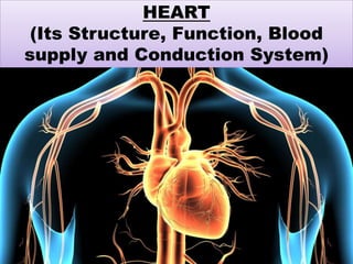
- 1. HEART (Its Structure, Function, Blood supply and Conduction System)
- 2. HEART The heart is a roughly cone-shaped hollow muscular organ found in all animals and human beings with a circulatory system, that is responsible for pumping blood throughout the blood vessels by repeated, rhythmic contractions. The term cardiac means "related to the heart" and comes from the Greek word, kardia, for "heart". The human heart is about the size of a fist and has a mass of between 250 and 300 grams. It is about 12 cm long, 9 cm wide, 6 cm thick and located slightly left of middle in the chest, anterior to the vertebral column and posterior to the sternum.
- 3. POSITION OF HEART The heart rest on the diaphragm near midline of thoracic cavity in mediastinum. It lies little more to the left than the right, apex is formed by the tip of the left ventricles & rest on diaphragm, the base of the heart is its posterior surface it is formed by atria. ORGANSASSOCIATED WITH THE HEART: Inferiorly: central tendon of the diaphragm venacava, Superiorly: the great blood vessels; aorta, superior pulmonary artery and vein. Posteriorly: oesophagus, trachea, bronchus, descending aorta Laterally: the lungs Anteriorly: the sternum, ribs and intercostal muscle.
- 5. STRUCTURE OFTHE HEART The heart is composed of three layers of tissue pericardium, myocardium and endocardium; Pericardium: The pericardium is a triple-layered fluid-filled sac that surrounds the heart. The outer layer of this sac is the fibrous pericardium. It is a strong layer of connective tissue. It adheres to the diaphragm inferiorly, and superiorly it is fused to the roots of the great vessels that leave and enter the heart. Deep to the fibrous pericardium is the double layered serous pericardium. The serous pericardium is a closed sac sandwiched between the fibrous pericardium and the heart. The outer layer is the parietal layer of the serous pericardium and adheres to the inner surface of the fibrous pericardium. The inner visceral layer of serous pericardium also called as epicardium.
- 7. Layers of the heart: the wall of heart consist of three layers: 1) Epicardium (external layer) 2) Myocardium (middle layer) 3) Endocardium (inner layer) 1. Epicardium: It is composed of two tissue layer (outer of visceral layer of serous pericardium) it is composed of mesothelium beneath this layer of delicate fibroelastic tissue and adipose tissue. Epicardium contains blood vessels, lymphatics and vessels that supply the myocardium.
- 8. Myocardium: The myocardium is the basic muscle that makes up the heart. This muscle is involuntary. The cardiac muscle structure consists of basic units of cardiac muscle cells known as myocytes. Coordinated contraction of the cardiac muscles is what makes the heart propel blood to various parts of the body. It is the function of the coronary arteries to supply blood and oxygen to the cardiac muscles. This is the thickest of all the layers of the heart. The cardiac muscles cannot afford to rest even for a single second So, it is absolutely essential that these muscles get blood supply and nutrition continuously, as any kind of disruption in the blood and nutrition supply to these muscles can result in death of a part of the cardiac muscle, which is known as myocardial infarction or heart attack. This could in turn lead to a complete cessation of functioning of the heart muscles, known as cardiac arrest.
- 9. Endocardium: The endocarium is the innermost, thin and smooth layer of epithelial tissue that lines the inner surface of all the heart chambers and valves. This layer is made of thin and flat cells that are in direct contact with the blood that flows in and out of the heart. Each heart valve is formed by a fold of endocardium with connective tissue between the two layers. However, rather than just being an inner lining of the heart, the endocardium also has an endocrine function. This is one of the only layers of the heart that has a single cell lining that secretes the hormone endocardin, which is responsible for prolonging myocardial contraction.
- 10. DIFFERENT VIEWS OF HEART
- 11. CHAMBERS OF HEART The heart is a hollow organ divided into four chambers: ⚫Right atrium (entry hall or chambers) ⚫Right ventricle (little bellies) ⚫Left atrium ⚫Left ventricle
- 12. FOUR CHAMBERS OF HEART
- 13. The heart is divided into a right and left side by the septum. After birth blood can not pass through the septum from one side to another side. Each side is divided by an atrioventricle valve into an upper chamber , the atrium and lower chamber, the ventricle. The atrioventricular valves are formed by double folds of endocardium. The heart consists of four chambers in which blood flows. Blood enters the right atrium and passes through the right ventricle. The right ventricle pumps the blood to the lungs where it becomes oxygenated. The oxygenated blood is brought back to the heart by the pulmonary veins which enter the left atrium. From the left atrium blood flows into the left ventricle. The left ventricle pumps the blood to the aorta which will distribute the oxygenated blood to all parts of the body.
- 16. 1. Right Atrium The right atrium receives blood from three veins: the superior vena cava, inferior vena cava and coronary sinus. Between right atrium and left atrium thin partition called interatrial septum. Blood passes from right atrium into the right ventricle through a valve called tricuspid valve also called as right atrio-ventricular valve.
- 17. 2. Right Ventricle It forms most of the anterior surface of the heart. Inside right ventricle contain series of ridges formed by raised bundles of cardiac muscle fibres called trabeculae carneae. The cusps of the tricuspid valve are connected to tendon like cords, the chordae tendineae & papillary muscles. Right ventricle is separated from left ventricle by partition called interventricular septum. Blood passes from right ventricle through pulmonary valve into large artery called pulmonary trunk.
- 18. 3. LeftAtrium It forms most of the base of the heart. It receives blood from the lungs through four pulmonary veins. Blood passes from the left atrium into the left ventricle through bicuspid (mitral) valve also called as left atrio- ventricular valve.
- 19. 4. Left Ventricle Left ventricle forms the apex of the heart. Blood passes from left ventricle through aortic valve into the ascending aorta. Some of the blood in aorta flows into coronary arteries. The remainder blood passes into arch of aorta & descending aorta (thoracic aorta & abdominal aorta).
- 20. HEARTVALVES: Every opening between the chambers and into the vessels is supplied with a valve that protects backward flow of blood. ⚫The twoAtrioventricular (AV) valves , which are between the atria and the ventricles; I. Bicuspid valve (Mitral valve) II. Tricuspid valve ⚫The two Semilunar (SL) valves, which are in the arteries leaving the heart; I. Aortic valve II. Pulmonary valve
- 21. Atrioventricular (AV) valves: These are small valves that prevent backflow from the ventricles into the atrium during systole. They are anchored to the wall of the ventricle by chordae tendineae, which prevent the valve from inverting. The chordae tendineae are attached to papillary muscles that cause tension to better hold the valve. Together, the papillary muscles and the chordae subvalvular apparatus. tendineae are known as the The closure of the AV valves is heard as the first heart sound (HS1).
- 22. I. Mitral valve: Also known as the "bicuspid valve" contains two flaps. It allows the blood to flow from the left atrium into the left ventricle. It is on the left side of the heart and has two cusps.
- 23. II. Tricuspid valve: The tricuspid valve is the three-flapped valve on the right side of the heart, between the right atrium and the right ventricle which stops the backflow of blood between the two. It has three cusps.
- 24. Semilunar valves: These are located at the base of both the pulmonary trunk (pulmonary artery) and the aorta, the two arteries taking blood out of the ventricles. These valves permit blood to be forced into the arteries, but prevent backflow of blood from the arteries into the ventricles. These valves do not have chordae tendineae, and are more similar to valves in veins than atrioventricularvalves.
- 25. I. Aortic valve: The aortic valve lies between the left ventricle and the aorta. The aortic valve has three cusps. During ventricular systole, pressure rises in the left ventricle. When the pressure in the left ventricle rises above the pressure in the aorta, the aortic valve opens, allowing blood to exit the left ventricle into the aorta. When ventricular systole ends, pressure in the left ventricle rapidly drops. When the pressure in the left ventricle decreases, the aortic pressure forces the aortic valve to close. The closure of the aortic valve cause second heart sound (HS2).
- 26. II. Pulmonary valve: The pulmonary valve (sometimes referred to as the pulmonic valve) is the semilunar valve of the heart that lies between the right ventricle and the pulmonary artery and has three cusps. The closure of the pulmonary valvecause second heart sound (HS2).
- 27. STRUCTURE OFV ALVE Heart valves are made up of flaps of thin, strong, tissue attached to the heart with fibrous cords called as cusps. They can only open in one direction. functions. Valves They to flow have two allow through blood them smoothly and prevent it from leaking back against this flow. Valves allow blood to flow in onedirection only.
- 28. 1. Chordae Tendineae: The chordae tendineae, or heart strings, are cord-like tendons that connect the papillary muscles to the tricuspid valve and the mitral valve in the heart. ventricle When the right of the heart contracts, the blood pressure pushes the tricuspid valve which closes and prevents a backflow of blood into the right atrium.
- 29. 2. Papillary Muscles: The chordae tendinae are connected to the ventricle via the papillary muscles, mitral valve related function is to the There are two ventricle. papillary muscles; medial and lateral muscles. papillary
- 31. The heart sound
- 32. Lub If you listen to your heartbeat, it makes a lubdub sound. The first sound (lub) happens when the mitral and tricuspid valves close. The next sound (dub) happens when the aortic & pulmonary valves close after the blood has been squeezed out of the heart. Dub
- 33. CONDUCTION SYSTEM OF HEART: The specialized heart cells of the cardiac conduction system generate and coordinate the transmission of electrical impulses to the myocardial cells. The result is sequential atrioventricular contraction, which provides for the most effective flow of blood, thereby optimizing cardiac output. Three physiologic characteristics of the cardiac conduction cells account for this coordination: ⚫Automaticity: ability to initiate an electrical impulse ⚫Excitability: ability to respond to an electrical impulse ⚫Conductivity: ability to transmit an electrical impulse from one cell toanother
- 34. The conductionsystem of heart has fourelements; ⚫Sinoatrial (SA) Node ⚫Atrioventricular (AV) Node ⚫Bundleof His ⚫Purkinje Fibers
- 36. ⚫Sinoatrial node (SA Node): The Sinoatrial node (SA Node) is the impulse-generating (pacemaker) tissue located in the right atrium of the heart. It is a group of cells positioned on the wall of the right atrium, near the entrance of the superior vena cava. These cells are modified cardiac myocytes. The SA node has a firing rate of 60 to 100 impulses per minute, but the rate can change in response to the metabolic demands of the body. The impulses cause electrical stimulation and subsequent contraction of the atria. The impulses are then conducted to theatrioventricular (AV) node. ⚫The Atrioventricular (AV) node: located in the right atrial wall near the tricuspid valve) consists of another group of specialized muscle cells similar to those of the SA node.
- 37. The AV node coordinates the incoming electrical impulses from the atria and, after a slight delay (allowing the atria time to contract and complete ventricular filling), relays the impulse to the ventricles. This impulse is then conducted through a bundle of specialized conduction cells (bundle of His) that travel in the septum separating the left and right ventricles. ⚫The bundle of His: divides into the right bundle branch (conducting impulses to the right ventricle) and the left bundle branch (conducting impulses to the left ventricle). To transmit impulses to the largest chamber of the heart, the left bundle branch bifurcates into the left anterior and left bundle branches to reach the terminal point in posterior bundle branches. Impulses travel through the the conduction system, called the Purkinje fibers. This is the point at which the myocardial cells are stimulated, causing ventricularcontraction.