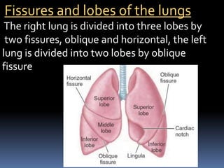
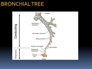
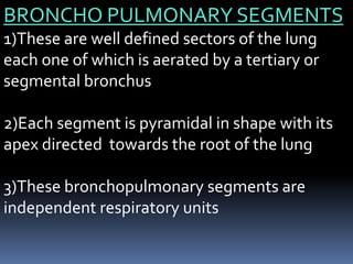
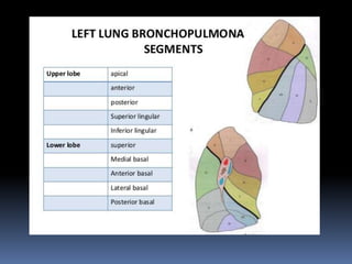
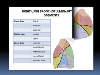
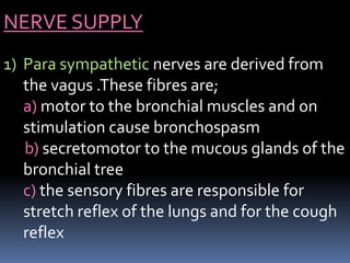
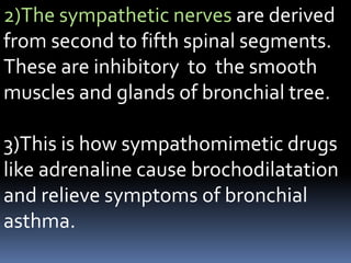
The lungs are a pair of cone-shaped respiratory organs located in the thoracic cavity. Each lung has an apex, base, and borders. The right lung has three lobes separated by two fissures, while the left lung has two lobes separated by one fissure. The root of each lung contains structures like the principal bronchus, pulmonary artery and veins, and nerves that enter and exit the hilum. Bronchopulmonary segments are independent respiratory units supplied by segmental bronchi, arteries, and multiple veins. The lungs receive arterial blood supply and have venous drainage pathways. They are innervated by both parasympathetic and sympathetic nerves.
This presentation is an overview of the description of the 4 stages of the cardiac cycle (atrial diastole, atrial systole, ventricular systole, ventricular diastole) as well as explaining the mechanism of the cardiac cycle.
The lungs are a pair of spongy, air-filled organs located on either side of the chest (thorax). The trachea (windpipe) conducts inhaled air into the lungs through its tubular branches, called bronchi. The bronchi then divide into smaller and smaller branches (bronchioles), finally becoming microscopic.
The bronchioles eventually end in clusters of microscopic air sacs called alveoli. In the alveoli, oxygen from the air is absorbed into the blood. Carbon dioxide, a waste product of metabolism, travels from the blood to the alveoli, where it can be exhaled. Between the alveoli is a thin layer of cells called the interstitium, which contains blood vessels and cells that help support the alveoli.
in this presentation the complete anatomy of the lungs is explained, which is very easy to understand. it is very useful for the students of medical field and other students who are appearing in the competitive exams like neet, cet etc.
Explore natural remedies for syphilis treatment in Singapore. Discover alternative therapies, herbal remedies, and lifestyle changes that may complement conventional treatments. Learn about holistic approaches to managing syphilis symptoms and supporting overall health.
Anti ulcer drugs and their Advance pharmacology ||
Anti-ulcer drugs are medications used to prevent and treat ulcers in the stomach and upper part of the small intestine (duodenal ulcers). These ulcers are often caused by an imbalance between stomach acid and the mucosal lining, which protects the stomach lining.
||Scope: Overview of various classes of anti-ulcer drugs, their mechanisms of action, indications, side effects, and clinical considerations.
- Video recording of this lecture in English language: https://youtu.be/lK81BzxMqdo
- Video recording of this lecture in Arabic language: https://youtu.be/Ve4P0COk9OI
- Link to download the book free: https://nephrotube.blogspot.com/p/nephrotube-nephrology-books.html
- Link to NephroTube website: www.NephroTube.com
- Link to NephroTube social media accounts: https://nephrotube.blogspot.com/p/join-nephrotube-on-social-media.html
MANAGEMENT OF ATRIOVENTRICULAR CONDUCTION BLOCK.pdfJim Jacob Roy
Cardiac conduction defects can occur due to various causes.
Atrioventricular conduction blocks ( AV blocks ) are classified into 3 types.
This document describes the acute management of AV block.
Flu Vaccine Alert in Bangalore Karnatakaaddon Scans
As flu season approaches, health officials in Bangalore, Karnataka, are urging residents to get their flu vaccinations. The seasonal flu, while common, can lead to severe health complications, particularly for vulnerable populations such as young children, the elderly, and those with underlying health conditions.
Dr. Vidisha Kumari, a leading epidemiologist in Bangalore, emphasizes the importance of getting vaccinated. "The flu vaccine is our best defense against the influenza virus. It not only protects individuals but also helps prevent the spread of the virus in our communities," he says.
This year, the flu season is expected to coincide with a potential increase in other respiratory illnesses. The Karnataka Health Department has launched an awareness campaign highlighting the significance of flu vaccinations. They have set up multiple vaccination centers across Bangalore, making it convenient for residents to receive their shots.
To encourage widespread vaccination, the government is also collaborating with local schools, workplaces, and community centers to facilitate vaccination drives. Special attention is being given to ensuring that the vaccine is accessible to all, including marginalized communities who may have limited access to healthcare.
Residents are reminded that the flu vaccine is safe and effective. Common side effects are mild and may include soreness at the injection site, mild fever, or muscle aches. These side effects are generally short-lived and far less severe than the flu itself.
Healthcare providers are also stressing the importance of continuing COVID-19 precautions. Wearing masks, practicing good hand hygiene, and maintaining social distancing are still crucial, especially in crowded places.
Protect yourself and your loved ones by getting vaccinated. Together, we can help keep Bangalore healthy and safe this flu season. For more information on vaccination centers and schedules, residents can visit the Karnataka Health Department’s official website or follow their social media pages.
Stay informed, stay safe, and get your flu shot today!
These simplified slides by Dr. Sidra Arshad present an overview of the non-respiratory functions of the respiratory tract.
Learning objectives:
1. Enlist the non-respiratory functions of the respiratory tract
2. Briefly explain how these functions are carried out
3. Discuss the significance of dead space
4. Differentiate between minute ventilation and alveolar ventilation
5. Describe the cough and sneeze reflexes
Study Resources:
1. Chapter 39, Guyton and Hall Textbook of Medical Physiology, 14th edition
2. Chapter 34, Ganong’s Review of Medical Physiology, 26th edition
3. Chapter 17, Human Physiology by Lauralee Sherwood, 9th edition
4. Non-respiratory functions of the lungs https://academic.oup.com/bjaed/article/13/3/98/278874
Report Back from SGO 2024: What’s the Latest in Cervical Cancer?bkling
Are you curious about what’s new in cervical cancer research or unsure what the findings mean? Join Dr. Emily Ko, a gynecologic oncologist at Penn Medicine, to learn about the latest updates from the Society of Gynecologic Oncology (SGO) 2024 Annual Meeting on Women’s Cancer. Dr. Ko will discuss what the research presented at the conference means for you and answer your questions about the new developments.
ARTIFICIAL INTELLIGENCE IN HEALTHCARE.pdfAnujkumaranit
Artificial intelligence (AI) refers to the simulation of human intelligence processes by machines, especially computer systems. It encompasses tasks such as learning, reasoning, problem-solving, perception, and language understanding. AI technologies are revolutionizing various fields, from healthcare to finance, by enabling machines to perform tasks that typically require human intelligence.
TEST BANK for Operations Management, 14th Edition by William J. Stevenson, Ve...kevinkariuki227
TEST BANK for Operations Management, 14th Edition by William J. Stevenson, Verified Chapters 1 - 19, Complete Newest Version.pdf
Tom Selleck Health: A Comprehensive Look at the Iconic Actor’s Wellness Journeygreendigital
Tom Selleck, an enduring figure in Hollywood. has captivated audiences for decades with his rugged charm, iconic moustache. and memorable roles in television and film. From his breakout role as Thomas Magnum in Magnum P.I. to his current portrayal of Frank Reagan in Blue Bloods. Selleck's career has spanned over 50 years. But beyond his professional achievements. fans have often been curious about Tom Selleck Health. especially as he has aged in the public eye.
Follow us on: Pinterest
Introduction
Many have been interested in Tom Selleck health. not only because of his enduring presence on screen but also because of the challenges. and lifestyle choices he has faced and made over the years. This article delves into the various aspects of Tom Selleck health. exploring his fitness regimen, diet, mental health. and the challenges he has encountered as he ages. We'll look at how he maintains his well-being. the health issues he has faced, and his approach to ageing .
Early Life and Career
Childhood and Athletic Beginnings
Tom Selleck was born on January 29, 1945, in Detroit, Michigan, and grew up in Sherman Oaks, California. From an early age, he was involved in sports, particularly basketball. which played a significant role in his physical development. His athletic pursuits continued into college. where he attended the University of Southern California (USC) on a basketball scholarship. This early involvement in sports laid a strong foundation for his physical health and disciplined lifestyle.
Transition to Acting
Selleck's transition from an athlete to an actor came with its physical demands. His first significant role in "Magnum P.I." required him to perform various stunts and maintain a fit appearance. This role, which he played from 1980 to 1988. necessitated a rigorous fitness routine to meet the show's demands. setting the stage for his long-term commitment to health and wellness.
Fitness Regimen
Workout Routine
Tom Selleck health and fitness regimen has evolved. adapting to his changing roles and age. During his "Magnum, P.I." days. Selleck's workouts were intense and focused on building and maintaining muscle mass. His routine included weightlifting, cardiovascular exercises. and specific training for the stunts he performed on the show.
Selleck adjusted his fitness routine as he aged to suit his body's needs. Today, his workouts focus on maintaining flexibility, strength, and cardiovascular health. He incorporates low-impact exercises such as swimming, walking, and light weightlifting. This balanced approach helps him stay fit without putting undue strain on his joints and muscles.
Importance of Flexibility and Mobility
In recent years, Selleck has emphasized the importance of flexibility and mobility in his fitness regimen. Understanding the natural decline in muscle mass and joint flexibility with age. he includes stretching and yoga in his routine. These practices help prevent injuries, improve posture, and maintain mobilit
2. INTRODUCTION
1)Lungs are a pair of respiratory organs situated in
the thoracic cavity.
2)Each lung innervates the corresponding pleural
cavity
3)In the young ,lungs are brown or grey in color.
Gradually, they become mottled black because of the
deposition of inhaled carbon particles
4)The right lung weighs about 700g.It is about 50-
100g heavier than the left lung.
3. FEATURES
1)Each lung is conical in shape
2)It has a) apex at the upper end
b) Base resting on the diaphragm
c)Three borders,i.e.,anterior,posterior,
and inferior
d)Two surfaces,i.e.costal and medial
4. 1) The apex lies above the level of first rib.It reaches
2.5 cm above the medial one third of clavicle,just
medial to supraclavicular fossa.
2) The base rests on the diaphragm which separates
the right lung from the right lobe of the liver and the
left lung from the left lobe of the liver,fundus of
stomach and the spleen
3) The anterior border of the left lung shows a wide
cardiac notch below the level of the fourth costal
cartilage.The heart and pericardium are uncovered by
the lung in the region of this notch.
5. 4) The posterior border corresponds to the medial
margins of the heads of the ribs.It extends from the
level of 7th cervical spine to the 10th thoracic spine.
5) Inferior border separates the base from costal
and medial surfaces.
6) The costal surface is large and convex.It is in contact
with the costal pleura and the overlying thoracic wall.
7) The medial surface is divided into a vertebral part
and mediastinal part.The mediastinal part shows a
cardiac impression ,the hilum
6. Fissures and lobes of the lungs
The right lung is divided into three lobes by
two fissures, oblique and horizontal, the left
lung is divided into two lobes by oblique
fissure
7. 1) The oblique fissure cuts into whole thickness
of the lung,except at the hilum
2) Due to the oblique plane of the fissure ,the
lower lobe is more posterior and the upper
and the middle lobe more anterior
3) In the right lung, the horizontal fissure
passes from the anterior border upto the
oblique fissure and separates a wedge
shaped middle lobe from the upper lobe.
8. 4)The tongue shaped projection of the left lung
below the cardiac notch is called lingula. It
corresponds to middle lobe of the right lung
5)The lungs expand maximally in the inferior
direction because movements of the thoracic wall
and diaphragm are maximal toward the base of the
lung
6)The presence of the the oblique fissure of the each
lung allows a more uniform expansion of the whole
lung
9. ROOT OFTHE LUNG
1)The root of the lung is a short ,broad
pedicle which connects the medial surface
of the lung to the mediastinum
2)It is formed by the structures which either
enter or come out of the lung at the hilum
3)The roots of lungs lie opposite the bodies
of fifth,sixth and seventh thoracic vertebrae
10. CONTENTS OF ROOT OFTHE LUNG
1)Principal bronchus on the left side, and eparterial
and hyparterial bronchi on right side
2)One pulmonary artery
3)Two pulmonary veins, superior and inferior
4)Bronchial arteries ,one on the right side and two on
the left side
5)Bronchial veins
6)Anterior and posterior pulmonary plexuses of
nerves
7)Lymphatics of lung
8)Bronchopulmonary lymph nodes
9)Areolar tissue
12. BRONCHO PULMONARY SEGMENTS
1)These are well defined sectors of the lung
each one of which is aerated by a tertiary or
segmental bronchus
2)Each segment is pyramidal in shape with its
apex directed towards the root of the lung
3)These bronchopulmonary segments are
independent respiratory units
13.
14.
15. 1)The branches of the pulmonary artery
accompany the bronchi ,the artery lies
dorsolateral to the bronchus ,thus each
segment has its own separate artery
2)The pulmonary veins do not accompany the
bronchi or the pulmonary artery ,they run in
inter segmental planes
3)Thus ,each segment has more than one vein
and each vein drains more than one segment
16. 1) Usually the infection of a segment
remains restricted to it although some
infections like tuberculosis may spread
from one segment to the other
2) However, the segments are no barriers
to the spread of bronchogenic
carcinoma.
17. ARTERIAL SUPPLY
1) on the right side there is one bronchial artery which
arises from either the third posterior intercostal
artery or from the upper left bronchial artery
2)On the left side there are two bronchial arteries
both of which arise from the descending thoracic
aorta
3)There are pre capillary anastomoses between
bronchial and pulmonary arteries. these connections
enlarge when any one of them is obstructed in
disease
18. VENOUS DRAINAGE
1) Usually there are two bronchial veins on each
side ,the right bronchial vein drain into the
azygous vein
2) The left bronchial vein drains either into the left
superior intercostal vein or into the hemi
azygous vein
3) The greater part of the venous blood from the
lung is drained by the pulmonary veins.
19. NERVE SUPPLY
1) Para sympathetic nerves are derived from
the vagus .These fibres are;
a) motor to the bronchial muscles and on
stimulation cause bronchospasm
b) secretomotor to the mucous glands of the
bronchial tree
c) the sensory fibres are responsible for
stretch reflex of the lungs and for the cough
reflex
20. 2)The sympathetic nerves are derived
from second to fifth spinal segments.
These are inhibitory to the smooth
muscles and glands of bronchial tree.
3)This is how sympathomimetic drugs
like adrenaline cause brochodilatation
and relieve symptoms of bronchial
asthma.