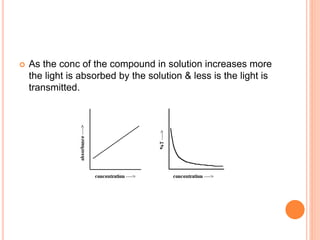
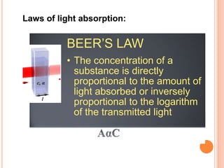
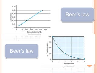
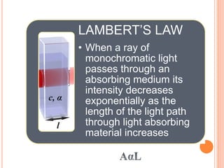
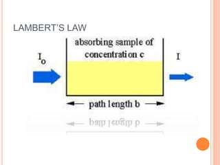
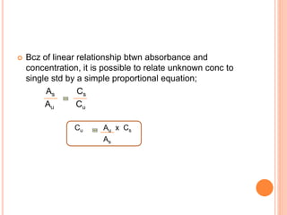
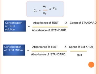
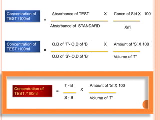
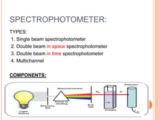
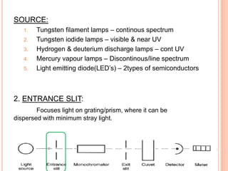
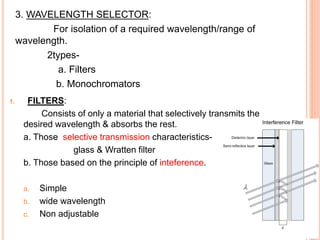
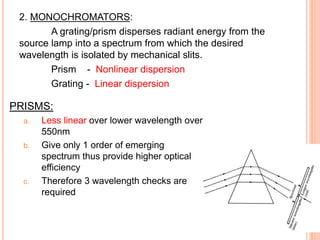
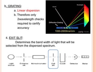
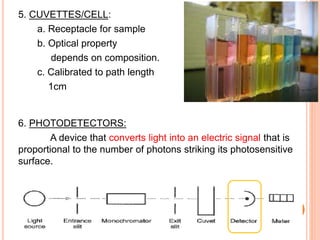
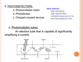
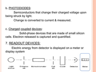
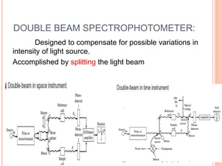
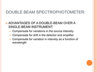
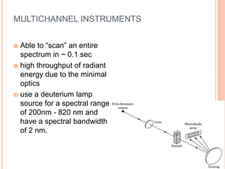
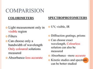
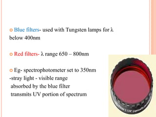
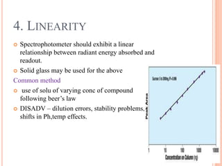
PHOTOMETRY
Presenter: Dr. Anurag Yadav
Moderator: Mr. Arun kumar
 What is light?
 How light interact with matter.
Is form of energy E=hv V=1/λ
Energy is inversely related to wavelength.
Matter react to the light either by
-Absorbing
-Emiting
EMISSION PROCESS
 When electron return to ground state, the energy is
dissipated as radiant energy.
 Used in flame photometry & flurometric methods.
ABSORPTION SPECTROSCOPY
 Io impinging on & passing through a
square cell that contain solution of compound that absorb
radiant energy of certain wavelength.
The intensity Is is less than Io.
The transmittance in solution is defined as proportion of
the incident light that is transmitted
T = Is/Io
 As the conc of the compound in solution increases more
the light is absorbed by the solution & less is the light is
transmitted.
T = IE /Io
% T= IE /Io X 100%
A = - log T
A = log 1/T
To convert T to %
A = log 1/T X 100%/100%= log 100%/%T
Rearranging
A = log 100%- log%T
EQUATION
A = 2 – log %T
BEER’S LAW
• The concentration of a
substance is directly
proportional to the amount of
light absorbed or inversely
proportional to the logarithm
of the transmitted light
Laws of light absorption:
Beer’s law
Beer’s law
LAMBERT’S LAW
• When a ray of
monochromatic light
passes through an
absorbing medium its
intensity decreases
exponentially as the
length of the light path
through light absorbing
material increases
LAMBERT’S LAW
 Bcz of linear relationship btwn absorbance and
concentration, it is possible to relate unknown conc to
single std by a simple proportional equation;
As Cs
Au Cu
Cu Au x Cs
As
CT =
AT
AS
X CS
Concentration
of TEST
solution
Absorbance of TEST
Absorbance of STANDARD
Concn of STANDARDX
=
Concentration
of TEST /100ml
Absorbance of TEST
Absorbance of STANDARD
Concn of Std X 100X
=
Xml
Concentration of
TEST /100ml
Absorbance of TEST
Absorbance of STANDARD
X
=
Xml
Concn of Std X 100
Concentration of
TEST /100ml
O.D of ‘T’- O.D of ‘B’
O.D of ‘S’- O.D of ‘B’
X
=
Volume of ‘T’
Amount of ‘S’ X 100
Concentration of
TEST /100ml
T - B
S - B
X=
Volume of ‘T’
Amount of ‘S’ X 100
LIMITATIONS:
 Very elevated conc cant be measured.
 Incident radiant energy is not monochromatic.
 Solvent absorption is significant compared to solute
absorbance.
 Radiant energy is transmitted by other mechanism (
stray light).
 The sides of the cell are not parallel.
Note on Stray Light:
- Is radiant energy that reaches the detector at
wavelength other than those indicated by
monochromator setting.
- All radiant energy that reaches detector with/without
having passed through the sample is recorded.
- As the amount of the stray light increases, deviation
from the Beer’s Law also increases.
1. Can be due to Light leaks – excluded by covering cell
compartment
2. Fluorescence- that increases signal to the detector and
causes apparent decrease in A
 Most spectrophotometers are equipped with stray light filters
SPECTROPHOTOMETER:
DEFINITIONS
PHOTOMETER:
If a filter is used as a wavelength selector,
monochromatic light at only discrete wavelength is
avialable & the instrument is called photometer.
SPECTROPHOTOMETER:
If a monochromater is used( prism/grating) as a
wavelength selector, the inst can provide monochromatic
light over a continous range of wavelengths & is called
spectrophotometer.
SPECTROPHOTOMETER:
TYPES:
1. Single beam spectrophotometer
2. Double beam in space spectrophotometer
3. Double beam in time spectrophotometer
4. Multichannel
COMPONENTS:
SOURCE:
1. Tungsten filament lamps – continous spectrum
2. Tungsten iodide lamps – visible & near UV
3. Hydrogen & deuterium discharge lamps – cont UV
4. Mercury vapour lamps – Discontinous/line spectrum
5. Light emitting diode(LED’s) – 2types of semiconductors
2. ENTRANCE SLIT:
Focuses light on grating/prism, where it can be
dispersed with minimum stray light.
3. WAVELENGTH SELECTOR:
For isolation of a required wavelength/range of
wavelength.
2types-
a. Filters
b. Monochromators
1. FILTERS:
Consists of only a material that selectively transmits the
desired wavelength & absorbs the rest.
a. Those selective transmission characteristics-
glass & Wratten filter
b. Those based on the principle of inteference.
a. Simple
b. wide wavelength
c. Non adjustable
2. MONOCHROMATORS:
A grating/prism disperses radiant energy from the
source lamp into a spectrum from which the desired
wavelength is isolated by mechanical slits.
Prism - Nonlinear dispersion
Grating - Linear dispersion
PRISMS:
a. Less linear over lower wavelength over
550nm
b. Give only 1 order of emerging
spectrum thus provide higher optical
efficiency
c. Therefore 3 wavelength checks are
required
b. GRATING:
a. Linear dispersion
b. Therefore only
2wavelength checks
required to certify
accuracy
4. EXIT SLIT:
Determines the band width of light that will be
selected from the dispersed spectrum.
1000-2000line/mm
5. CUVETTES/CELL:
a. Receptacle for sample
b. Optical property
depends on composition.
c. Calibrated to path length
1cm
6. PHOTODETECTORS:
A device that converts light into an electric signal that is
proportional to the number of photons striking its photosensitive
surface.
6. PHOTODETECTORS:
a. Photomultiplier tubes
b. Photodiodes
c. Charged coupled devices
a. Photomultiplier tubes:
An electron tube that is capable of significantly
amplifying a current.
Ideal detector :
high sensitivity,
high signal/noise,
constant response for λs,
and fast response time.
b. PHOTODIODES:
Semiconductors that change their charged voltage upon
being struck by light.
Change is converted to current & measured.
c. Charged coupled devices:
Solid-phase devices that are made of small silicon
cells. Electron released is captured and quantified.
7. READOUT DEVICES:
Electric energy from detector is displayed on a meter or
display system
DOUBLE BEAM SPECTROPHOTOMETER:
Designed to compensate for possible variations in
intensity of light source.
Accomplished by splitting the light beam
DOUBLE BEAM SPECTROPHOTOMETER:
 ADVANTAGES OF A DOUBLE-BEAM OVER A
SINGLE-BEAM INSTRUMENT:
 Compensate for variations in the source intensity.
 Compensate for drift in the detector and amplifier.
 Compensate for variation in intensity as a function of
wavelength
MULTICHANNEL INSTRUMENTS
 Able to “scan” an entire
spectrum in ~ 0.1 sec
 high throughput of radiant
energy due to the minimal
optics
 use a deuterium lamp
source for a spectral range
of 200nm - 820 nm and
have a spectral bandwidth
of 2 nm.
COMPARISION
COLORIMETERS
 Light measurement only in
visible region
 Filters
 Can choose only a
bandwidth of wavelength
Only coloured solutions
measured
 Absorbance-less accurate
SPECTROPHOTOMETERS
 UV, visible, IR
 Diffraction gratings, prisms
 Can choose exact
wavelength, Colourless
solution can also be
measured
 Absorbance –more accurate
 Kinetic studies and spectrum
can be better studied
 Wavelength accuracy: mercury vapor lamp, dueterium
lamp,(strong emmision lines), holmium oxide (strong
absorbtion lines).
 Linearity of detector response: solutions of varying
concentrations of compound(Beer’s law) Eg: oxyhb at
415nm, cobalt ammonium sulphate at 512nm
 Stray radiation : by LiCO3 below 250nm, NaBr below
240nm
 Photometric accuracy: pottasium dichromate soln,
cobalt ammonium sulphate soln
QUALITY CONTROL CHECK FOR
SPECTROPHOTOMETER
 NIST formerly NBS provide the SRM –useful
for calibration/verification of performance of the
instrument
 Eg- SRM 930e –verifies and calibrates T and A –
visible range of spectrophotometer.
 IRMM –provide reference material for
verification of performance of the instrument.
 listed in the IRMM BCR ref material catalogue.
QUALITY CONTROL CHECK FOR
SPECTROPHOTOMETER
 satisfactory if its close to λmax of chromogen and if
its reproducible.
 H and Du lamps have built in sources of checking
accuracy
 Prisms(2) and gratings(3) – continuous choice of λ.
 Rare earth glass filters like holmium oxide and
didymium- narrow and wide spectral band widths
1. WAVELENGTH CALIBRATION
 Holmium oxide –
 280-360nm ,
 show sharp absorption
peaks at defined λ
 Another method is by use of solutions.
Disadvantage
absorption peaks are broad and causes spectral
shifts due to
1. contamination
2. aging or
3. preparation errors
 Hg vapor lamp that shows a no of sharp, well
defined emissions lines bet 250 - 580nm .
 Can be calculated from manufactures
specifications.
 Interference filters – 1-2 nm are available and
can be used to check spectral bandwidth of 8nm
or more
2. SPECTRAL BANDWIDTH
 Increases at extreme ends of spectral range
where detector response is lowest.
 Methods to detect
1. filters or solu that’s highly transmitting over a
portion of the spectrum but opaque below an
abrupt cutoff λ
 Egs
1. Li2 CO3 below 250nm
2. NaBr – below 240nm
3. Acetone- below 320nm
3.STRAY RADIATION
 Stray light can also be due to
1. Light leaks – excluded by covering cell
compartment
2. Fluorescence- that increases signal to the
detector and causes apparent decrease in A
 Most spectrophotometers are equipped with stray
light filters
 Blue filters- used with Tungsten lamps for λ
below 400nm
 Red filters- λ range 650 – 800nm
 Eg- spectrophotometer set to 350nm
-stray light - visible range
absorbed by the blue filter
transmits UV portion of spectrum
 If solu / filters –transmitting no radiant energy at measurement
λ
measured T = amount of stray light
T X 100% = % of stray radiation.
If stray light > 1% - instrument malfunction
Liquid cut off filters- UV range where there is more stray light
problem
 UV stray light filters
 Spectrophotometer should exhibit a linear
relationship between radiant energy absorbed and
readout.
 Solid glass may be used for the above
Common method
 use of solu of varying conc of compound
following beer’s law
 DISADV – dilution errors, stability problems,
shifts in Ph,temp effects.
4. LINEARITY
 Absorbance std – constant stable A with no variation to
spectral band width / light beam.
 NIST –set of 3 neutral density glass filters with known A at 4
λ for each filter.
 They are not always stable- need recalibration by NIST
periodically
Standards for checking accuracy
 - potassium dichromate
 -cobalt amm sulfate
 -nitrate solu
5. PHOTOMETRIC ACCURACY
 Background interferences – min by including blank or taking
A at 2-3 λ.
 Bichromatic-A is measured at 2 λ
1. corresponds to peak A
2. at a point at the base of the peak serves as baseline.
 Diff in A is related to conc – gives a blank ref point for each
sample.
 Another method to correct background interference- measure
A at 2 λ equidistant from peak and latter is averaged to get a
baseline and that’s subtracted from the peak A –
CORRECTED A
6. MULTIPLE WAVELENGTH
READINGS
 In cases of spectral overlap –
extinction coeff of each component at
each λ should be known.
 Eg – in blood Hb (red Hb ,oxy Hb
,carboxyHb ,meth Hb , sulfHb)
 ext coeff is known the matrix eq can
be set up to calculate each component
– principle used in COOXIMETERS
 Visible Spectrophotometer Application
- Niacin, Pyridoxine, Vitamin B12, Metal Determination (Fe),
Fat-quality Determination, Enzyme Activity (glucose oxidase)
 UV Spectrophotometer Application
-Protein, Amino Acids (aromatic), Pantothenic Acid, Glucose
Determination, enzyme Activity (Hexokinase)
APPLICTIONS OF SPECTROPHOTOMETER
Photometry by Dr. Anurag Yadav

Photometry by Dr. Anurag Yadav

  • 1.
    PHOTOMETRY Presenter: Dr. AnuragYadav Moderator: Mr. Arun kumar
  • 2.
     What islight?  How light interact with matter. Is form of energy E=hv V=1/λ Energy is inversely related to wavelength. Matter react to the light either by -Absorbing -Emiting
  • 3.
    EMISSION PROCESS  Whenelectron return to ground state, the energy is dissipated as radiant energy.  Used in flame photometry & flurometric methods.
  • 4.
    ABSORPTION SPECTROSCOPY  Ioimpinging on & passing through a square cell that contain solution of compound that absorb radiant energy of certain wavelength. The intensity Is is less than Io. The transmittance in solution is defined as proportion of the incident light that is transmitted T = Is/Io
  • 5.
     As theconc of the compound in solution increases more the light is absorbed by the solution & less is the light is transmitted.
  • 6.
    T = IE/Io % T= IE /Io X 100% A = - log T A = log 1/T To convert T to % A = log 1/T X 100%/100%= log 100%/%T Rearranging A = log 100%- log%T EQUATION A = 2 – log %T
  • 7.
    BEER’S LAW • Theconcentration of a substance is directly proportional to the amount of light absorbed or inversely proportional to the logarithm of the transmitted light Laws of light absorption:
  • 8.
  • 9.
    LAMBERT’S LAW • Whena ray of monochromatic light passes through an absorbing medium its intensity decreases exponentially as the length of the light path through light absorbing material increases
  • 10.
  • 11.
     Bcz oflinear relationship btwn absorbance and concentration, it is possible to relate unknown conc to single std by a simple proportional equation; As Cs Au Cu Cu Au x Cs As
  • 12.
    CT = AT AS X CS Concentration ofTEST solution Absorbance of TEST Absorbance of STANDARD Concn of STANDARDX = Concentration of TEST /100ml Absorbance of TEST Absorbance of STANDARD Concn of Std X 100X = Xml
  • 13.
    Concentration of TEST /100ml Absorbanceof TEST Absorbance of STANDARD X = Xml Concn of Std X 100 Concentration of TEST /100ml O.D of ‘T’- O.D of ‘B’ O.D of ‘S’- O.D of ‘B’ X = Volume of ‘T’ Amount of ‘S’ X 100 Concentration of TEST /100ml T - B S - B X= Volume of ‘T’ Amount of ‘S’ X 100
  • 14.
    LIMITATIONS:  Very elevatedconc cant be measured.  Incident radiant energy is not monochromatic.  Solvent absorption is significant compared to solute absorbance.  Radiant energy is transmitted by other mechanism ( stray light).  The sides of the cell are not parallel.
  • 15.
    Note on StrayLight: - Is radiant energy that reaches the detector at wavelength other than those indicated by monochromator setting. - All radiant energy that reaches detector with/without having passed through the sample is recorded. - As the amount of the stray light increases, deviation from the Beer’s Law also increases.
  • 16.
    1. Can bedue to Light leaks – excluded by covering cell compartment 2. Fluorescence- that increases signal to the detector and causes apparent decrease in A  Most spectrophotometers are equipped with stray light filters
  • 17.
    SPECTROPHOTOMETER: DEFINITIONS PHOTOMETER: If a filteris used as a wavelength selector, monochromatic light at only discrete wavelength is avialable & the instrument is called photometer. SPECTROPHOTOMETER: If a monochromater is used( prism/grating) as a wavelength selector, the inst can provide monochromatic light over a continous range of wavelengths & is called spectrophotometer.
  • 18.
    SPECTROPHOTOMETER: TYPES: 1. Single beamspectrophotometer 2. Double beam in space spectrophotometer 3. Double beam in time spectrophotometer 4. Multichannel COMPONENTS:
  • 19.
    SOURCE: 1. Tungsten filamentlamps – continous spectrum 2. Tungsten iodide lamps – visible & near UV 3. Hydrogen & deuterium discharge lamps – cont UV 4. Mercury vapour lamps – Discontinous/line spectrum 5. Light emitting diode(LED’s) – 2types of semiconductors 2. ENTRANCE SLIT: Focuses light on grating/prism, where it can be dispersed with minimum stray light.
  • 20.
    3. WAVELENGTH SELECTOR: Forisolation of a required wavelength/range of wavelength. 2types- a. Filters b. Monochromators 1. FILTERS: Consists of only a material that selectively transmits the desired wavelength & absorbs the rest. a. Those selective transmission characteristics- glass & Wratten filter b. Those based on the principle of inteference. a. Simple b. wide wavelength c. Non adjustable
  • 21.
    2. MONOCHROMATORS: A grating/prismdisperses radiant energy from the source lamp into a spectrum from which the desired wavelength is isolated by mechanical slits. Prism - Nonlinear dispersion Grating - Linear dispersion PRISMS: a. Less linear over lower wavelength over 550nm b. Give only 1 order of emerging spectrum thus provide higher optical efficiency c. Therefore 3 wavelength checks are required
  • 22.
    b. GRATING: a. Lineardispersion b. Therefore only 2wavelength checks required to certify accuracy 4. EXIT SLIT: Determines the band width of light that will be selected from the dispersed spectrum. 1000-2000line/mm
  • 23.
    5. CUVETTES/CELL: a. Receptaclefor sample b. Optical property depends on composition. c. Calibrated to path length 1cm 6. PHOTODETECTORS: A device that converts light into an electric signal that is proportional to the number of photons striking its photosensitive surface.
  • 24.
    6. PHOTODETECTORS: a. Photomultipliertubes b. Photodiodes c. Charged coupled devices a. Photomultiplier tubes: An electron tube that is capable of significantly amplifying a current. Ideal detector : high sensitivity, high signal/noise, constant response for λs, and fast response time.
  • 25.
    b. PHOTODIODES: Semiconductors thatchange their charged voltage upon being struck by light. Change is converted to current & measured. c. Charged coupled devices: Solid-phase devices that are made of small silicon cells. Electron released is captured and quantified. 7. READOUT DEVICES: Electric energy from detector is displayed on a meter or display system
  • 26.
    DOUBLE BEAM SPECTROPHOTOMETER: Designedto compensate for possible variations in intensity of light source. Accomplished by splitting the light beam
  • 27.
    DOUBLE BEAM SPECTROPHOTOMETER: ADVANTAGES OF A DOUBLE-BEAM OVER A SINGLE-BEAM INSTRUMENT:  Compensate for variations in the source intensity.  Compensate for drift in the detector and amplifier.  Compensate for variation in intensity as a function of wavelength
  • 28.
    MULTICHANNEL INSTRUMENTS  Ableto “scan” an entire spectrum in ~ 0.1 sec  high throughput of radiant energy due to the minimal optics  use a deuterium lamp source for a spectral range of 200nm - 820 nm and have a spectral bandwidth of 2 nm.
  • 29.
    COMPARISION COLORIMETERS  Light measurementonly in visible region  Filters  Can choose only a bandwidth of wavelength Only coloured solutions measured  Absorbance-less accurate SPECTROPHOTOMETERS  UV, visible, IR  Diffraction gratings, prisms  Can choose exact wavelength, Colourless solution can also be measured  Absorbance –more accurate  Kinetic studies and spectrum can be better studied
  • 30.
     Wavelength accuracy:mercury vapor lamp, dueterium lamp,(strong emmision lines), holmium oxide (strong absorbtion lines).  Linearity of detector response: solutions of varying concentrations of compound(Beer’s law) Eg: oxyhb at 415nm, cobalt ammonium sulphate at 512nm  Stray radiation : by LiCO3 below 250nm, NaBr below 240nm  Photometric accuracy: pottasium dichromate soln, cobalt ammonium sulphate soln QUALITY CONTROL CHECK FOR SPECTROPHOTOMETER
  • 31.
     NIST formerlyNBS provide the SRM –useful for calibration/verification of performance of the instrument  Eg- SRM 930e –verifies and calibrates T and A – visible range of spectrophotometer.  IRMM –provide reference material for verification of performance of the instrument.  listed in the IRMM BCR ref material catalogue. QUALITY CONTROL CHECK FOR SPECTROPHOTOMETER
  • 32.
     satisfactory ifits close to λmax of chromogen and if its reproducible.  H and Du lamps have built in sources of checking accuracy  Prisms(2) and gratings(3) – continuous choice of λ.  Rare earth glass filters like holmium oxide and didymium- narrow and wide spectral band widths 1. WAVELENGTH CALIBRATION
  • 33.
     Holmium oxide–  280-360nm ,  show sharp absorption peaks at defined λ
  • 34.
     Another methodis by use of solutions. Disadvantage absorption peaks are broad and causes spectral shifts due to 1. contamination 2. aging or 3. preparation errors
  • 35.
     Hg vaporlamp that shows a no of sharp, well defined emissions lines bet 250 - 580nm .  Can be calculated from manufactures specifications.  Interference filters – 1-2 nm are available and can be used to check spectral bandwidth of 8nm or more 2. SPECTRAL BANDWIDTH
  • 36.
     Increases atextreme ends of spectral range where detector response is lowest.  Methods to detect 1. filters or solu that’s highly transmitting over a portion of the spectrum but opaque below an abrupt cutoff λ  Egs 1. Li2 CO3 below 250nm 2. NaBr – below 240nm 3. Acetone- below 320nm 3.STRAY RADIATION
  • 37.
     Stray lightcan also be due to 1. Light leaks – excluded by covering cell compartment 2. Fluorescence- that increases signal to the detector and causes apparent decrease in A  Most spectrophotometers are equipped with stray light filters
  • 38.
     Blue filters-used with Tungsten lamps for λ below 400nm  Red filters- λ range 650 – 800nm  Eg- spectrophotometer set to 350nm -stray light - visible range absorbed by the blue filter transmits UV portion of spectrum
  • 39.
     If solu/ filters –transmitting no radiant energy at measurement λ measured T = amount of stray light T X 100% = % of stray radiation. If stray light > 1% - instrument malfunction Liquid cut off filters- UV range where there is more stray light problem  UV stray light filters
  • 40.
     Spectrophotometer shouldexhibit a linear relationship between radiant energy absorbed and readout.  Solid glass may be used for the above Common method  use of solu of varying conc of compound following beer’s law  DISADV – dilution errors, stability problems, shifts in Ph,temp effects. 4. LINEARITY
  • 41.
     Absorbance std– constant stable A with no variation to spectral band width / light beam.  NIST –set of 3 neutral density glass filters with known A at 4 λ for each filter.  They are not always stable- need recalibration by NIST periodically Standards for checking accuracy  - potassium dichromate  -cobalt amm sulfate  -nitrate solu 5. PHOTOMETRIC ACCURACY
  • 42.
     Background interferences– min by including blank or taking A at 2-3 λ.  Bichromatic-A is measured at 2 λ 1. corresponds to peak A 2. at a point at the base of the peak serves as baseline.  Diff in A is related to conc – gives a blank ref point for each sample.  Another method to correct background interference- measure A at 2 λ equidistant from peak and latter is averaged to get a baseline and that’s subtracted from the peak A – CORRECTED A 6. MULTIPLE WAVELENGTH READINGS
  • 43.
     In casesof spectral overlap – extinction coeff of each component at each λ should be known.  Eg – in blood Hb (red Hb ,oxy Hb ,carboxyHb ,meth Hb , sulfHb)  ext coeff is known the matrix eq can be set up to calculate each component – principle used in COOXIMETERS
  • 44.
     Visible SpectrophotometerApplication - Niacin, Pyridoxine, Vitamin B12, Metal Determination (Fe), Fat-quality Determination, Enzyme Activity (glucose oxidase)  UV Spectrophotometer Application -Protein, Amino Acids (aromatic), Pantothenic Acid, Glucose Determination, enzyme Activity (Hexokinase) APPLICTIONS OF SPECTROPHOTOMETER