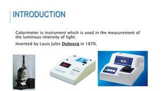
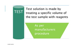
The document provides information about a colorimeter. It begins with an introduction to colorimeters, noting they measure light intensity and were invented in 1870. It then describes the basic principle that colorimeters measure light absorbed by colored solutions according to Beer's and Lambert's laws. The document outlines the typical parts of a colorimeter like its electronics and sample compartment. It discusses preparing blank, standard, and test solutions and explains Beer's and Lambert's laws. The document concludes by covering common colorimeter applications in clinical labs and industries like using them to estimate biochemical compounds in various samples.
It is the most common analytical technique used in biochemical estimation in clinical laboratory.
It involves the quantitative estimation of color.
A substance to be estimated colorimetrically, must be colored or it should be capable of forming chromogens (colored complexes) through the addition of reagents.
A spectrophotometer is an instrument containing a monochromator, a device which produces a light beam containing wavelengths in a narrow band around a selected wavelength, and a means of measuring the ratio of that beam's intensity as it enters and leaves a cuvette 99 This describes a single-beam photometer.
Colorimeter and spectrophotometer, Mass Spectrometerprachann
It contains a brief knowledge on Introduction, Principle, Laws, Flow representation, Instrumentation, Applications
and Mass spectrometer
- Principle
- Instrumentation
A spectrophotometer is an instrument that measures the amount of light absorbed by a sample. Spectrophotometer techniques are used to measure the concentration of solutes in solution by measuring the amount of the light that is absorbed by the solution in a cuvette placed in the spectrophotometer .
I am HAFIZ M WASEEM FROM mailsi vehari
BSc in science college Multan Pakistan
MSC university of education Lahore Pakistan
i love Pakistan and my teachers
• Chromatographic methods will separate ionic species, inorganic or organic, and molecular species ranging in size from the lightest and smallest, helium and hydrogen, to particulate matter such as single cells.
• Chromatography will separate several hundreds of components of unknown identity and unknown concentrations, leaving the components unchanged.
recent microbial techniques & advancement in identifying, cultivating,& handl...Karunanidhan3
I tried to include all techniques & diseases that are included in Pharm D 2nd year microbiology syllabus as per PCI. Do suggest if i have to improve my writing skills, on officialkarunanidhan@gmail.com
Ion exchange cromatography and affinity chromatographyKAUSHAL SAHU
Introduction.
Bygone days.
Basic terms related to chromatography.
Different type of chromatography techniques.
Ion exchange chromatography:
Principle of ion exchange chromatography.
Resin selection in ion exchange chromatography.
Commonly used ion exchangers.
The applications of ion exchange chromatography.
Merits and demerits of ion exchange chromatography.
Affinity chromatography:
Why use affinity chromatography?
Steps involved.
An example illustrating about the technique.
Choice of ligand.
The applications of affinity chromatography.
Merits and demerits of affinity chromatography.
Conclusion.
Bibliography.
Cancer cell metabolism: special Reference to Lactate PathwayAADYARAJPANDEY1
Normal Cell Metabolism:
Cellular respiration describes the series of steps that cells use to break down sugar and other chemicals to get the energy we need to function.
Energy is stored in the bonds of glucose and when glucose is broken down, much of that energy is released.
Cell utilize energy in the form of ATP.
The first step of respiration is called glycolysis. In a series of steps, glycolysis breaks glucose into two smaller molecules - a chemical called pyruvate. A small amount of ATP is formed during this process.
Most healthy cells continue the breakdown in a second process, called the Kreb's cycle. The Kreb's cycle allows cells to “burn” the pyruvates made in glycolysis to get more ATP.
The last step in the breakdown of glucose is called oxidative phosphorylation (Ox-Phos).
It takes place in specialized cell structures called mitochondria. This process produces a large amount of ATP. Importantly, cells need oxygen to complete oxidative phosphorylation.
If a cell completes only glycolysis, only 2 molecules of ATP are made per glucose. However, if the cell completes the entire respiration process (glycolysis - Kreb's - oxidative phosphorylation), about 36 molecules of ATP are created, giving it much more energy to use.
IN CANCER CELL:
Unlike healthy cells that "burn" the entire molecule of sugar to capture a large amount of energy as ATP, cancer cells are wasteful.
Cancer cells only partially break down sugar molecules. They overuse the first step of respiration, glycolysis. They frequently do not complete the second step, oxidative phosphorylation.
This results in only 2 molecules of ATP per each glucose molecule instead of the 36 or so ATPs healthy cells gain. As a result, cancer cells need to use a lot more sugar molecules to get enough energy to survive.
introduction to WARBERG PHENOMENA:
WARBURG EFFECT Usually, cancer cells are highly glycolytic (glucose addiction) and take up more glucose than do normal cells from outside.
Otto Heinrich Warburg (; 8 October 1883 – 1 August 1970) In 1931 was awarded the Nobel Prize in Physiology for his "discovery of the nature and mode of action of the respiratory enzyme.
WARNBURG EFFECT : cancer cells under aerobic (well-oxygenated) conditions to metabolize glucose to lactate (aerobic glycolysis) is known as the Warburg effect. Warburg made the observation that tumor slices consume glucose and secrete lactate at a higher rate than normal tissues.
Introduction:
RNA interference (RNAi) or Post-Transcriptional Gene Silencing (PTGS) is an important biological process for modulating eukaryotic gene expression.
It is highly conserved process of posttranscriptional gene silencing by which double stranded RNA (dsRNA) causes sequence-specific degradation of mRNA sequences.
dsRNA-induced gene silencing (RNAi) is reported in a wide range of eukaryotes ranging from worms, insects, mammals and plants.
This process mediates resistance to both endogenous parasitic and exogenous pathogenic nucleic acids, and regulates the expression of protein-coding genes.
What are small ncRNAs?
micro RNA (miRNA)
short interfering RNA (siRNA)
Properties of small non-coding RNA:
Involved in silencing mRNA transcripts.
Called “small” because they are usually only about 21-24 nucleotides long.
Synthesized by first cutting up longer precursor sequences (like the 61nt one that Lee discovered).
Silence an mRNA by base pairing with some sequence on the mRNA.
Discovery of siRNA?
The first small RNA:
In 1993 Rosalind Lee (Victor Ambros lab) was studying a non- coding gene in C. elegans, lin-4, that was involved in silencing of another gene, lin-14, at the appropriate time in the
development of the worm C. elegans.
Two small transcripts of lin-4 (22nt and 61nt) were found to be complementary to a sequence in the 3' UTR of lin-14.
Because lin-4 encoded no protein, she deduced that it must be these transcripts that are causing the silencing by RNA-RNA interactions.
Types of RNAi ( non coding RNA)
MiRNA
Length (23-25 nt)
Trans acting
Binds with target MRNA in mismatch
Translation inhibition
Si RNA
Length 21 nt.
Cis acting
Bind with target Mrna in perfect complementary sequence
Piwi-RNA
Length ; 25 to 36 nt.
Expressed in Germ Cells
Regulates trnasposomes activity
MECHANISM OF RNAI:
First the double-stranded RNA teams up with a protein complex named Dicer, which cuts the long RNA into short pieces.
Then another protein complex called RISC (RNA-induced silencing complex) discards one of the two RNA strands.
The RISC-docked, single-stranded RNA then pairs with the homologous mRNA and destroys it.
THE RISC COMPLEX:
RISC is large(>500kD) RNA multi- protein Binding complex which triggers MRNA degradation in response to MRNA
Unwinding of double stranded Si RNA by ATP independent Helicase
Active component of RISC is Ago proteins( ENDONUCLEASE) which cleave target MRNA.
DICER: endonuclease (RNase Family III)
Argonaute: Central Component of the RNA-Induced Silencing Complex (RISC)
One strand of the dsRNA produced by Dicer is retained in the RISC complex in association with Argonaute
ARGONAUTE PROTEIN :
1.PAZ(PIWI/Argonaute/ Zwille)- Recognition of target MRNA
2.PIWI (p-element induced wimpy Testis)- breaks Phosphodiester bond of mRNA.)RNAse H activity.
MiRNA:
The Double-stranded RNAs are naturally produced in eukaryotic cells during development, and they have a key role in regulating gene expression .
(May 29th, 2024) Advancements in Intravital Microscopy- Insights for Preclini...Scintica Instrumentation
Intravital microscopy (IVM) is a powerful tool utilized to study cellular behavior over time and space in vivo. Much of our understanding of cell biology has been accomplished using various in vitro and ex vivo methods; however, these studies do not necessarily reflect the natural dynamics of biological processes. Unlike traditional cell culture or fixed tissue imaging, IVM allows for the ultra-fast high-resolution imaging of cellular processes over time and space and were studied in its natural environment. Real-time visualization of biological processes in the context of an intact organism helps maintain physiological relevance and provide insights into the progression of disease, response to treatments or developmental processes.
In this webinar we give an overview of advanced applications of the IVM system in preclinical research. IVIM technology is a provider of all-in-one intravital microscopy systems and solutions optimized for in vivo imaging of live animal models at sub-micron resolution. The system’s unique features and user-friendly software enables researchers to probe fast dynamic biological processes such as immune cell tracking, cell-cell interaction as well as vascularization and tumor metastasis with exceptional detail. This webinar will also give an overview of IVM being utilized in drug development, offering a view into the intricate interaction between drugs/nanoparticles and tissues in vivo and allows for the evaluation of therapeutic intervention in a variety of tissues and organs. This interdisciplinary collaboration continues to drive the advancements of novel therapeutic strategies.
This pdf is about the Schizophrenia.
For more details visit on YouTube; @SELF-EXPLANATORY;
https://www.youtube.com/channel/UCAiarMZDNhe1A3Rnpr_WkzA/videos
Thanks...!
Slide 1: Title Slide
Extrachromosomal Inheritance
Slide 2: Introduction to Extrachromosomal Inheritance
Definition: Extrachromosomal inheritance refers to the transmission of genetic material that is not found within the nucleus.
Key Components: Involves genes located in mitochondria, chloroplasts, and plasmids.
Slide 3: Mitochondrial Inheritance
Mitochondria: Organelles responsible for energy production.
Mitochondrial DNA (mtDNA): Circular DNA molecule found in mitochondria.
Inheritance Pattern: Maternally inherited, meaning it is passed from mothers to all their offspring.
Diseases: Examples include Leber’s hereditary optic neuropathy (LHON) and mitochondrial myopathy.
Slide 4: Chloroplast Inheritance
Chloroplasts: Organelles responsible for photosynthesis in plants.
Chloroplast DNA (cpDNA): Circular DNA molecule found in chloroplasts.
Inheritance Pattern: Often maternally inherited in most plants, but can vary in some species.
Examples: Variegation in plants, where leaf color patterns are determined by chloroplast DNA.
Slide 5: Plasmid Inheritance
Plasmids: Small, circular DNA molecules found in bacteria and some eukaryotes.
Features: Can carry antibiotic resistance genes and can be transferred between cells through processes like conjugation.
Significance: Important in biotechnology for gene cloning and genetic engineering.
Slide 6: Mechanisms of Extrachromosomal Inheritance
Non-Mendelian Patterns: Do not follow Mendel’s laws of inheritance.
Cytoplasmic Segregation: During cell division, organelles like mitochondria and chloroplasts are randomly distributed to daughter cells.
Heteroplasmy: Presence of more than one type of organellar genome within a cell, leading to variation in expression.
Slide 7: Examples of Extrachromosomal Inheritance
Four O’clock Plant (Mirabilis jalapa): Shows variegated leaves due to different cpDNA in leaf cells.
Petite Mutants in Yeast: Result from mutations in mitochondrial DNA affecting respiration.
Slide 8: Importance of Extrachromosomal Inheritance
Evolution: Provides insight into the evolution of eukaryotic cells.
Medicine: Understanding mitochondrial inheritance helps in diagnosing and treating mitochondrial diseases.
Agriculture: Chloroplast inheritance can be used in plant breeding and genetic modification.
Slide 9: Recent Research and Advances
Gene Editing: Techniques like CRISPR-Cas9 are being used to edit mitochondrial and chloroplast DNA.
Therapies: Development of mitochondrial replacement therapy (MRT) for preventing mitochondrial diseases.
Slide 10: Conclusion
Summary: Extrachromosomal inheritance involves the transmission of genetic material outside the nucleus and plays a crucial role in genetics, medicine, and biotechnology.
Future Directions: Continued research and technological advancements hold promise for new treatments and applications.
Slide 11: Questions and Discussion
Invite Audience: Open the floor for any questions or further discussion on the topic.
2. PRESENTATION OUTLINE
INTODUCTION AND BRIEF HISTORY
PRINCIPLE
PARTS, COMPONENTS AND SCHEMATICS
CUVETTES AND TEST PROCEDURE
BEER-LAMBERT’S LAW
ADVANTAGES AND DISADVANTAGES
APPLICATION
CARE, USE AND MAINTENANCE
REFERENCES
4. COLORIMETER
It is the most common analytical technique
used in biochemical estimation in clinical
laboratory
It involves the quantitative estimation of color
A substance to be estimated colometrically,
must be colored or capable of forming
chromogens (colored complexes) through the
addition of reagents.
The color of light is the function of its
wavelength
5. COLORIMETER PRINCIPLE
When a monochromatic light passes
through a coloured solution, some specific
wavelengths of light are absorbed which is
related to colour intensity.
The amount of light absorbed or
transmitted by a colour solution is in
accordance with two law i.e. Beer’s &
Lambert’s Law.
10. SAMPLE HOLDER/ CUVETTE
•Cuvettes are rectangular cell , square cell or circular
one.
•Made up of optical glass for visible wavelength
(quartz or fused silica for UV).
•Optical path (length) of cuvette is always1cm.
•Capacity may be 3ml/2ml/1ml depending upon the
thickness of the wall of the cuvette.
•For accurate and precise reading cuvette must be
transparent, clean, devoid of any scratches and there
should be no bubble adhering to the inner surface of
the filled cuvette.
11. PREPARATION OF SOLUTION FOR
INVESTIGATIONS
In colorimetric estimation it is necessary to prepare 3
solutions
5/19/2017 8:46 PM
BLANK(B)
STANDARD(S)
TEST(T)
12. BLANK
To eliminate the effect of light
absorption by the reagent used
Water BLANK
Reagent BLANK
5/19/2017 8:46 PM
13. STANDARD
Solution of known concentration of the
substance
Known concentration
So concentration of
unknown can be
calculated
5/19/2017 8:46 PM
14. TEST
Test solution is made by
treating a specific volume of
the test sample with reagents
As per
manufacturers
procedure
5/19/2017 8:46 PM
15. Observed Color of
Compound
Color of Light
Absorbed
Approximate
Wavelength of Light
Absorbed
Green Red 700 nm
Blue-green Orange-red 600 nm
Violet Yellow 550 nm
Red-violet Yellow-green 530 nm
Red Green 500 nm
Orange Blue 450 nm
Yellow Violet 400 nm
17. LAMBERT’S LAW
•When a ray of monochromatic light passes
through an absorbing medium its intensity
decreases exponentially as the length of the
light path through light absorbing material
increases
BEER’S LAW
•The concentration of a substance is directly
proportional to the amount of light absorbed
or inversely proportional to the logarithm of
the transmitted light
5/19/2017 8:46 PM
19. RELATIONSHIP BETWEEN
WAVELENGTH & COLOUR
Wavelength between 400nm to 700nm form the visible
spectrum of light
visible band of light in electromagnetic spectrum
21. COLORIMETER
Disadvantages
Cannot be used for colorless compounds.
It does not work in UV and IR regions.
We cannot set specific wavelength, as we have to set a range as a
parameter.
Similar colors from interfering substances can produce errors in
results .
22. APPLICATION
It is widely used in hospital & laboratory for estimation of
biochemical samples , like plasma, serum, cerebrospinal fluid (
csf ) , urine.
It is also used to quantitative estimation of serum components
as well as glucose, proteins and other various biochemical
compound.
They are used by the food industry and by manufacturers of
paints and textiles.
23. They are used to test for water quality, by screening for chemicals
such as chlorine, fluoride, cyanide, dissolved oxygen, iron,
molybdenum, zinc and hydrazine.
They are also used to determine the concentrations of plant nutrients
(such as phosphorus, nitrate and ammonia) in the soil or hemoglobin
in the blood and to identify substandard and counterfeit drugs.
24. USE, CARE AND MAINTENANCE OF
A COLORIMETER:
Read the user manual carefully.
Use the correct type of cuvette in the colorimeter as recommended by the manufacturer.
Make sure that the cuvette is clean and it’s optical surfaces are dry and free from finger marks
and scratches.
Clean the outside of the cuvette with tissue paper to remove any marks from the optical
surfaces.
To prolong the life of the lamp, switch off the colorimeter after use.
At the end of the day, disconnect It from the main switch and cover the colorimeter with its
protective cover.
25. Reference
Instrumental method of chemical analysis,chatwal.
Instrumental method of analysis, N.Grey, M.Calvin.
Pharmaceutical analysis, Ashutoshkar.
Instrumental method of analysis, B.K.Sharma
Principles and application of ultaviolet and visible
spectroscopy, A.Rajasekaran.
WWW.Pharmatwiter.com
WIKIPEDIA
Light source: tungsten filament lamp
Slit :It is adjustable which allows only a beam of light to pass through. It prevents unwanted or stray light
Condensing lens: Light after passing through a slit falls on a condenser which gives parallel beam of light.
Filters : are usually made of colored glass. it is used for selecting narrow wavelength .they absorb light of unwanted wavelength and allow only monochromatic light to pass through
For e.g.: , a green filter absorbs all colours,except green light which is allowed to pass through.light transmitted through a green filter has a wavelength from 500-560nm.Filters used is always complimentary to the colour of the solution
Cuvette
-may be square,rectangular or round shape with fixed diameter and having uniform surface
-made up of plastic ,glass material
-solution in the cuvette absorbs a part of the light and the remaining is allowed to fall on the detector
Detector (photocell): The detectors are photosensitive elements which converts light energy into electrical signal .the electrical signal is directly proportional to the intensity of light falling on the detector
Output : the electrical signal generated in a photocell is measured by a galvanometer which displays transmission and optical density