Objectives
Upon completion ofthis chapter the student will be able to:
Explain the composition of blood
Describe the morphology and functions of the formed
elements of blood
Discuss the functions of plasma
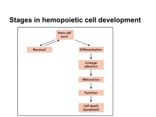
Define hemopoiesis and explain the process of blood cell
origin and development
Indicate the sites of hemopoiesis in infancy, childhood and
adulthood
List at least three hemopoietic growth factors
Name the cells in the development order that will mature into
erythrocytes, thrombocytes and the five leukocytes
3.
Objectives cont’d
Discusshow hemopoiesis is regulated
Describe the morphology of the red blood cell, white blood cell, and
platelet precursors
Define extramedullary hemopoiesis
Differentiate between intramedulary and extramedulary hemopoiesis
Define erythropoiesis
Explain how erythropiesis is regulated and list the effects of the
hormone erythropoietin on erythropoiesis
Define megaloblastic erythropoiesis
Define ineffective erythropoiesis
Define myeloid erythroid ratio
4.
Outline
Composition ofBlood
Characteristics of Blood
Formation of blood cells
Hemopoiesis
The Hemopoietic Microenvironment
Regulation of Hemopoiesis
Maturational characteristics of hemopoietic cells
5.
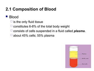
2.1 Composition ofBlood
Blood
is the only fluid tissue
constitutes 6-8% of the total body weight
consists of cells suspended in a fluid called plasma.
about 45% cells; 55% plasma
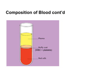
Composition cont’d
Plasma
part of the extracellular fluid
a complex solution of proteins, salts and numerous
metabolic substances
acts as a transport medium carrying its constituents to
specialized organs of the body.
Consists of:
about 91.5% water
about 8.5% solutes of which about 7% are proteins
Out of the 7% protein:
54% albumin
38% globulins
7% fibrinogen
8.
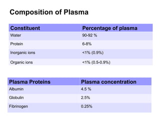
Composition of Plasma
ConstituentPercentage of plasma
Water 90-92 %
Protein 6-8%
Inorganic ions <1% (0.9%)
Organic ions <1% (0.5-0.9%)
Plasma Proteins Plasma concentration
Albumin 4.5 %
Globulin 2.5%
Fibrinogen 0.25%
9.
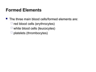
Formed Elements
Thethree main blood cells/formed elements are:
red blood cells (erythrocytes)
white blood cells (leucocytes)
platelets (thrombocytes)
10.
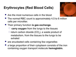
Erythrocytes (Red BloodCells)
Are the most numerous cells in the blood
The normal RBC count is approximately 4.5 to 6 million
cells per microliter.
Their primary function is gas exchange.
carry oxygen from the lungs to the tissues
return carbon dioxide (CO2), a waste product of
metabolism, from the tissues to the lungs to be
exhaled
are anucleated cells containing few organelles
a large proportion of their cytoplasm consists of the iron
containing oxygen transport molecule hemoglobin.
11.
Erythrocytes cont’d
shapedlike biconcave disks approximately 7 to 8µ m in
diameter with a thickness of 1.7-2.4m
The biconcave disk shape gives red blood cells (RBCs) the
flexibility to squeeze their way through capillaries and other
small blood vessels.
In stained smears, RBCs look like a circle with a central
hole, or central pallor, which is approximately one-third the
diameter of the cell
normally survives in the blood stream for approximately 120
days
after finishing its life span, it is removed by the phagocytic
cells of the reticuloendothelial system, broken down and
some of its constituents re utilized for the formation of new
cells.
12.
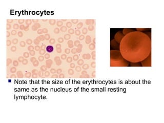
Erythrocytes
Note thatthe size of the erythrocytes is about the
same as the nucleus of the small resting
lymphocyte.
13.
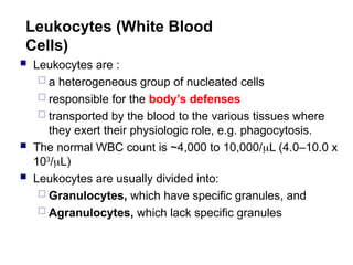
Leukocytes (White Blood
Cells)
Leukocytes are :
a heterogeneous group of nucleated cells
responsible for the body’s defenses
transported by the blood to the various tissues where
they exert their physiologic role, e.g. phagocytosis.
The normal WBC count is ~4,000 to 10,000/L (4.0–10.0 x
103
/L)
Leukocytes are usually divided into:
Granulocytes, which have specific granules, and
Agranulocytes, which lack specific granules
14.
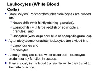
Leukocytes (White Blood
Cells)
Granulocytes/ Polymorphonuclear leukocytes are divided
into:
Neutrophils (with faintly staining granules),
Eosinophils (with large reddish or eosinophilic
granules), and
Basophils (with large dark blue or basophilic granules).
Agranulocytes/mononuclear leukocytes are divided into:
Lymphocytes and
Monocytes.
Although they are called white blood cells, leukocytes
predominantly function in tissues.
They are only in the blood transiently, while they travel to
their site of action.
15.
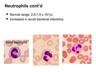
Neutrophils
are themost common type of WBCs in adults
The segmented neutrophils “segs,” also called
polymorphonuclear neutrophil leukocytes
[PMNs or “polys”]
are the primary defense against bacterial
infection
Their size ranges from 10-12m in diameter.
They are capable of amoeboid movement.
There are 2-5 lobes to their nucleus that stain
purple violet.
The cytoplasm stains light pink with pinkish dust
like granules.
16.
Neutrophils cont’d
Normalrange: 2.0-7.5 x 10³
/l.
Increased in acute bacterial infections.
Band Neutrphil
17.
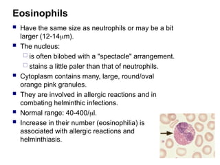
Eosinophils
Have thesame size as neutrophils or may be a bit
larger (12-14m).
The nucleus:
is often bilobed with a "spectacle" arrangement.
stains a little paler than that of neutrophils.
Cytoplasm contains many, large, round/oval
orange pink granules.
They are involved in allergic reactions and in
combating helminthic infections.
Normal range: 40-400/l.
Increase in their number (eosinophilia) is
associated with allergic reactions and
helminthiasis.
18.
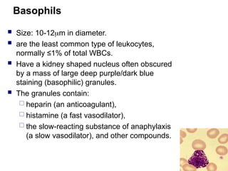
Basophils
Size: 10-12min diameter.
are the least common type of leukocytes,
normally ≤1% of total WBCs.
Have a kidney shaped nucleus often obscured
by a mass of large deep purple/dark blue
staining (basophilic) granules.
The granules contain:
heparin (an anticoagulant),
histamine (a fast vasodilator),
the slow-reacting substance of anaphylaxis
(a slow vasodilator), and other compounds.
19.
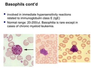
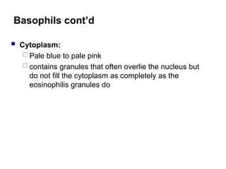
Basophils cont’d
involvedin immediate hypersensitivity reactions
related to immunoglobulin class E (IgE)
Normal range: 20-200/l. Basophilia is rare except in
cases of chronic myeloid leukemia.
20.
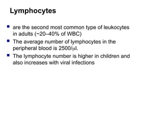
Lymphocytes
are thesecond most common type of leukocytes
in adults (~20–40% of WBC)
The average number of lymphocytes in the
peripheral blood is 2500/l.
The lymphocyte number is higher in children and
also increases with viral infections
21.
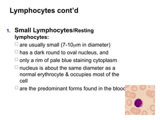
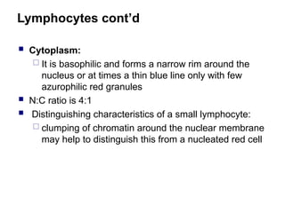
Lymphocytes cont’d
1. SmallLymphocytes/Resting
lymphocytes:
are usually small (7-10m in diameter)
has a dark round to oval nucleus, and
only a rim of pale blue staining cytoplasm
nucleus is about the same diameter as a
normal erythrocyte & occupies most of the
cell
are the predominant forms found in the blood.
22.
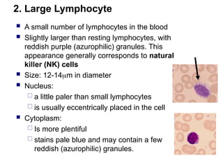
2. Large Lymphocyte
A small number of lymphocytes in the blood
Slightly larger than resting lymphocytes, with
reddish purple (azurophilic) granules. This
appearance generally corresponds to natural
killer (NK) cells
Size: 12-14m in diameter
Nucleus:
a little paler than small lymphocytes
is usually eccentrically placed in the cell
Cytoplasm:
Is more plentiful
stains pale blue and may contain a few
reddish (azurophilic) granules.
23.
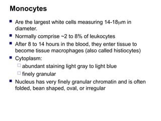
Monocytes
Are thelargest white cells measuring 14-18m in
diameter.
Normally comprise ~2 to 8% of leukocytes
After 8 to 14 hours in the blood, they enter tissue to
become tissue macrophages (also called histiocytes)
Cytoplasm:
abundant staining light gray to light blue
finely granular
Nucleus has very finely granular chromatin and is often
folded, bean shaped, oval, or irregular
24.
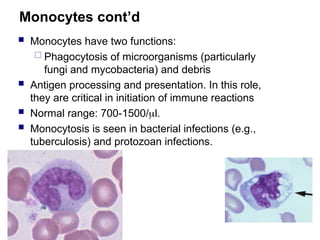
Monocytes cont’d
Monocyteshave two functions:
Phagocytosis of microorganisms (particularly
fungi and mycobacteria) and debris
Antigen processing and presentation. In this role,
they are critical in initiation of immune reactions
Normal range: 700-1500/l.
Monocytosis is seen in bacterial infections (e.g.,
tuberculosis) and protozoan infections.
25.
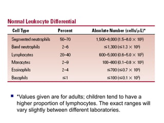
*Values givenare for adults; children tend to have a
higher proportion of lymphocytes. The exact ranges will
vary slightly between different laboratories.
26.
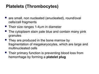
Platelets (Thrombocytes)
aresmall, non nucleated (anucleated), round/oval
cells/cell fragments
Their size ranges 1-4m in diameter
The cytoplasm stain pale blue and contain many pink
granules
They are produced in the bone marrow by
fragmentation of megakaryocytes, which are large and
multinucleated cells
Their primary function is preventing blood loss from
hemorrhage by forming a platelet plug
27.
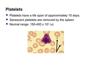
Platelets
Platelets havea life span of approximately 10 days.
Senescent platelets are removed by the spleen
Normal range: 150-400 x 103
/l.
28.
2.2. Characteristics ofBlood
1. Temperature
Roughly 38°C (100.4 °F)
2. Viscosity
Five times that of H2O due to interactions among
dissolved proteins, formed elements, & surrounding
H2O molecules
Sticky, cohesive, and resistant to flow
3. pH
Ranges from 7.35- 7.45, averaging 7.4
29.
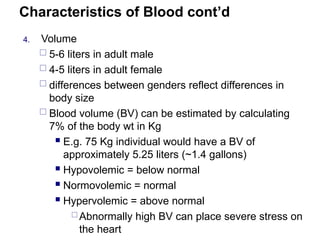
Characteristics of Bloodcont’d
4. Volume
5-6 liters in adult male
4-5 liters in adult female
differences between genders reflect differences in
body size
Blood volume (BV) can be estimated by calculating
7% of the body wt in Kg
E.g. 75 Kg individual would have a BV of
approximately 5.25 liters (~1.4 gallons)
Hypovolemic = below normal
Normovolemic = normal
Hypervolemic = above normal
Abnormally high BV can place severe stress on
the heart
30.
2.3. Function ofBlood
Transportation
O2 to tissues & CO2 from tissues to lung
Nutrients from GIT to cells
Heat and waste products from cells for excretion
Hormones from endocrine glands to other body cells
Regulation
pH
Temperature
Osmotic pressure (influence water and ion content of
cells)
31.
Function of Bloodcont’d
Protection
From bleeding (by the clotting mechanism)
Immunity (phagocytes, lymphocytes, antibodies,
complement proteins, etc)
32.
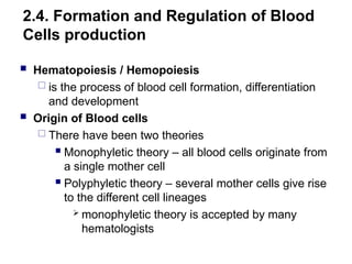
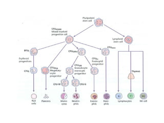
2.4. Formation andRegulation of Blood
Cells production
Hematopoiesis / Hemopoiesis
is the process of blood cell formation, differentiation
and development
Origin of Blood cells
There have been two theories
Monophyletic theory – all blood cells originate from
a single mother cell
Polyphyletic theory – several mother cells give rise
to the different cell lineages
monophyletic theory is accepted by many
hematologists
33.
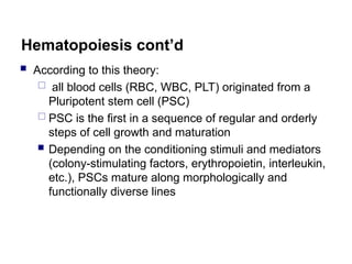
Hematopoiesis cont’d
Accordingto this theory:
all blood cells (RBC, WBC, PLT) originated from a
Pluripotent stem cell (PSC)
PSC is the first in a sequence of regular and orderly
steps of cell growth and maturation
Depending on the conditioning stimuli and mediators
(colony-stimulating factors, erythropoietin, interleukin,
etc.), PSCs mature along morphologically and
functionally diverse lines
34.
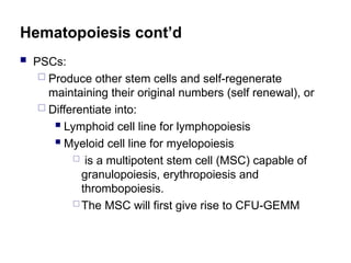
Hematopoiesis cont’d
PSCs:
Produce other stem cells and self-regenerate
maintaining their original numbers (self renewal), or
Differentiate into:
Lymphoid cell line for lymphopoiesis
Myeloid cell line for myelopoiesis
is a multipotent stem cell (MSC) capable of
granulopoiesis, erythropoiesis and
thrombopoiesis.
The MSC will first give rise to CFU-GEMM
35.
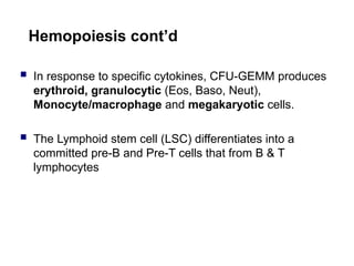
Hemopoiesis cont’d
Inresponse to specific cytokines, CFU-GEMM produces
erythroid, granulocytic (Eos, Baso, Neut),
Monocyte/macrophage and megakaryotic cells.
The Lymphoid stem cell (LSC) differentiates into a
committed pre-B and Pre-T cells that from B & T
lymphocytes
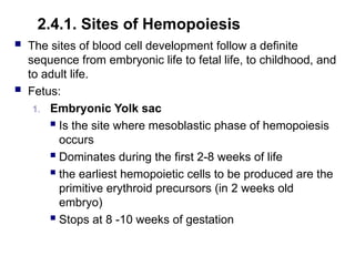
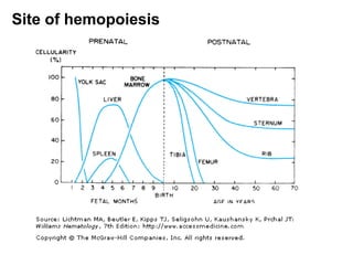
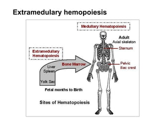
2.4.1. Sites ofHemopoiesis
The sites of blood cell development follow a definite
sequence from embryonic life to fetal life, to childhood, and
to adult life.
Fetus:
1. Embryonic Yolk sac
Is the site where mesoblastic phase of hemopoiesis
occurs
Dominates during the first 2-8 weeks of life
the earliest hemopoietic cells to be produced are the
primitive erythroid precursors (in 2 weeks old
embryo)
Stops at 8 -10 weeks of gestation
40.
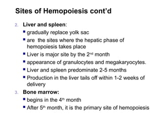
Sites of Hemopoiesiscont’d
2. Liver and spleen:
gradually replace yolk sac
are the sites where the hepatic phase of
hemopoiesis takes place
Liver is major site by the 2nd
month
appearance of granulocytes and megakaryocytes.
Liver and spleen predominate 2-5 months
Production in the liver tails off within 1-2 weeks of
delivery
3. Bone marrow:
begins in the 4th
month
After 5th
month, it is the primary site of hemopoiesis
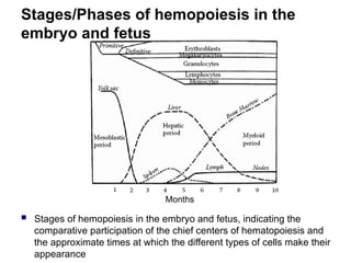
Stages/Phases of hemopoiesisin the
embryo and fetus
Stages of hemopoiesis in the embryo and fetus, indicating the
comparative participation of the chief centers of hematopoiesis and
the approximate times at which the different types of cells make their
appearance
Months
43.
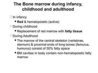
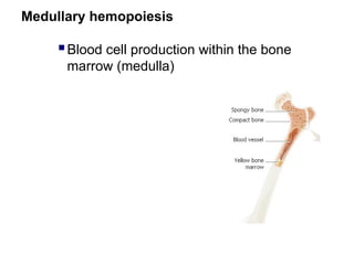
The Bone marrowduring infancy,
childhood and adulthood
In infancy
Red & hematopoietic (active)
During childhood
Replacement of red marrow with fatty tissue
During Adulthood
The marrow of the central skeleton (vertebrae,
sternum) & proximal ends of long bones (femurus,
humurus) consist of 50% fatty space
BM cavities in body contain non-hematopoietic fatty
marrow
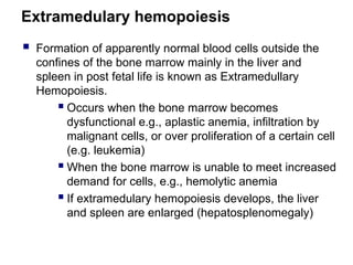
Extramedulary hemopoiesis
Formationof apparently normal blood cells outside the
confines of the bone marrow mainly in the liver and
spleen in post fetal life is known as Extramedullary
Hemopoiesis.
Occurs when the bone marrow becomes
dysfunctional e.g., aplastic anemia, infiltration by
malignant cells, or over proliferation of a certain cell
(e.g. leukemia)
When the bone marrow is unable to meet increased
demand for cells, e.g., hemolytic anemia
If extramedulary hemopoiesis develops, the liver
and spleen are enlarged (hepatosplenomegaly)
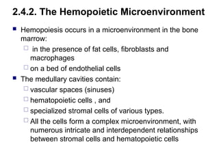
2.4.2. The HemopoieticMicroenvironment
Hemopoiesis occurs in a microenvironment in the bone
marrow:
in the presence of fat cells, fibroblasts and
macrophages
on a bed of endothelial cells
The medullary cavities contain:
vascular spaces (sinuses)
hematopoietic cells , and
specialized stromal cells of various types.
All the cells form a complex microenvironment, with
numerous intricate and interdependent relationships
between stromal cells and hematopoietic cells
49.
Hemopoietic Microenvironment cont’d
an extracellular matrix of fibronectin, collagen and
laminin combines with these cells to provide a setting in
which stem cells can grow and divide.
50.
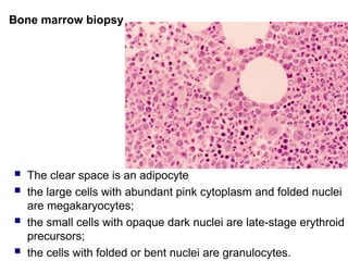
Bone marrow biopsy
The clear space is an adipocyte
the large cells with abundant pink cytoplasm and folded nuclei
are megakaryocytes;
the small cells with opaque dark nuclei are late-stage erythroid
precursors;
the cells with folded or bent nuclei are granulocytes.
51.
Bone Marrow Microenvironmentcont’d
Hemopoietic Cords (parenchyma) are the
extravascular portions of the bone marrow and the site of
blood cell production
Sinuses (vascular spaces) of the marrow are lined
with specialized endothelial cells, which prevent the
premature escape of immature cells into the peripheral
blood.
The basal lamina is incomplete, allowing mature cells to
pass through the wall of the sinuses.
52.
Bone Marrow Microenvironmentcont’d
Stromal Cells compose the supportive tissues of the bone
marrow. Some of these cells produce hemopoietic growth factors.
Examples include:
Adventitial (reticular) cells:
Are modified fibroblasts that produce the reticulin framework of
the bone marrow
Macrophages:
Produce hemopoietic growth factors
store iron for hemoglobin production, and
carry out phagocytosis of debris
Adipocytes: Store energy in the form of fat
53.
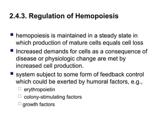
2.4.3. Regulation ofHemopoiesis
hemopoiesis is maintained in a steady state in
which production of mature cells equals cell loss
Increased demands for cells as a consequence of
disease or physiologic change are met by
increased cell production.
system subject to some form of feedback control
which could be exerted by humoral factors, e.g.,
erythropoietin
colony-stimulating factors
growth factors
54.
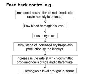
Feed back controle.g.
Increased destruction of red blood cells
(as in hemolytic anemia)
Low blood hemoglobin level
Tissue hypoxia
stimulation of increased erythropoietin
production by the kidneys
Increase in the rate at which committed
progenitor cells divide and differentiate
Hemoglobin level brought to normal
55.
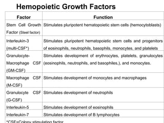
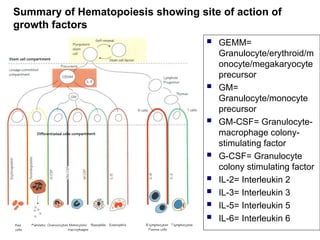
Hemopoietic Growth Factors
FactorFunction
Stem Cell Growth
Factor (Steel factor)
Stimulates pluripotent hematopoietic stem cells (hemocytoblasts)
Interleukin-3
(multi-CSF*)
Stimulates pluripotent hematopoietic stem cells and progenitors
of eosinophils, neutrophils, basophils, monocytes, and platelets
Granulocyte-
Macrophage CSF
(GM-CSF)
Stimulates development of erythrocytes, platelets, granulocytes
(eosinophils, neutrophils, and basophiles,), and monocytes.
Macrophage CSF
(M-CSF)
Stimulates development of monocytes and macrophages
Granulocyte CSF
(G-CSF)
Stimulates development of neutrophils
Interleukin-5 Stimulates development of eosinophils
Interleukin-7 Stimulates development of B lymphocytes
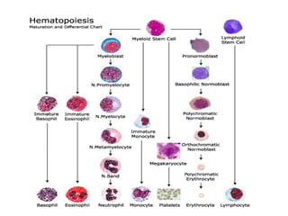
2.4.4. Maturation Characteristics
Blood cells go through maturation stages in the bone
marrow and are released into the blood at maturity to
perform their function
In any cell series, a progression of cells exists between
the most immature ‘blast’ cell and the mature cells
Sometimes, it is difficult to know what stage is
represented by a particular cell
The general rule is to identify the cell as the most
mature form.
59.
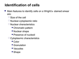
Identification of cells
Main features to identify cells on a Wright’s- stained smear
are:
Size of the cell
Nuclear-cytoplasmic ratio
Nuclear characteristics
Chromatin pattern
Nuclear shape
Presence of nucleoli
Cytoplasmic characteristics
Color
Granulation
Vacuoles
Shape
60.
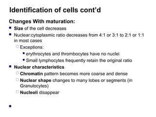
Identification of cellscont’d
Changes With maturation:
Size of the cell decreases
Nuclear:cytoplasmic ratio decreases from 4:1 or 3:1 to 2:1 or 1:1
in most cases
Exceptions:
erythrocytes and thrombocytes have no nuclei
Small lymphocytes frequently retain the original ratio
Nuclear characteristics
Chromatin pattern becomes more coarse and dense
Nuclear shape changes to many lobes or segments (in
Granulocytes)
Nucleoli disappear
61.
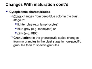
Changes With maturationcont’d
Cytoplasmic characteristics
Color changes from deep blue color in the blast
stage to:
lighter blue (e.g. lymphocytes)
blue-gray (e.g. moncytes) or
pink (e.g. RBC)
Granulation: in the granulocytic series changes
from no granules in the blast stage to non-specific
granules then to specific granules
62.
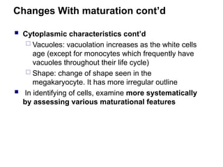
Changes With maturationcont’d
Cytoplasmic characteristics cont’d
Vacuoles: vacuolation increases as the white cells
age (except for monocytes which frequently have
vacuoles throughout their life cycle)
Shape: change of shape seen in the
megakaryocyte. It has more irregular outline
In identifying of cells, examine more systematically
by assessing various maturational features
63.
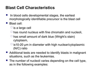
Blast Cell Characteristics
In blood cells developmental stages, the earliest
morphologically identifiable precursor is the blast cell
Blast cell:
is a large cell
has round nucleus with fine chromatin and nucleoli,
has small amount of dark blue (Wright’s stain)
cytoplasm,
is10-20 μm in diameter with high nuclear/cytoplasmic
(N/C) ratio
Additional tests are needed to identify blasts in malignant
situations, such as the leukemias.
The number of nucleoli varies depending on the cell type,
as in the following examples:
64.
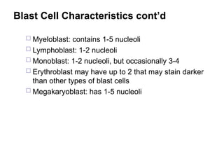
Blast Cell Characteristicscont’d
Myeloblast: contains 1-5 nucleoli
Lymphoblast: 1-2 nucleoli
Monoblast: 1-2 nucleoli, but occasionally 3-4
Erythroblast may have up to 2 that may stain darker
than other types of blast cells
Megakaryoblast: has 1-5 nucleoli
65.
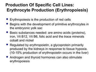
Production Of SpecificCell Lines:
Erythrocyte Production (Erythropoiesis)
Erythropoiesis is the production of red cells
Begins with the development of primitive erythrocytes in
the embryonic yolk sac
Basic substances needed: are amino acids (proteins),
iron, Vit B12, Vit B6, folic acid and the trace minerals
cobalt and nickel
Regulated by erythropoietin, a glycoprotein primarily
produced by the kidneys in response to tissue hypoxia.
(10-15% production of erythropoietin occurs in the liver)
Androgen and thyroid hormones can also stimulate
erythropoiesis
66.
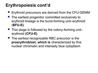
Erythropoiesis cont’d
Erythroidprecursors are derived from the CFU-GEMM
The earliest progenitor committed exclusively to
erythroid lineage is the burst-forming unit–erythroid
(BFU-E)
This stage is followed by the colony-forming unit–
erythroid (CFU-E)
The earliest recognizable RBC precursor is the
proerythroblast, which is characterized by fine
nuclear chromatin and intensely blue cytoplasm
67.
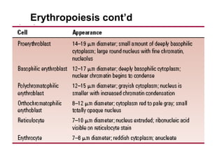
Pronormoblast/Proerythroblast
(Rubriblast)
Pronormoblast isthe earliest morphologically
recognizable red cell precursor.
Size: 20-25m in diameter.
Nucleus:
large, round to oval
contains 0-2 light bluish, indistinct nucleoli
The chromatin forms a delicate network giving the
nucleus a reticular appearance.
Cytoplasm:
there is a narrow (about 2m) rim of dark marine blue
cytoplasm
There may be a perinuclear halo
The N:C ratio is about 4:1
68.
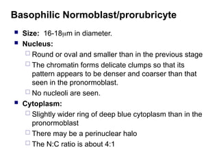
Basophilic Normoblast/prorubricyte
Size:16-18m in diameter.
Nucleus:
Round or oval and smaller than in the previous stage
The chromatin forms delicate clumps so that its
pattern appears to be denser and coarser than that
seen in the pronormoblast.
No nucleoli are seen.
Cytoplasm:
Slightly wider ring of deep blue cytoplasm than in the
pronormoblast
There may be a perinuclear halo
The N:C ratio is about 4:1
69.
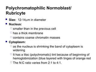
Polychromatophilic Normoblast/
Rubricyte
Size:12-14m in diameter
Nucleus:
smaller than in the previous cell
has a thick membrane
contains coarse chromatin masses
Cytoplasm:
as the nucleus is shrinking the band of cytoplasm is
widening
It has a lilac (polychromatic) tint because of beginning of
hemoglobinization (blue layered with tinges of orange red
The N:C ratio varies from 2:1 to 4:1.
70.
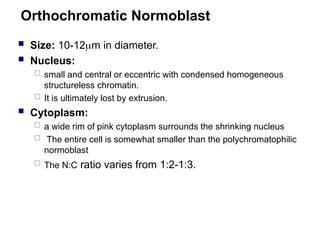
Orthochromatic Normoblast
Size:10-12m in diameter.
Nucleus:
small and central or eccentric with condensed homogeneous
structureless chromatin.
It is ultimately lost by extrusion.
Cytoplasm:
a wide rim of pink cytoplasm surrounds the shrinking nucleus
The entire cell is somewhat smaller than the polychromatophilic
normoblast
The N:C ratio varies from 1:2-1:3.
71.
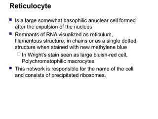
Reticulocyte
Is alarge somewhat basophilic anuclear cell formed
after the expulsion of the nucleus
Remnants of RNA visualized as reticulum,
filamentous structure, in chains or as a single dotted
structure when stained with new methylene blue
In Wright’s stain seen as large bluish-red cell,
Polychromatophilic macrocytes
This network is responsible for the name of the cell
and consists of precipitated ribosomes.
72.
Reticulocyte cont’d
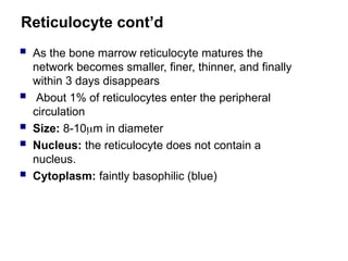
Asthe bone marrow reticulocyte matures the
network becomes smaller, finer, thinner, and finally
within 3 days disappears
About 1% of reticulocytes enter the peripheral
circulation
Size: 8-10m in diameter
Nucleus: the reticulocyte does not contain a
nucleus.
Cytoplasm: faintly basophilic (blue)
73.
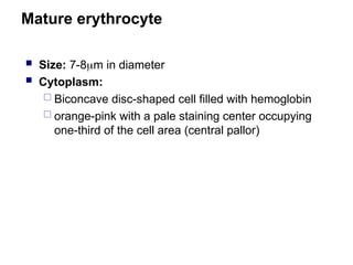
Mature erythrocyte
Size:7-8m in diameter
Cytoplasm:
Biconcave disc-shaped cell filled with hemoglobin
orange-pink with a pale staining center occupying
one-third of the cell area (central pallor)
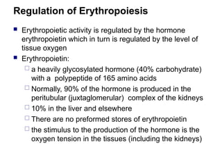
Regulation of Erythropoiesis
Erythropoietic activity is regulated by the hormone
erythropoietin which in turn is regulated by the level of
tissue oxygen
Erythropoietin:
a heavily glycosylated hormone (40% carbohydrate)
with a polypeptide of 165 amino acids
Normally, 90% of the hormone is produced in the
peritubular (juxtaglomerular) complex of the kidneys
10% in the liver and elsewhere
There are no preformed stores of erythropoietin
the stimulus to the production of the hormone is the
oxygen tension in the tissues (including the kidneys)
76.
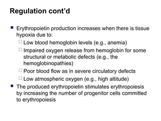
Regulation cont’d
Erythropoietinproduction increases when there is tissue
hypoxia due to:
Low blood hemoglobin levels (e.g., anemia)
Impaired oxygen release from hemoglobin for some
structural or metabolic defects (e.g., the
hemoglobinopathies)
Poor blood flow as in severe circulatory defects
Low atmospheric oxygen (e.g., high altitude)
The produced erythropoietin stimulates erythropoiesis
by increasing the number of progenitor cells committed
to erythropoiesis
77.
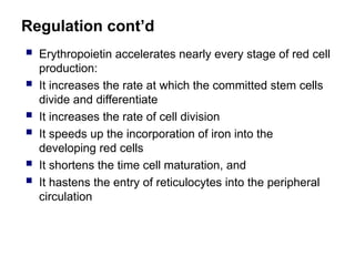
Regulation cont’d
Erythropoietinaccelerates nearly every stage of red cell
production:
It increases the rate at which the committed stem cells
divide and differentiate
It increases the rate of cell division
It speeds up the incorporation of iron into the
developing red cells
It shortens the time cell maturation, and
It hastens the entry of reticulocytes into the peripheral
circulation
78.
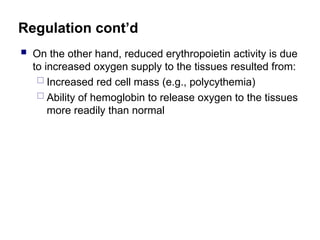
Regulation cont’d
Onthe other hand, reduced erythropoietin activity is due
to increased oxygen supply to the tissues resulted from:
Increased red cell mass (e.g., polycythemia)
Ability of hemoglobin to release oxygen to the tissues
more readily than normal
79.
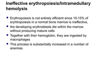
Ineffective erythropoiesis/Intramedullary
hemolysis
Erythropoiesisis not entirely efficient since 10-15% of
eryhtropoiesis in a normal bone marrow is ineffective,
the developing erythroblasts die within the marrow
without producing mature cells
Together with their hemoglobin, they are ingested by
macrophages
This process is substantially increased in a number of
anemias.
80.
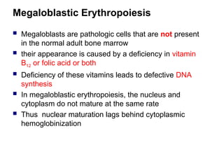
Megaloblastic Erythropoiesis
Megaloblastsare pathologic cells that are not present
in the normal adult bone marrow
their appearance is caused by a deficiency in vitamin
B12 or folic acid or both
Deficiency of these vitamins leads to defective DNA
synthesis
In megaloblastic erythropoiesis, the nucleus and
cytoplasm do not mature at the same rate
Thus nuclear maturation lags behind cytoplasmic
hemoglobinization
81.
Megaloblastic Erythropoiesis cont’d
This nuclear lag appears to be caused by
interference with DNA synthesis while RNA and
protein synthesis continue at a normal rate
The end stage of megaloblastic maturation is the
megalocyte which is abnormally large in size (9-
12m in diameter).
82.
Formation of whiteblood cells
(Leukopoiesis)
Granulopoiesis and Monocytopoiesis
Neutrophils and monocytes arise form a common
committed progenitor
The myeloblast is the earliest recognizable precursor in
the granulocytic series
on division the myeloblast gives rise to promyelocyte
The promyelocyte contain abundant dark “azurophilic”
primary granules that overlie both nucleus and
cytoplasm
with subsequent cell divisions these primary granules
become progressively diluted by the secondary, less
conspicuous “neutrophilic” granules that are
characteristic of the mature cells.
83.
Granulopoiesis cont’d
Thisconcomitant cell division and maturation
sequence continues form promyelocytes to early
myelocytes, late myelocytes, and then
metamyelocytes
As the metamyelocyte matures the nucleus becomes
more attenuated and the cell is then called a “band” or
“stab” form
Subsequent segmentation of the nucleus gives rise to
the mature neutrophil or polymorphonuclear leucocyte.
84.
Granulopoiesis cont’d
Theaverage interval from the initiation of
granulopoiesis to the entry of the mature neutrophil
into the circulation is 10 to 13 days.
The mature neutrophil remains in the circulation for
only about 10 to 14 hours before entering the tissue,
where it soon dies after performing its phagocytic
function.
85.
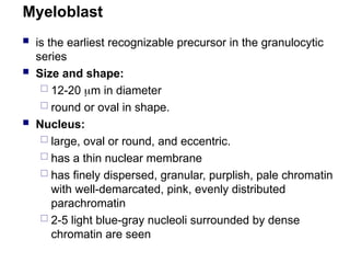
Myeloblast
is theearliest recognizable precursor in the granulocytic
series
Size and shape:
12-20 m in diameter
round or oval in shape.
Nucleus:
large, oval or round, and eccentric.
has a thin nuclear membrane
has finely dispersed, granular, purplish, pale chromatin
with well-demarcated, pink, evenly distributed
parachromatin
2-5 light blue-gray nucleoli surrounded by dense
chromatin are seen
86.
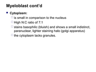
Myeloblast cont’d
Cytoplasm:
is small in comparison to the nucleus
High N:C ratio of 7:1
stains basophilic (bluish) and shows a small indistinct,
paranuclear, lighter staining halo (golgi apparatus)
the cytoplasm lacks granules.
87.
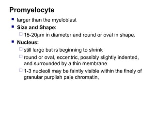
Promyelocyte
larger thanthe myeloblast
Size and Shape:
15-20m in diameter and round or oval in shape.
Nucleus:
still large but is beginning to shrink
round or oval, eccentric, possibly slightly indented,
and surrounded by a thin membrane
1-3 nucleoli may be faintly visible within the finely of
granular purplish pale chromatin,
88.
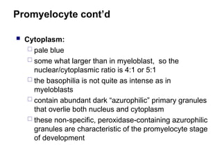
Promyelocyte cont’d
Cytoplasm:
pale blue
some what larger than in myeloblast, so the
nuclear/cytoplasmic ratio is 4:1 or 5:1
the basophilia is not quite as intense as in
myeloblasts
contain abundant dark “azurophilic” primary granules
that overlie both nucleus and cytoplasm
these non-specific, peroxidase-containing azurophilic
granules are characteristic of the promyelocyte stage
of development
89.
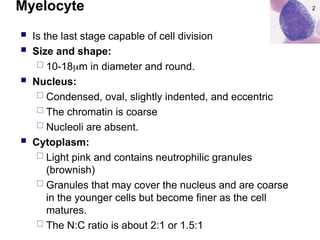
Myelocyte
Is thelast stage capable of cell division
Size and shape:
10-18m in diameter and round.
Nucleus:
Condensed, oval, slightly indented, and eccentric
The chromatin is coarse
Nucleoli are absent.
Cytoplasm:
Light pink and contains neutrophilic granules
(brownish)
Granules that may cover the nucleus and are coarse
in the younger cells but become finer as the cell
matures.
The N:C ratio is about 2:1 or 1.5:1
90.
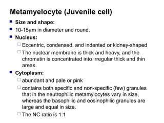
Metamyelocyte (Juvenile cell)
Size and shape:
10-15m in diameter and round.
Nucleus:
Eccentric, condensed, and indented or kidney-shaped
The nuclear membrane is thick and heavy, and the
chromatin is concentrated into irregular thick and thin
areas.
Cytoplasm:
abundant and pale or pink
contains both specific and non-specific (few) granules
that in the neutrophilic metamylocytes vary in size,
whereas the basophilic and eosinophilic granules are
large and equal in size.
The NC ratio is 1:1
91.
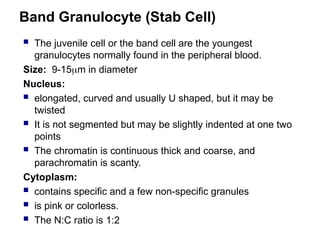
Band Granulocyte (StabCell)
The juvenile cell or the band cell are the youngest
granulocytes normally found in the peripheral blood.
Size: 9-15m in diameter
Nucleus:
elongated, curved and usually U shaped, but it may be
twisted
It is not segmented but may be slightly indented at one two
points
The chromatin is continuous thick and coarse, and
parachromatin is scanty.
Cytoplasm:
contains specific and a few non-specific granules
is pink or colorless.
The N:C ratio is 1:2
92.
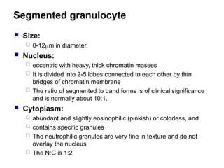
Segmented granulocyte
Size:
0-12m in diameter.
Nucleus:
eccentric with heavy, thick chromatin masses
It is divided into 2-5 lobes connected to each other by thin
bridges of chromatin membrane
The ratio of segmented to band forms is of clinical significance
and is normally about 10:1.
Cytoplasm:
abundant and slightly eosinophilic (pinkish) or colorless, and
contains specific granules
The neutrophilic granules are very fine in texture and do not
overlay the nucleus
The N:C is 1:2
93.
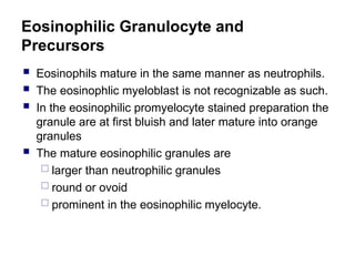
Eosinophilic Granulocyte and
Precursors
Eosinophils mature in the same manner as neutrophils.
The eosinophlic myeloblast is not recognizable as such.
In the eosinophilic promyelocyte stained preparation the
granule are at first bluish and later mature into orange
granules
The mature eosinophilic granules are
larger than neutrophilic granules
round or ovoid
prominent in the eosinophilic myelocyte.
94.
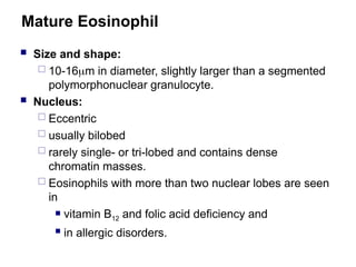
Mature Eosinophil
Sizeand shape:
10-16m in diameter, slightly larger than a segmented
polymorphonuclear granulocyte.
Nucleus:
Eccentric
usually bilobed
rarely single- or tri-lobed and contains dense
chromatin masses.
Eosinophils with more than two nuclear lobes are seen
in
vitamin B12 and folic acid deficiency and
in allergic disorders.
95.
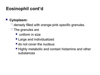
Eosinophil cont’d
Cytoplasm:
densely filled with orange-pink specific granules.
The granules are
uniform in size
Large and individualized
do not cover the nucleus
Highly metabolic and contain histamine and other
substances
96.
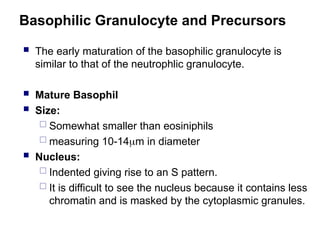
Basophilic Granulocyte andPrecursors
The early maturation of the basophilic granulocyte is
similar to that of the neutrophlic granulocyte.
Mature Basophil
Size:
Somewhat smaller than eosiniphils
measuring 10-14m in diameter
Nucleus:
Indented giving rise to an S pattern.
It is difficult to see the nucleus because it contains less
chromatin and is masked by the cytoplasmic granules.
97.
Basophils cont’d
Cytoplasm:
Pale blue to pale pink
contains granules that often overlie the nucleus but
do not fill the cytoplasm as completely as the
eosinophilis granules do
98.
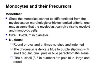
Monocytes and theirPrecursors
Monoblast
Since the monoblast cannot be differentiated from the
myeloblast on morphologic or histochemical criteria, one
may assume that the myeloblast can give rise to myeloid
and monocytic cells.
Size: 15-20m in diameter.
Nucleus:
Round or oval and at times notched and indented
The chromatin is delicate blue to purple stippling with
small regular, pink, pale or blue parachromatin areas
The nucleoli (3-5 in number) are pale blue, large and
round
99.
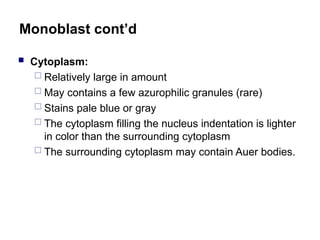
Monoblast cont’d
Cytoplasm:
Relatively large in amount
May contains a few azurophilic granules (rare)
Stains pale blue or gray
The cytoplasm filling the nucleus indentation is lighter
in color than the surrounding cytoplasm
The surrounding cytoplasm may contain Auer bodies.
100.
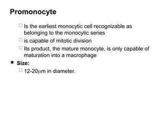
Promonocyte
Is theearliest monocytic cell recognizable as
belonging to the monocytic series
is capable of mitotic division
Its product, the mature monocyte, is only capable of
maturation into a macrophage
Size:
12-20m in diameter.
101.
Promonocyte cont’d
Nucleus:
Large
ovoid to round, convoluted, grooved, and indented
The chromatin forms a loose open network containing
a few larger clumps
there may be two or more nucleoli.
Cytoplasm:
sparse, gray-blue, contains fine azurophilic granules
N:C ratio is about 3:1
102.
Monocyte
Size:
12-20min diameter.
Nucleus:
Eccentric or central
Takes different shapes from brainy convolutions to
lobulated and S shaped (often lobulated)
The chromatin network consists of fine, pale, loose,
linear threads producing small areas of thickening
at their junctions
No nucleolus is seen
The overall impression is that of a pale nucleus
quite variable in shape.
103.
Monocyte cont’d
Cytoplasm:
Abundant, opaque, gray-blue with moderate
granules
unevenly stained and may be vacuolated
N:C ratio 1:1
104.
Lymphopoiesis
The precursorof the lymphocyte is believed to be the
primitive mulipotential stem cell that also gives rise to the
pluirpotenital myeloid stem cell for the granulocytic,
erythyroid, and megakaryocytic cell lines
Lymphoid precursor cells travel to specific sites
There, they differentiate into cells capable of either
expressing cell-mediated immune responses or
secreting immunoglobulins
The influence for the former type of differentiation in
humans is the thymus gland;
the resulting cells are defined as thymus-dependent
lymphocytes, or T cells.
105.
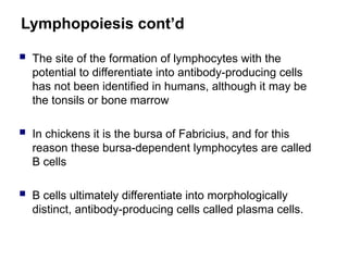
Lymphopoiesis cont’d
Thesite of the formation of lymphocytes with the
potential to differentiate into antibody-producing cells
has not been identified in humans, although it may be
the tonsils or bone marrow
In chickens it is the bursa of Fabricius, and for this
reason these bursa-dependent lymphocytes are called
B cells
B cells ultimately differentiate into morphologically
distinct, antibody-producing cells called plasma cells.
106.
Lymphocytes and Precursors
Lymphoblast
Size:
10-20m in diameter.
Nucleus:
Central, round or oval
the chromatin has a stippled pattern
The nuclear membrane is distinct and one or two pink
nucleoli are present and are usually well outlined
Cytoplasm:
Non-granular and sky blue
may have a deep blue border
It forms a thin perinuclear ring.
N:C ratio 4:1
107.
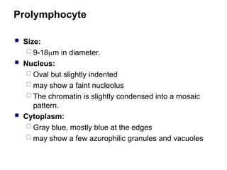
Prolymphocyte
Size:
9-18min diameter.
Nucleus:
Oval but slightly indented
may show a faint nucleolus
The chromatin is slightly condensed into a mosaic
pattern.
Cytoplasm:
Gray blue, mostly blue at the edges
may show a few azurophilic granules and vacuoles
108.
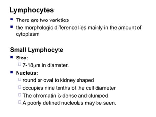
Lymphocytes
There aretwo varieties
the morphologic difference lies mainly in the amount of
cytoplasm
Small Lymphocyte
Size:
7-18m in diameter.
Nucleus:
round or oval to kidney shaped
occupies nine tenths of the cell diameter
The chromatin is dense and clumped
A poorly defined nucleolus may be seen.
109.
Lymphocytes cont’d
Cytoplasm:
It is basophilic and forms a narrow rim around the
nucleus or at times a thin blue line only with few
azurophilic red granules
N:C ratio is 4:1
Distinguishing characteristics of a small lymphocyte:
clumping of chromatin around the nuclear membrane
may help to distinguish this from a nucleated red cell
110.
Large Lymphocyte
Size:
9-12m in diameter
Nucleus:
the dense, oval, or slightly indented nucleus is centrally
or eccentricity located
Its chromatin is dense and clumped.
Cytoplasm:
Abundant
gray to pale blue, unevenly stained, and streaked at
times
A few azurophilic granules are contained in 30-60% of
the cells.
These are large granular lymphocytes (LGLs).
111.
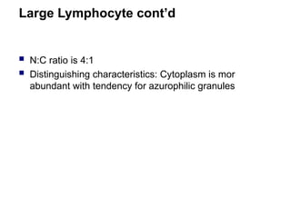
Large Lymphocyte cont’d
N:C ratio is 4:1
Distinguishing characteristics: Cytoplasm is mor
abundant with tendency for azurophilic granules
112.
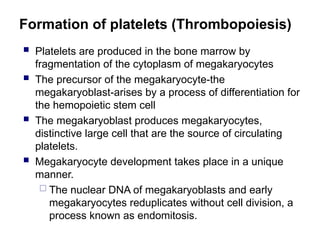
Formation of platelets(Thrombopoiesis)
Platelets are produced in the bone marrow by
fragmentation of the cytoplasm of megakaryocytes
The precursor of the megakaryocyte-the
megakaryoblast-arises by a process of differentiation for
the hemopoietic stem cell
The megakaryoblast produces megakaryocytes,
distinctive large cell that are the source of circulating
platelets.
Megakaryocyte development takes place in a unique
manner.
The nuclear DNA of megakaryoblasts and early
megakaryocytes reduplicates without cell division, a
process known as endomitosis.
113.
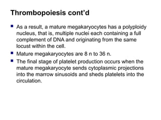
Thrombopoiesis cont’d
Asa result, a mature megakaryocytes has a polyploidy
nucleus, that is, multiple nuclei each containing a full
complement of DNA and originating from the same
locust within the cell.
Mature megakaryocytes are 8 n to 36 n.
The final stage of platelet production occurs when the
mature megakaryocyte sends cytoplasmic projections
into the marrow sinusoids and sheds platelets into the
circulation.
114.
Thrombopoiesis cont’d
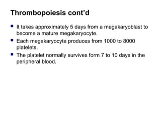
Ittakes approximately 5 days from a megakaryoblast to
become a mature megakaryocyte.
Each megakaryocyte produces from 1000 to 8000
platelets.
The platelet normally survives form 7 to 10 days in the
peripheral blood.
115.
Morphology of thePlatelets and their
Precursors
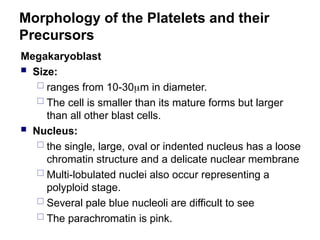
Megakaryoblast
Size:
ranges from 10-30m in diameter.
The cell is smaller than its mature forms but larger
than all other blast cells.
Nucleus:
the single, large, oval or indented nucleus has a loose
chromatin structure and a delicate nuclear membrane
Multi-lobulated nuclei also occur representing a
polyploid stage.
Several pale blue nucleoli are difficult to see
The parachromatin is pink.
116.
Megakaryoblast cont’d
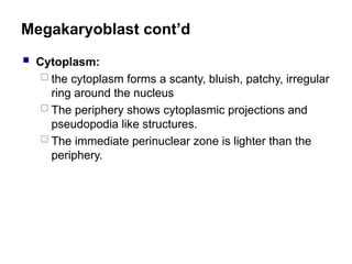
Cytoplasm:
the cytoplasm forms a scanty, bluish, patchy, irregular
ring around the nucleus
The periphery shows cytoplasmic projections and
pseudopodia like structures.
The immediate perinuclear zone is lighter than the
periphery.
117.
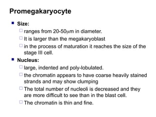
Promegakaryocyte
Size:
rangesfrom 20-50m in diameter.
It is larger than the megakaryoblast
in the process of maturation it reaches the size of the
stage III cell.
Nucleus:
large, indented and poly-lobulated.
the chromatin appears to have coarse heavily stained
strands and may show clumping
The total number of nucleoli is decreased and they
are more difficult to see than in the blast cell.
The chromatin is thin and fine.
118.
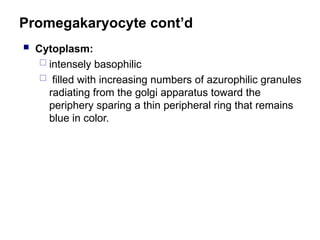
Promegakaryocyte cont’d
Cytoplasm:
intensely basophilic
filled with increasing numbers of azurophilic granules
radiating from the golgi apparatus toward the
periphery sparing a thin peripheral ring that remains
blue in color.
119.
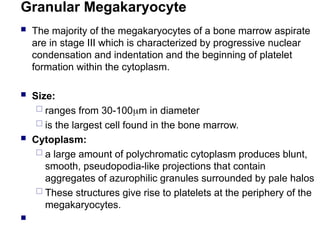
Granular Megakaryocyte
Themajority of the megakaryocytes of a bone marrow aspirate
are in stage III which is characterized by progressive nuclear
condensation and indentation and the beginning of platelet
formation within the cytoplasm.
Size:
ranges from 30-100m in diameter
is the largest cell found in the bone marrow.
Cytoplasm:
a large amount of polychromatic cytoplasm produces blunt,
smooth, pseudopodia-like projections that contain
aggregates of azurophilic granules surrounded by pale halos
These structures give rise to platelets at the periphery of the
megakaryocytes.
120.
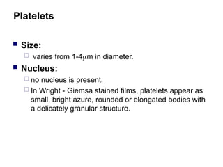
Platelets
Size:
variesfrom 1-4m in diameter.
Nucleus:
no nucleus is present.
In Wright - Giemsa stained films, platelets appear as
small, bright azure, rounded or elongated bodies with
a delicately granular structure.
121.
Review Questions/Summary
1. Whatis hemopoiesis and how is the process regulated?
2. What are the hemopoietic tissues during fetal life, in infancy,
in childhood and in adulthood?
3. What are the effects of the hormone erythropoietin on red
cell development and maturation.
4. Describe the microenvironment briefly.
5. Explain megaloblastic erythropoiesis.
6. Describe general Characteristic feature of cells during
maturation (nuclear , cytoplasmic, etc )
7. State the composition of blood.
8. State the main functions of blood.
9. List main characteristics of blood.
10. What is extramedulary hemopoiesis and when does it occur?
Editor's Notes
#5 Instructor note: discuss difference between serum and plasma: Serum is the liquid remaining
after blood clots; it is essentially the same as plasma, except that the clotting
factors and fibrinogen have been removed.
#7 Out of the 7% protein, 54% albumin, 38% globulin, 7% fibrinogen
#22 Instructor note: small lymphocye is shown for comparison
#24 Instructor: note the vacoules in the monos (right)
#26 • The normal platelet number is ~150,000 to 350,000 cells/L. (PLT count is low in Ethiopian Tsegaye et al 1999
• Platelets have different types of granules, designated alpha granules and
dense bodies. Platelet granules contain clotting factors, adenosine
diphosphate (ADP) and adenosine triphosphate (ATP), calcium, sero- tonin, and catecholamines; many of these stimulate platelet aggregation or are important in the coagulation cascade.
#27 Note: Platelets are shown by the arrow; the big cell in the film is a basophil.
#29 BV can be estimated for an individual of either gender by calculating 7% of the body weight in Kg
Ex. 75 Kg ind. Would have a BV of approximately 5.25 liters (~1.4 gallons)
Abnormally high BV can place severe stress on the heart (pushing extra fluid through circulatory system stresses heart)
#30 Function of plasma components
Water – is a medium for carrying other constituents i.e it is used as a solvent
Inorganic substances – include Na- , K+, Cl- , Ca2+, Respiratory gases, etc.
keep water in the extra cellular environment
act as buffers ( for stabilizing blood pH )
function in membrane excitability
used as metabolic sources, etc
Organic Substances
A. Glucose, Amino acid and fatty acid
-Use plasma as means of transportation to their site of
utilization
- are used as building materials for larger molecules
- used as energy sources, etc
B. Proteins
Used to bind other plasma constituents such as lipids, hormones, vitamins, metals etc,
some are used as enzymes or enzyme precursors
Fibrinogen is the precursor for fibrin and is used in hemostasis
Albumin is important for colloid osmotic pressure
Globulin -
- is associated with the transport of Bilirubin, lipid and steroids
- act as a substrate for formation of other substances
- for transport of Fe and Cu in plasma
- for antibody production
#31 Function of plasma components
Water – is a medium for carrying other constituents i.e it is used as a solvent
Inorganic substances – include Na- , K+, Cl- , Ca2+, Respiratory gases, etc.
keep water in the extra cellular environment
act as buffers ( for stabilizing blood pH )
function in membrane excitability
used as metabolic sources, etc
Organic Substances
A. Glucose, Amino acid and fatty acid
-Use plasma as means of transportation to their site of
utilization
- are used as building materials for larger molecules
- used as energy sources, etc
B. Proteins
Used to bind other plasma constituents such as lipids, hormones, vitamins, metals etc,
some are used as enzymes or enzyme precursors
Fibrinogen is the precursor for fibrin and is used in hemostasis
Albumin is important for colloid osmotic pressure
Globulin -
- is associated with the transport of Bilirubin, lipid and steroids
- act as a substrate for formation of other substances
- for transport of Fe and Cu in plasma
- for antibody production
#32 Although many questions remain unanswered, a hypothetical scheme of hemopoiesis based on a monophyletic theory is accepted by many hematologists. According to this theory, the main blood cell groups including the red blood cells, white blood cells and platelets are derived from a Pluripotent stem cell. This stem cell is the first in a sequence of regular and orderly steps of cell growth and maturation.
#34 Stem cells are unique in that one daughter retains all the characteristics of a stem cell while the other daughter cell has the capability of dividing and differentiating into mature cells.
#35 Proliferation of the pluripotent stem cell may be regulated by a humoral factor, hemopoietin-1 which is not yet well characterized. Mix-CFU proliferation is regulated, in part, by a humoral factor, multi-CSF, also referred to in the literature as IL-3 (interleukin-3) or hemopoietin-2. Multi-CSF may also stimulate proliferation of the pluripotent stem cell. Proliferation of the mix-CFU leads to an increase in the population of the restricted precursors, BFU-E, GM-CFU, Eo-CFU, Baso-CFU, and MK-CFU.
#37 Instructor note: note that this is a simplified scheme of blood cell origin and development. Complete stages of development should be discussed
#41 Reference: Williams Hematology. Expansion and recession of hematopoietic activity in extramedullary and medullary sites. For details regarding the nature of yolk sac and hepatic hematopoiesis, see "Sites of Hematopoiesis: Embryogenesis and Early Stem Cell Development." Chapter 6 provides a more comprehensive treatment of this topic (see Fig. 6-1).
#42 Instructor note: the previous figure focuses on site of hemopoiesis. This figure shows the three main phases and cell types produced including the lymph node.
(From Rothstein, G., in Wintrobe’s Clinical Hematology, Lee, G.R. et al., Eds., Williams & Wilkins, Baltimore, 1993, p. 80
#57 Instructor note: this figure summarize hemopoiesis including growth factors. Production of specific cell lines will follow
#67 Size 18-20 m in diameter; N:C 4:1 in some reference books (Betty Ciesla 2007)
#68 Cytoplasm: corn flower blue with indistinct areas of clearing. N:C 4:1 in some reference books (Betty Ciesla 2007) size also varies
#69 N:C 2:1 in some reference books (Betty Ciesla 2007) size also varies
“The dawn of hemoglobinization”
#74 Instructor note: different reference books may give different size ranges
e.g. Ref: From PDQ hematology 2002 P20
#82 Monocytes evolve to macrophages when they enter the tissues
#85 Color atlas 2004: Myeloblasts are the least mature cells in the granulocyte lineage. Mononuclear,
round-to-ovoid cells, they may be distinguished from proerythroblasts
by the finer, “grainy” reticular structure of their nuclei and the
faintly basophilic cytoplasm. On first impression, they may look like large
or even small lymphocytes (micromyeloblasts), but the delicate structure
of their nuclei always gives them away asmyeloblasts. In some areas, condensed
chromatin may start to look like nucleoli. Sporadically, the cytoplasm
contains azurophilic granules.
#86 Color atlas 2004: Myeloblasts are the least mature cells in the granulocyte lineage. Mononuclear,
round-to-ovoid cells, they may be distinguished from proerythroblasts
by the finer, “grainy” reticular structure of their nuclei and the
faintly basophilic cytoplasm. On first impression, they may look like large
or even small lymphocytes (micromyeloblasts), but the delicate structure
of their nuclei always gives them away asmyeloblasts. In some areas, condensed
chromatin may start to look like nucleoli. Sporadically, the cytoplasm
contains azurophilic granules.
#87 which contain abundant dark “azurophilic” primary granules that overlie both nucleus and cytoplasm
#90 Metamyelocytes are no longer capable of cell division
#95 Distinguishing characteristics of eosinophils: Granules are uniformly round, large, and individualized. If stain is less than adequate, observe granules carefully for their crystalloid nature
#106 Distinguishing feature for a lymphoblast: Nucleoli is surrounded by a dark rim of chromatin

![Neutrophils
are the most common type of WBCs in adults
The segmented neutrophils “segs,” also called
polymorphonuclear neutrophil leukocytes
[PMNs or “polys”]
are the primary defense against bacterial
infection
Their size ranges from 10-12m in diameter.
They are capable of amoeboid movement.
There are 2-5 lobes to their nucleus that stain
purple violet.
The cytoplasm stains light pink with pinkish dust
like granules.](https://image.slidesharecdn.com/hemaichapter2compositionformationfunction-250828084600-b93ebd36/85/Hema-I-Chapter-2_composition-formation-function-ppt-15-320.jpg)