Downloaded 10 times


![Procedure –
– 2ml of venous blood is collected and mixed with double
oxalate (ammonium oxalate and potassium oxalate) or EDTA
powder in the proportion of 1.5mg/ml
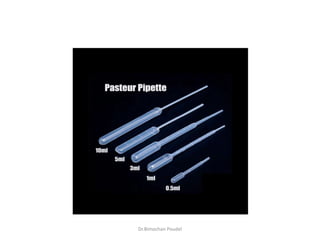
– Blood is drawn into Pasteur pipette and introduced in the
Wintrobes tube from the bottom to 0 or 10 mark above
– Place the Wintrobes tube in the centrifuge machine and other
Wintrobes tube filled with water on the opposite side so as to
balance it.
– Centrifuge the tube at the speed of 3000rpm for 30 minutes
– After 30 minutes stop the centrifuge, take out the tube and
note the readings
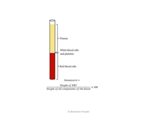
– Calculation –
• Hematocrit = [ Height of RBC’s in mm/Height of RBC and plasma] X
100
Dr.Bimochan Poudel](https://image.slidesharecdn.com/packedcellvolumeestimation-220731171025-f881a7d5/85/Packed-Cell-volume-estimation-pptx-9-320.jpg)


The document discusses packed cell volume (PCV) as a measure of the volume of blood occupied by red blood cells, detailing various methods for its estimation, particularly the Wintrobe method. It describes the procedure for using the Wintrobe tube and centrifuge, along with the interpretation of results based on the separation of blood components. Clinical implications of varying PCV levels are outlined, noting causes for both increased and decreased measurements.


