Downloaded 697 times
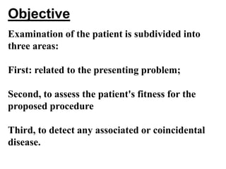
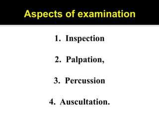
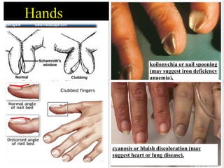
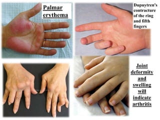
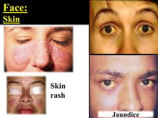
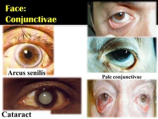
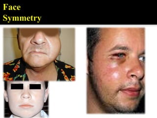
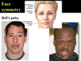
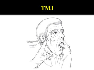
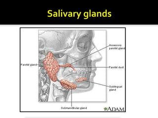
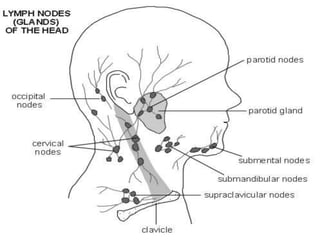
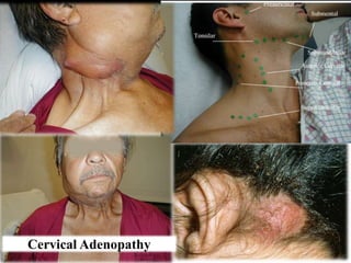
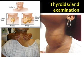
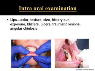
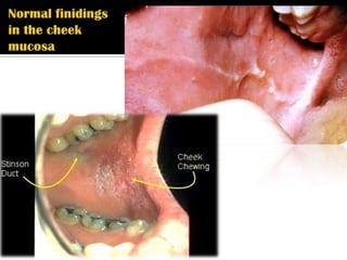
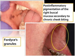
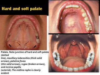
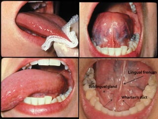
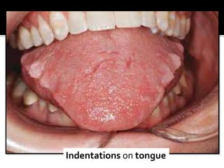
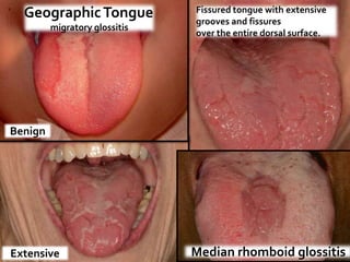
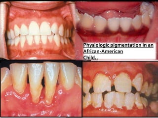
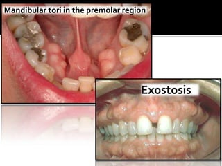
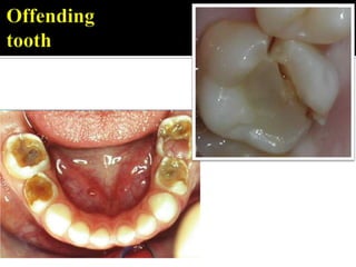

The document outlines an examination process with three objectives: assessing the presenting problem, fitness for procedures, and detecting associated diseases. It describes examining the general appearance, hands, face, neck, and performing inspection, palpation, percussion, and auscultation. Specific areas of examination include the nails, skin, conjunctivae, oral cavity, palate, tongue, and mandible. Common findings and conditions are listed.


![PERI-PROSTHETIC FRACTURE NAIL-PLATE CONSTRUCT [NPC].pptx](https://cdn.slidesharecdn.com/ss_thumbnails/drarunkumardrmohamedashrafperiprostheticfrasturenail-plateconstructnpc-260209164459-7e9d15a1-thumbnail.jpg?width=640&height=640&fit=bounds)


![ONFH[AVN HIP] -TRIPLE REGIME -A NOVAL SURGICAL CONCEPT .pptx](https://cdn.slidesharecdn.com/ss_thumbnails/onfhavnhip2026koaconcalicutdrgokuldevdrmashraf-260210064517-213ec005-thumbnail.jpg?width=640&height=640&fit=bounds)


