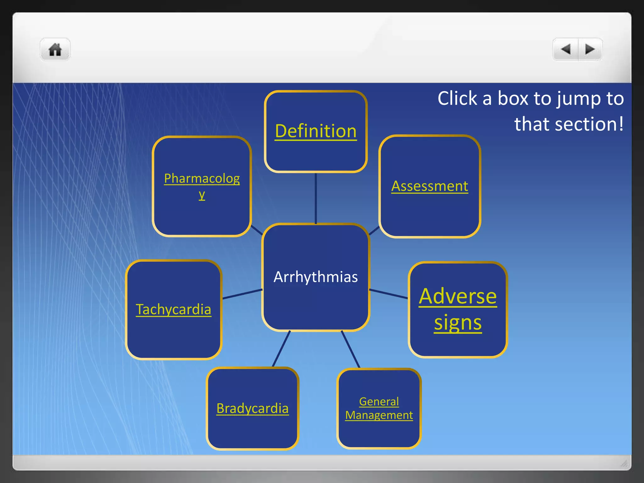
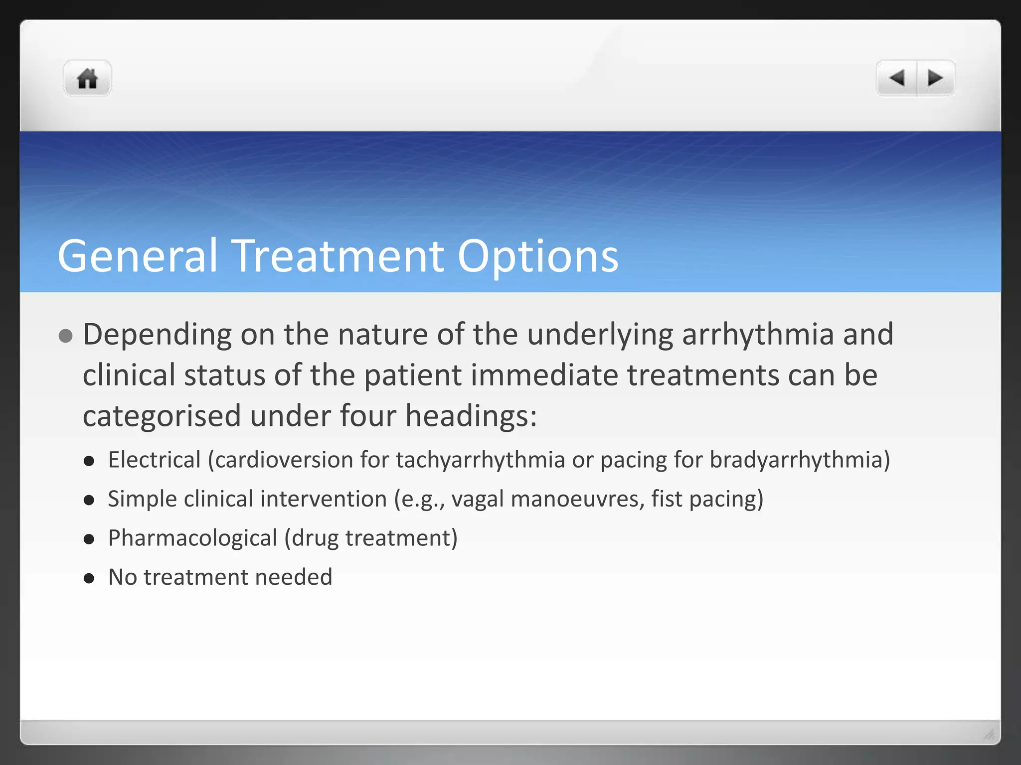
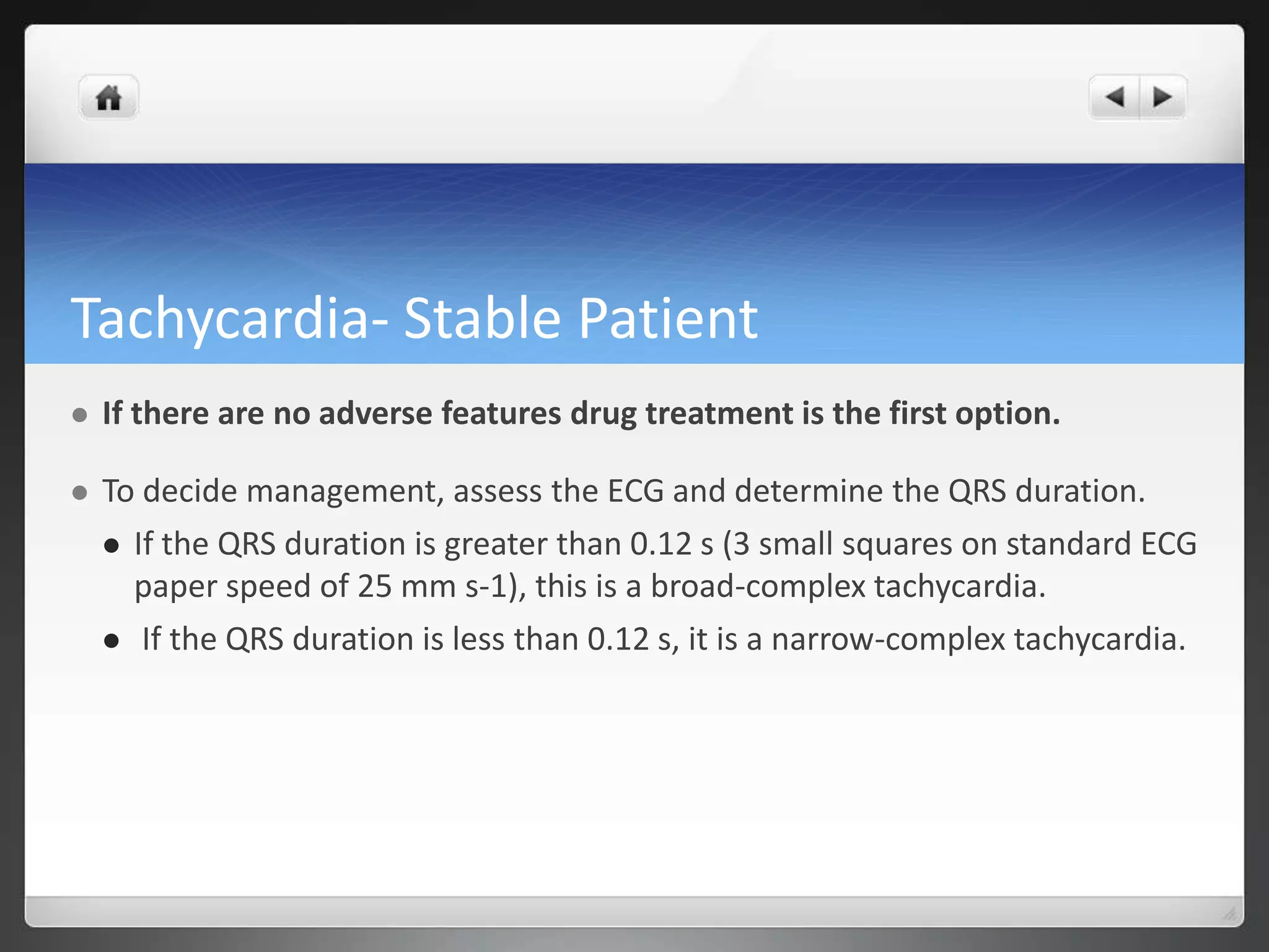
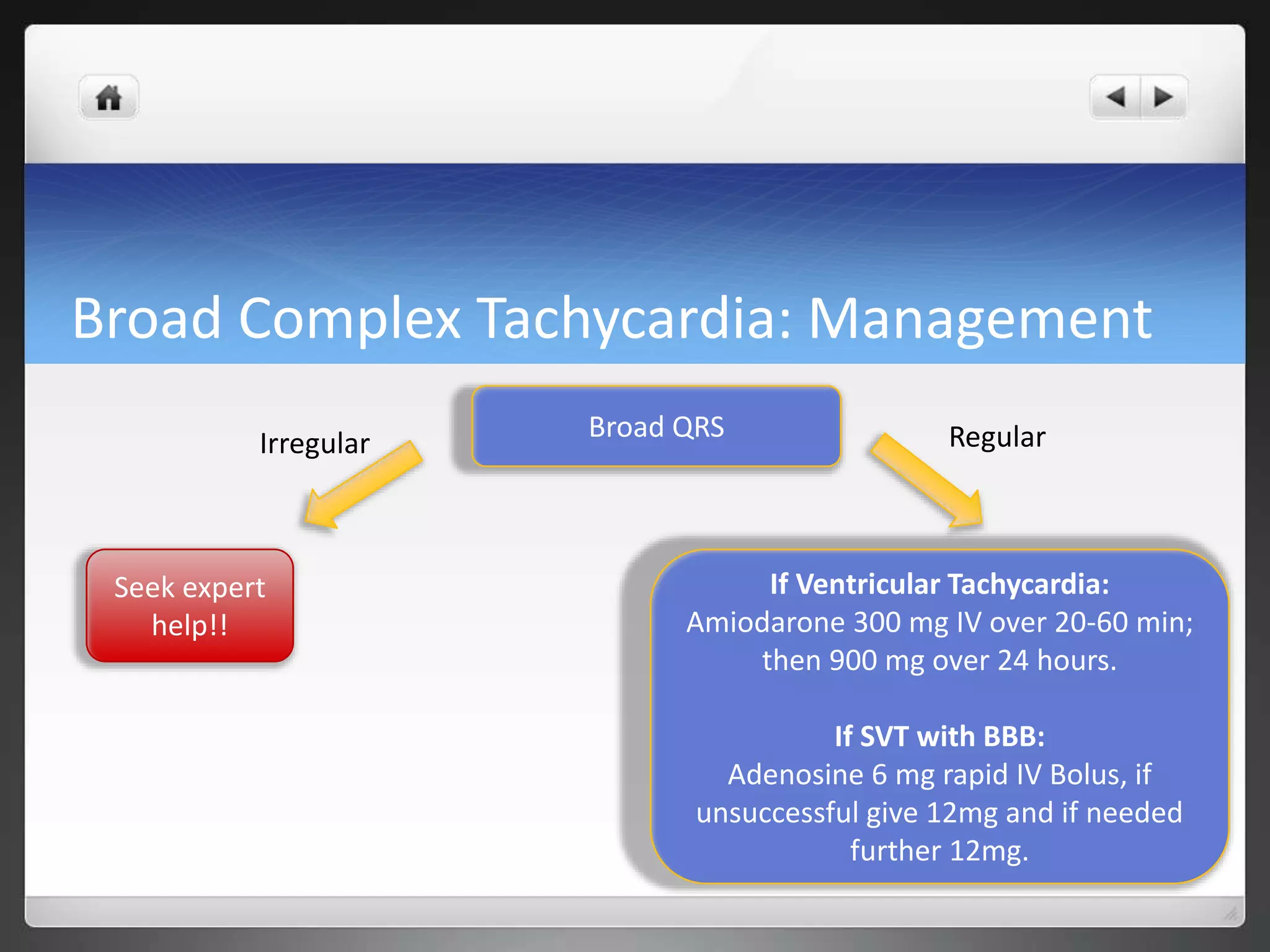
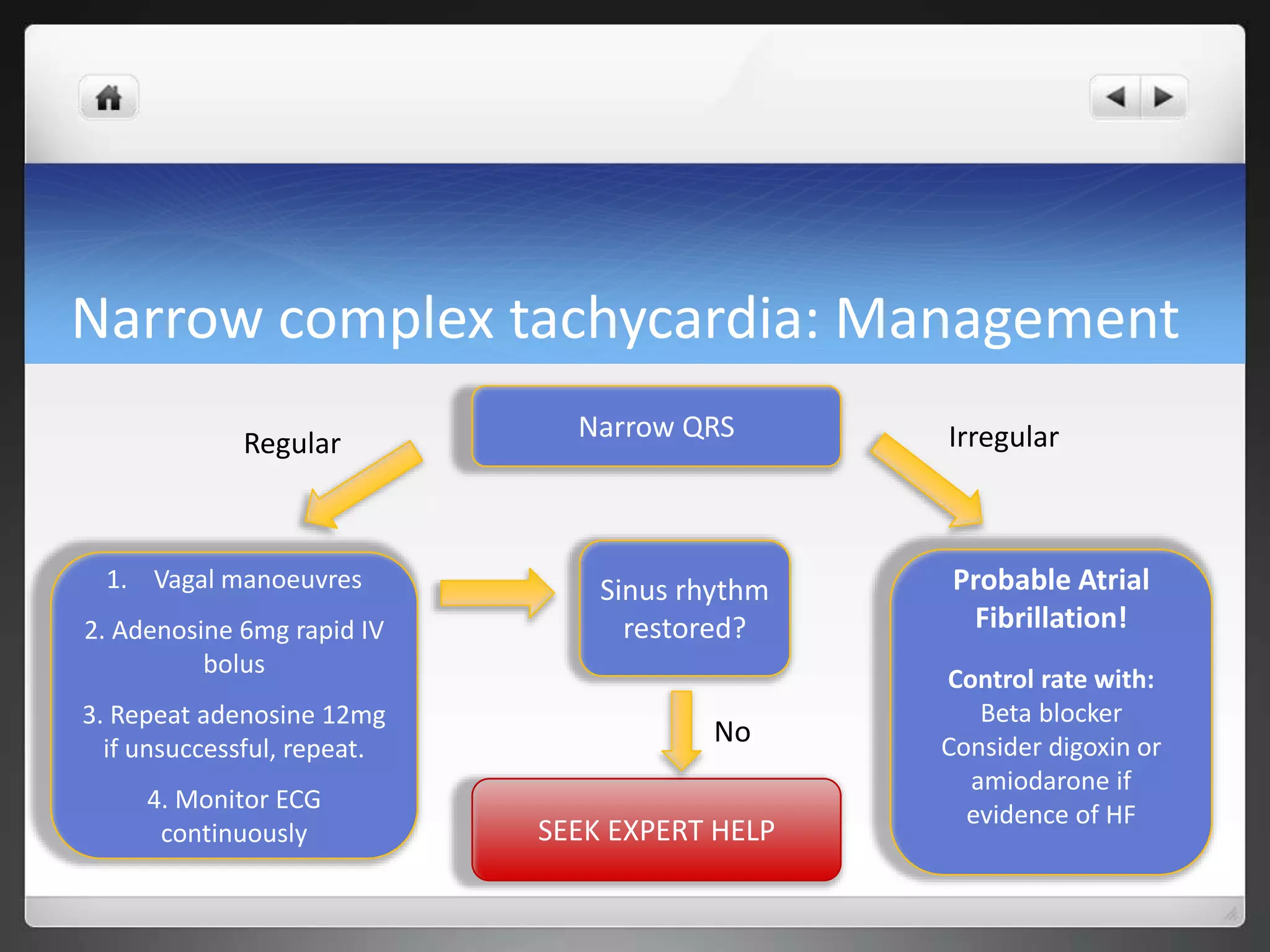
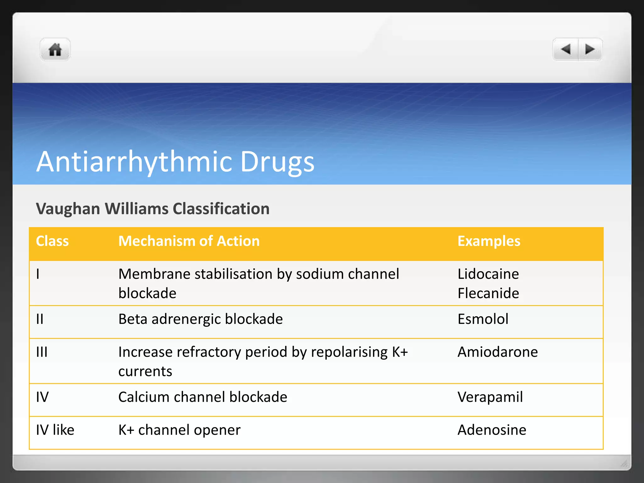
Peninsula Handbook of Clinical
Medicine
Management of Peri-Arrest Arrhythmias
Click a box to jump to
that section!
Arrhythmias
Definition
Assessment
Adverse
signs
General
ManagementBradycardia
Tachycardia
Pharmacolog
y
Definition of Arrhythmia
 An arrhythmia is an abnormal rate and/or rhythm of the heartbeat
occurring as a result of a problem with the electrical conducting system of
the heart.
 The term ‘peri – arrest arrhythmias’ are used to describe cardiac rhythm
disorders that may precede cardiac arrest or follow initial resuscitation from
a cardiac arrest. Effective treatment of such arrhythmias may prevent
cardiac arrest.
Assessment
 Assess the patient using the ABCDE approach.
 In all cases, give oxygen and insert an intravenous cannula and assess the
patient for adverse features.
 Whenever possible, record a 12-lead ECG to help determine rhythm
abnormality.
 Correct any electrolyte abnormalities
 e.g. K+, Mg++, Ca++
Adverse Features
 The following are adverse features which may indicate that the
patient is unstable as a result of the arrythmia. Their presence effects
the choice of treatment:
 Shock – hypotension (systolic blood pressure < 90 mmHg), pallor, sweating,
cold, clammy extremities, confusion or impaired consciousness.
 Syncope – transient loss of consciousness due to global reduction in blood
flow to the brain.
 Myocardial ischaemia – typical ischaemic chest pain and/or evidence of
myocardial ischaemia on 12-lead ECG.
 Heart failure – pulmonary oedema and/or raised jugular venous pressure
(with or without peripheral oedema and liver enlargement).
General Treatment Options
 Depending on the nature of the underlying arrhythmia and
clinical status of the patient immediate treatments can be
categorised under four headings:
 Electrical (cardioversion for tachyarrhythmia or pacing for bradyarrhythmia)
 Simple clinical intervention (e.g., vagal manoeuvres, fist pacing)
 Pharmacological (drug treatment)
 No treatment needed
Types of Arrythmia
 BRADYCARDIA
A bradycardia is defined as a ventricular rate below 60 beats / min..
 BROAD COMPLEX TACHYCARDIA
In the context of resuscitation, a broad complex tachycardia will almost
invariably be ventricular in origin. If there is no palpable pulse, the patient
should be treated as for ventricular fibrillation
 NARROW COMPLEX TACHYCARDIA
Supraventricular in origin. Can be a trigger for ventricular fibrillation in
vulnerable patients. Atrial fibrillation, on the other hand, occurs commonly in
the early period after resuscitation.
Bradycardia
Bradycardia
 Bradycardia is defined as a heart rate of < 60 min-1. It may be:
 physiological (e.g., in athletes)
 cardiac in origin (e.g., atrioventricular block or sinus node disease)
 non-cardiac in origin (e.g., vasovagal, hypothermia, hypothyroidism,
hyperkalaemia)
 drug-induced (e.g., beta blockade, diltiazem, digoxin, amiodarone)
Bradycardia: Management
 Assess for presence of adverse signs
 If adverse signs are present, give atropine, 500 mcg, intravenously and, if necessary,
repeat every 3-5 min to a total of 3 mg.
 If bradycardia with adverse signs persist despite atropine, consider cardiac pacing.
 in some clinical settings second-line drugs may be appropriate before the use of cardiac
pacing.
 Intravenous glucagon if a beta-blocker or calcium channel blocker overdose.
 Digoxin-specific antibody fragments for digoxin toxicity.
 Theophylline (100-200 mg by slow intravenous injection) for bradycardia complicating acute
inferior wall myocardial infarction, spinal cord injury or cardiac transplantation.
Bradycardia: Management
Adverse features?
Satisfactory
response?
Atropine
500 mcg IV
Interim measures:
Atropine 500 mcg IV
Isoprenaline 5 mcg min IV
Adrenaline 2-10mcg min IV
Risk of asystole?
Observe
SEEK EXPERT HELP
Transcutaneous pacing!
YES NO
YES
NO
Tachycardias
Stable vs Unstable Tachycardia
Broad Complex Tachycardia
Narrow Complex Tachycardia
Tachycardia- Unstable Patient
 If the patient is unstable and deteriorating (i.e., has adverse features caused
by the tachycardia) synchronised cardioversion is the treatment of choice.
 If cardioversion fails to restore sinus rhythm, and the patient remains
unstable, give amiodarone 300 mg IV over 10 - 20 min and re-attempt
electrical cardioversion.
 The loading dose of amiodarone can be followed by an infusion of 900 mg
over 24 h.
Tachycardia- Stable Patient
 If there are no adverse features drug treatment is the first option.
 To decide management, assess the ECG and determine the QRS duration.
 If the QRS duration is greater than 0.12 s (3 small squares on standard ECG
paper speed of 25 mm s-1), this is a broad-complex tachycardia.
 If the QRS duration is less than 0.12 s, it is a narrow-complex tachycardia.
Broad vs. Narrow Complex
Broad Complex Tachycardia
 Broad complex tachycardias are usually ventricular in origin, unless bundle
branch block present in which case could be supraventricular rhythm with
aberrant conduction.
 If patient unstable assume the rhythm is ventricular in origin and attempt
synchronized cardioversion.
 If the patient is stable determine whether rhythm is regular or irregular:
 Regular: likely ventricular tachycardia or supraventricular tachycardia and BBB
 Irregular: Likely atrial fibrillation with BBB but should seek expert help for
assessment and treatment or irregular broad complex tachycardias.
Broad Complex Tachycardia: Management
Broad QRS
Seek expert
help!!
If Ventricular Tachycardia:
Amiodarone 300 mg IV over 20-60 min;
then 900 mg over 24 hours.
If SVT with BBB:
Adenosine 6 mg rapid IV Bolus, if
unsuccessful give 12mg and if needed
further 12mg.
Irregular Regular
Narrow Complex Tachycardia
Examine the ECG to determine if the rhythm is regular or irregular.
 Regular narrow-complex tachycardias include:
 Sinus tachycardia;
 AV nodal re-entry tachycardia (AVNRT) – the commonest type of regular narrow-
complex tachyarrhythmia;
 AV re-entry tachycardia (AVRT) – due to Wolff-Parkinson-White syndrome;
 Atrial flutter with regular AV conduction (usually 2:1).
 An irregular narrow-complex tachycardia is most likely to be AF or
sometimes atrial flutter with variable AV conduction (‘variable block’).
Narrow complex tachycardia: Management
Narrow QRS
Sinus rhythm
restored?
1. Vagal manoeuvres
2. Adenosine 6mg rapid IV
bolus
3. Repeat adenosine 12mg
if unsuccessful, repeat.
4. Monitor ECG
continuously
Probable Atrial
Fibrillation!
Control rate with:
Beta blocker
Consider digoxin or
amiodarone if
evidence of HF
SEEK EXPERT HELP
Regular Irregular
No
Pharmacology
Antiarrhythmic Drugs
Vaughan Williams Classification
Class Mechanism of Action Examples
I Membrane stabilisation by sodium channel
blockade
Lidocaine
Flecanide
II Beta adrenergic blockade Esmolol
III Increase refractory period by repolarising K+
currents
Amiodarone
IV Calcium channel blockade Verapamil
IV like K+ channel opener Adenosine
Amioadarone
 Class III antiarrythmic
 Indication: Effective in both supraventricular and ventricular arrythmias
 Slows ventricular rate in atrial fibrillation
 Treat ventricular fibrillation and ventricular tachycardia
 Mechanism of action:
 Prolongs the action potential, increases the refractoriness of all cardiac tissue.
 Also blocks Na+ channels (Class I action)
 Has some anti-adrenergic effecs (Class II action)
 Prolongs QT interval- reflects global prolongation of repolarisation
 When given via IV, effects AV node to cause a delay in nodal conduction and accessory
pathway conduction.
Atropine
Type: Antimuscarinic cholinoceptor blocking drug
 Indication:
 Symptomatic bradycardia
 Asystole or Pulseless electrical activity (PEA)
 Mechanism of Action:
 Atropine is a parasympatholytic drug that enhances both the sinus node
automaticity and the AV conduction via its direct vagolytic action.
Atropine
 Adverse effects:
 Rebound tachycardia
 Pupil dilation
 Paradoxical rate slowing
 Type II second degree AV block
 Third degree AV block
 Arrhythmia
 Ventricular fibrillation
 Ventricular tachycardia
 Anticholinergic toxicity
 Mild sedation to Delirium
Digoxin
 Indications
 Atrial fibrillation to slow ventricular rate
 Heart failure, especially if AF is present
 Mechanism of action
 Increases the force of cardiac contraction (positive inotrope)
 Reduces heart rate by decreasing conductivity in the atrioventricular node
 Side effects:
 Non cardiac effects: anorexia, nausea, vomiting and diarrhoea.
 Visual disturbances: Blurring, colour vision changes, photophobia.
 Peripheral vasoconstriction
Useful Links and Resources
UK Resuscitation council- Per-arrest arrhythmias
http://www.resus.org.uk/GL05arch/periarst.pdf
British National Formulary
http://www.bnf.org/bnf/index.htm
At a glance Pharmacology
Rang and Dale’s Pharmacology

Peri-arrest Arrhythmias

  • 1.
    Peninsula Handbook ofClinical Medicine Management of Peri-Arrest Arrhythmias
  • 2.
    Click a boxto jump to that section! Arrhythmias Definition Assessment Adverse signs General ManagementBradycardia Tachycardia Pharmacolog y
  • 3.
    Definition of Arrhythmia An arrhythmia is an abnormal rate and/or rhythm of the heartbeat occurring as a result of a problem with the electrical conducting system of the heart.  The term ‘peri – arrest arrhythmias’ are used to describe cardiac rhythm disorders that may precede cardiac arrest or follow initial resuscitation from a cardiac arrest. Effective treatment of such arrhythmias may prevent cardiac arrest.
  • 4.
    Assessment  Assess thepatient using the ABCDE approach.  In all cases, give oxygen and insert an intravenous cannula and assess the patient for adverse features.  Whenever possible, record a 12-lead ECG to help determine rhythm abnormality.  Correct any electrolyte abnormalities  e.g. K+, Mg++, Ca++
  • 5.
    Adverse Features  Thefollowing are adverse features which may indicate that the patient is unstable as a result of the arrythmia. Their presence effects the choice of treatment:  Shock – hypotension (systolic blood pressure < 90 mmHg), pallor, sweating, cold, clammy extremities, confusion or impaired consciousness.  Syncope – transient loss of consciousness due to global reduction in blood flow to the brain.  Myocardial ischaemia – typical ischaemic chest pain and/or evidence of myocardial ischaemia on 12-lead ECG.  Heart failure – pulmonary oedema and/or raised jugular venous pressure (with or without peripheral oedema and liver enlargement).
  • 6.
    General Treatment Options Depending on the nature of the underlying arrhythmia and clinical status of the patient immediate treatments can be categorised under four headings:  Electrical (cardioversion for tachyarrhythmia or pacing for bradyarrhythmia)  Simple clinical intervention (e.g., vagal manoeuvres, fist pacing)  Pharmacological (drug treatment)  No treatment needed
  • 7.
    Types of Arrythmia BRADYCARDIA A bradycardia is defined as a ventricular rate below 60 beats / min..  BROAD COMPLEX TACHYCARDIA In the context of resuscitation, a broad complex tachycardia will almost invariably be ventricular in origin. If there is no palpable pulse, the patient should be treated as for ventricular fibrillation  NARROW COMPLEX TACHYCARDIA Supraventricular in origin. Can be a trigger for ventricular fibrillation in vulnerable patients. Atrial fibrillation, on the other hand, occurs commonly in the early period after resuscitation.
  • 8.
  • 9.
    Bradycardia  Bradycardia isdefined as a heart rate of < 60 min-1. It may be:  physiological (e.g., in athletes)  cardiac in origin (e.g., atrioventricular block or sinus node disease)  non-cardiac in origin (e.g., vasovagal, hypothermia, hypothyroidism, hyperkalaemia)  drug-induced (e.g., beta blockade, diltiazem, digoxin, amiodarone)
  • 10.
    Bradycardia: Management  Assessfor presence of adverse signs  If adverse signs are present, give atropine, 500 mcg, intravenously and, if necessary, repeat every 3-5 min to a total of 3 mg.  If bradycardia with adverse signs persist despite atropine, consider cardiac pacing.  in some clinical settings second-line drugs may be appropriate before the use of cardiac pacing.  Intravenous glucagon if a beta-blocker or calcium channel blocker overdose.  Digoxin-specific antibody fragments for digoxin toxicity.  Theophylline (100-200 mg by slow intravenous injection) for bradycardia complicating acute inferior wall myocardial infarction, spinal cord injury or cardiac transplantation.
  • 11.
    Bradycardia: Management Adverse features? Satisfactory response? Atropine 500mcg IV Interim measures: Atropine 500 mcg IV Isoprenaline 5 mcg min IV Adrenaline 2-10mcg min IV Risk of asystole? Observe SEEK EXPERT HELP Transcutaneous pacing! YES NO YES NO
  • 12.
    Tachycardias Stable vs UnstableTachycardia Broad Complex Tachycardia Narrow Complex Tachycardia
  • 13.
    Tachycardia- Unstable Patient If the patient is unstable and deteriorating (i.e., has adverse features caused by the tachycardia) synchronised cardioversion is the treatment of choice.  If cardioversion fails to restore sinus rhythm, and the patient remains unstable, give amiodarone 300 mg IV over 10 - 20 min and re-attempt electrical cardioversion.  The loading dose of amiodarone can be followed by an infusion of 900 mg over 24 h.
  • 14.
    Tachycardia- Stable Patient If there are no adverse features drug treatment is the first option.  To decide management, assess the ECG and determine the QRS duration.  If the QRS duration is greater than 0.12 s (3 small squares on standard ECG paper speed of 25 mm s-1), this is a broad-complex tachycardia.  If the QRS duration is less than 0.12 s, it is a narrow-complex tachycardia.
  • 15.
  • 16.
    Broad Complex Tachycardia Broad complex tachycardias are usually ventricular in origin, unless bundle branch block present in which case could be supraventricular rhythm with aberrant conduction.  If patient unstable assume the rhythm is ventricular in origin and attempt synchronized cardioversion.  If the patient is stable determine whether rhythm is regular or irregular:  Regular: likely ventricular tachycardia or supraventricular tachycardia and BBB  Irregular: Likely atrial fibrillation with BBB but should seek expert help for assessment and treatment or irregular broad complex tachycardias.
  • 17.
    Broad Complex Tachycardia:Management Broad QRS Seek expert help!! If Ventricular Tachycardia: Amiodarone 300 mg IV over 20-60 min; then 900 mg over 24 hours. If SVT with BBB: Adenosine 6 mg rapid IV Bolus, if unsuccessful give 12mg and if needed further 12mg. Irregular Regular
  • 18.
    Narrow Complex Tachycardia Examinethe ECG to determine if the rhythm is regular or irregular.  Regular narrow-complex tachycardias include:  Sinus tachycardia;  AV nodal re-entry tachycardia (AVNRT) – the commonest type of regular narrow- complex tachyarrhythmia;  AV re-entry tachycardia (AVRT) – due to Wolff-Parkinson-White syndrome;  Atrial flutter with regular AV conduction (usually 2:1).  An irregular narrow-complex tachycardia is most likely to be AF or sometimes atrial flutter with variable AV conduction (‘variable block’).
  • 19.
    Narrow complex tachycardia:Management Narrow QRS Sinus rhythm restored? 1. Vagal manoeuvres 2. Adenosine 6mg rapid IV bolus 3. Repeat adenosine 12mg if unsuccessful, repeat. 4. Monitor ECG continuously Probable Atrial Fibrillation! Control rate with: Beta blocker Consider digoxin or amiodarone if evidence of HF SEEK EXPERT HELP Regular Irregular No
  • 20.
  • 21.
    Antiarrhythmic Drugs Vaughan WilliamsClassification Class Mechanism of Action Examples I Membrane stabilisation by sodium channel blockade Lidocaine Flecanide II Beta adrenergic blockade Esmolol III Increase refractory period by repolarising K+ currents Amiodarone IV Calcium channel blockade Verapamil IV like K+ channel opener Adenosine
  • 22.
    Amioadarone  Class IIIantiarrythmic  Indication: Effective in both supraventricular and ventricular arrythmias  Slows ventricular rate in atrial fibrillation  Treat ventricular fibrillation and ventricular tachycardia  Mechanism of action:  Prolongs the action potential, increases the refractoriness of all cardiac tissue.  Also blocks Na+ channels (Class I action)  Has some anti-adrenergic effecs (Class II action)  Prolongs QT interval- reflects global prolongation of repolarisation  When given via IV, effects AV node to cause a delay in nodal conduction and accessory pathway conduction.
  • 23.
    Atropine Type: Antimuscarinic cholinoceptorblocking drug  Indication:  Symptomatic bradycardia  Asystole or Pulseless electrical activity (PEA)  Mechanism of Action:  Atropine is a parasympatholytic drug that enhances both the sinus node automaticity and the AV conduction via its direct vagolytic action.
  • 24.
    Atropine  Adverse effects: Rebound tachycardia  Pupil dilation  Paradoxical rate slowing  Type II second degree AV block  Third degree AV block  Arrhythmia  Ventricular fibrillation  Ventricular tachycardia  Anticholinergic toxicity  Mild sedation to Delirium
  • 25.
    Digoxin  Indications  Atrialfibrillation to slow ventricular rate  Heart failure, especially if AF is present  Mechanism of action  Increases the force of cardiac contraction (positive inotrope)  Reduces heart rate by decreasing conductivity in the atrioventricular node  Side effects:  Non cardiac effects: anorexia, nausea, vomiting and diarrhoea.  Visual disturbances: Blurring, colour vision changes, photophobia.  Peripheral vasoconstriction
  • 26.
    Useful Links andResources UK Resuscitation council- Per-arrest arrhythmias http://www.resus.org.uk/GL05arch/periarst.pdf British National Formulary http://www.bnf.org/bnf/index.htm At a glance Pharmacology Rang and Dale’s Pharmacology

Editor's Notes

  • #12  We need to make similar diagrams ourselves
  • #18  We need to make similar diagrams ourselves
  • #20  We need to make similar diagrams ourselves