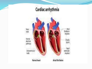
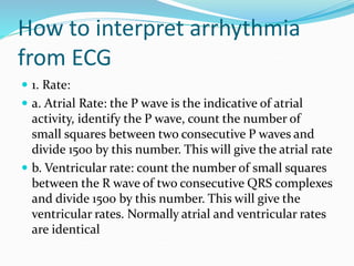
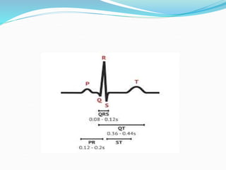
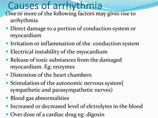
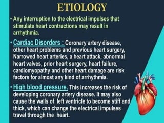
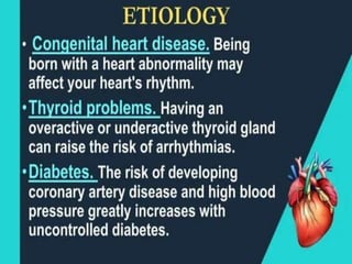
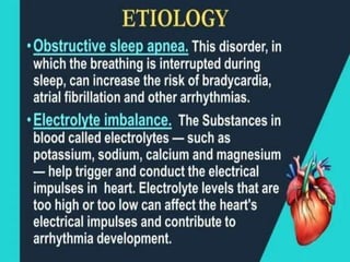
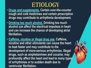
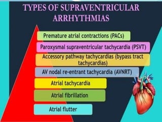
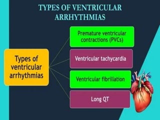
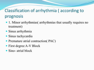
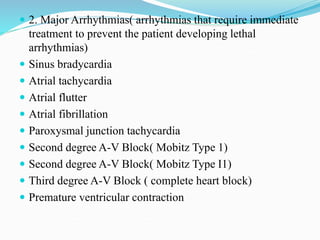
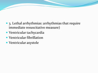
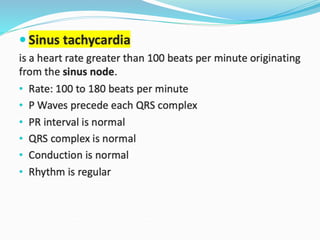
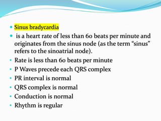
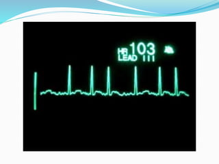
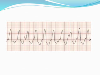
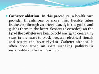
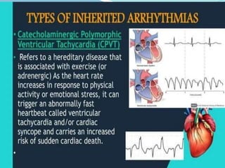
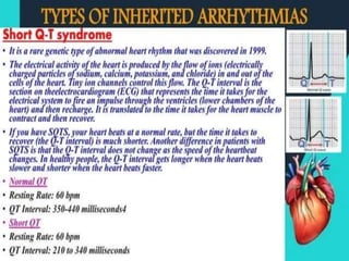
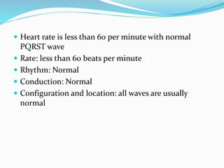
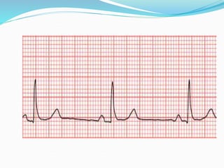
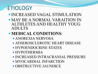
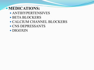
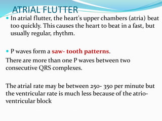
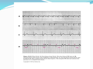
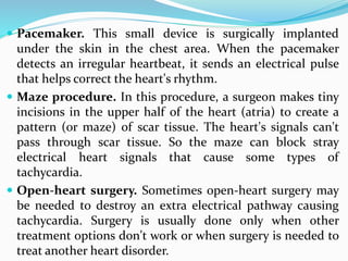
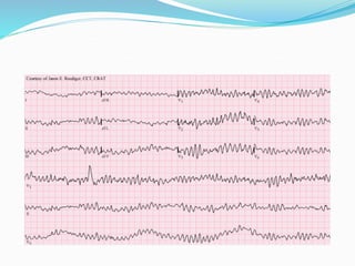
4. Definition
A heart arrhythmia is an irregular
heartbeat. Heart rhythm problems (heart
arrhythmias) occur when the electrical
signals that coordinate the heart's beats
don't work properly. The faulty signaling
causes the heart to beat too fast
(tachycardia), too slow (bradycardia) or
irregularly.
5.
6. How to interpret arrhythmia
from ECG
1. Rate:
a. Atrial Rate: the P wave is the indicative of atrial
activity, identify the P wave, count the number of
small squares between two consecutive P waves and
divide 1500 by this number. This will give the atrial rate
b. Ventricular rate: count the number of small squares
between the R wave of two consecutive QRS complexes
and divide 1500 by this number. This will give the
ventricular rates. Normally atrial and ventricular rates
are identical
7.
8. Causes of arrhythmia
One or more of the following factors may gives rise to
arrhythmia
Direct damage to a portion of conduction system or
myocardium
Irritation or inflammation of the conduction system
Electrical instability of the myocardium
Release of toxic substances from the damaged
myocardium. Eg: enzymes
Distention of the heart chambers
Stimulation of the autonomic nervous system{
sympathetic and parasympathetic nerves)
Blood gas abnormalities
Increased or decreased level of elctrolytes in the blood
Over dose of a cardiac drug eg: digoxin
9.
10.
11.
12.
13.
14.
15. Classification of arrhythmia ( according to
prognosis
1. Minor arrhythmias( arrhythmias that usually requires no
treatment)
Sinus arrhythmia
Sinus tachycardia
Premature atrial contraction( PAC)
First degree A-V Block
Sino- atrial block
16. 2. Major Arrhythmias( arrhythmias that require immediate
treatment to prevent the patient developing lethal
arrhythmias)
Sinus bradycardia
Atrial tachycardia
Atrial flutter
Atrial fibrillation
Paroxysmal junction tachycardia
Second degree A-V Block( Mobitz Type 1)
Second degree A-V Block( Mobitz Type I1)
Third degree A-V Block ( complete heart block)
Premature ventricular contraction
19. Causes of sinus tachycardia may include
exercise, anxiety, fever, drugs, anemia, heart
failure, hypovolemia, and shock.
Sinus tachycardia is often asymptomatic.
Management, however is directed at the
treatment of the primary cause.
Carotid sinus pressure (carotid massage) or
a beta-blocker may be used to reduce heart rate.
20.
21.
22.
23. Causes
drugs, vagal stimulation, hypo endocrine states,
hypothermia, or sinus node involvement in MI.
This arrhythmia may be normal in athletes as
they have quality stroke volume.
manifestations
syncope, fatigue, dizziness.
Management includes treating the underlying
cause and administering anticholinergic drugs like
atropine sulfate as prescribed.
24.
25. PREMATURE ATRIAL CONTRACTIONS
Premature atrial contractions (PACs) are extra heartbeats
that start in the upper chambers of your heart. When the
premature, or early, signal tells the heart to contract, there
may not be much blood in the heart at that moment. That
means there’s not much blood to pump out. A pause and a
strong beat may follow the extra heartbeat, making it feel
like a skipped beat.
Rate: varies
Rhythm: irregular; R-R interval of a premature beat is shorter
than normal.
Occurs when impulse is generated by an irritable area of tissue
in the atria.
26. Monitor the patient continously and watch for other
arrhythmias
Inform the doctor if more than 8- 10 PACs appear per
minute and follow medical orders
If digitalis overdose is suspected withhold the drug;
digoxin may be administered if there is CHF
IV potassium may be given if serum potassium level is
low
Oral quinidine (antiarrhythmic medications) may be
ordered for frequent PACs
Verapamil( calcium channel blockers) may be given if
PAC leads to atrial tachycardia
27.
28. Atrial flutter
is an abnormal rhythm that occurs in the atria of
the heart. Atrial flutter has an atrial rhythm that is
regular but has an atrial rate of 250 to 400
beats/minute.
It has sawtooth appearance.
QRS complexes are uniform in shape but often
irregular in rate.
29.
30. Management
if the patient is unstable with ventricular rate of
greater than 150 bpm, prepare for immediate
cardioversion.
If patient is stable, drug therapy may include
calcium channel blocker, beta-adrenergic
blockers, or antiarhythmics.
Anticoagulation may be necessary as there
would be pooling of blood in the atria
31.
32. Atrial fibrillation
is disorganized and uncoordinated twitching of
atrial musculature caused by overly rapid
production of atrial impulses.
• Atrial Rate: 350 to 600 bpm
• Ventricular Rate: 120 to 200 bpm
• P wave is not discernible with an irregular baseline
• PR interval is not measurable
• QRS complex is normal
• Rhythm is irregular and usually rapid unless
controlled
33. Atrial fibrillation may be asymptomatic
clinical manifestation
palpitations, dyspnea, and pulmonary edema.
Nursing goal is towards administration of
prescribed treatment to decrease ventricular
response, decrease atrial irritability and eliminate
the cause.
34.
35. svt
an electrical impulse outside the sinus node fires
repeatedly, often due to a short circuit — a circular
electrical pathway.
Electricity circles the atria again and again, causing
the upper chambers to contract more than 100 times
per minute.
36. Atrial tachycardia usually occurs for brief periods
and starts and stops spontaneously. That's
called paroxysmal AT.
If it continues, it is called persistent AT
Palpitations (a fluttering in the chest)
Fainting
Chest pain
Shortness of breath
Fatigue
Heart failure
37. causes
A "stretched" atrium resulting from high blood
pressure (hypertension) or from cardiomyopathy
A previous heart attack
Excessive use of alcohol, cocaine and other
stimulants
38. Treatment of any underlying conditions
Catheter ablation to destroy specific patches of heart
muscle that are incorrectly producing electrical
signals; usually performed at the same time as an
electrophysiological study
Medications to control the heart rhythm including
beta blockers, calcium channel blockers or
antiarrhythmic medications such as flecainide,
propafenone or amiodarone
39. PAROXYSMAL ATRIAL
TACHYCARDIA
Caused by an irritable area of tissue in the atria that
dominates the sinoatrial node and takes over as the
pacemaker. Paroxysmal supraventricular tachycardia
(PSVT) is a type of abnormal heart rhythm, or
arrhythmia. It occurs when a short circuit rhythm
develops in the upper chamber of the heart. This
results in a regular but rapid heartbeat that starts and
stops abruptly.
Usually preceded by premature atrial contractions.
Begin and end abruptly.
The rapid rate prevents adequate ventricular filling.
41. VENTRICULAR TACHYCARDIA
Ventricular tachycardia is a heart rhythm
problem (arrhythmia) caused by irregular
electrical signals in the lower chambers of the
heart (ventricles). This condition may also be
called V-tach or VT.
In ventricular tachycardia, the heart beats
faster, usually 100 or more beats a minute.
The rapid heartbeat prevents the heart
chambers from properly filling with blood.
45. Catheter ablation. In this procedure, a health care
provider threads one or more thin, flexible tubes
(catheters) through an artery, usually in the groin, and
guides them to the heart. Sensors (electrodes) on the
tip of the catheter use heat or cold energy to create tiny
scars in the heart to block irregular electrical signals
and restore the heart rhythm. Catheter ablation is
often done when an extra signaling pathway is
responsible for the fast heart rate.
46. Implantable cardioverter-defibrillator (ICD): An
ICD is device implanted under the skin near the collar
bone. It continuously monitor heart beat and deliver
precisely caliberated electrical shock when an irregular
rhythm is detected. The shock helps to restore regular
heart rhythm.
47.
48.
49.
50.
51. Heart Block
Heart block, also called AV block, is when the
electrical signal that controls the heartbeat is partially
or completely blocked. This makes the heart beat
slowly or skip beats and heart can’t pump blood
effectively. Symptoms include dizziness, fainting,
tiredness and shortness of breath. Pacemaker
implantation is a common treatment.
Heart block is a problem with heart's electrical system,
which makes the heart beat and controls heart rate
and rhythm. The condition is also called
atrioventricular (AV) block or a conduction disorder.
52. Normally, electrical signals travel from the upper
chambers of the heart (atria) to the lower chambers
(ventricles). The AV node is a cluster of cells that
connect the electrical activity – like a bridge – from the
top chambers of your heart to the bottom chambers. If
there is heart block, the electrical signal does not
travel through the AV node to the ventricles. The result
is a heart that doesn’t function effectively, means the
heart beats slowly or skips beats and it can’t pump
blood through its chambers and out to the body as a
normal heart would.
53. TYPES
Heart block can be first, second or third degree,
depending on the extent of electrical signal
impairment.
First-degree heart block: The electrical impulse still
reaches the ventricles, but moves more slowly than
normal through the AV node. The impulses are
delayed. This is the mildest type of heart block
54. Second-degree heart block is classified into two
categories: Type I and Type II. In second-degree
heart block, the impulses are intermittently
blocked.
Type I, also called Mobitz Type I or
Wenckebach’s AV block: This is a less serious
form of second-degree heart block. The electrical
signal gets slower and slower until your heart
actually skips a beat.
Type II, also called Mobitz Type II: While most
of the electrical signals reach the ventricles every
so often, some do not and your heartbeat
becomes irregular and slower than normal.
55. Mobitz I and Mobitz II are both subtypes
of a 2nd degree AV block. Mobitz I and
Mobitz II can be distinguished on an ECG by
the pattern in which P waves are blocked; in
Mobitz I, there is a progressive prolongation
of the PR interval until a P wave fails to
conduct.
Whereas in Mobitz II, PR intervals are
always the same length, but are followed by
a pattern of one or more non-conducted P
waves.
56. Mobitz I and Mobitz II also differ in the
severity of the conduction block. Mobitz I is
a benign rhythm that generally reflects a
block at the AV node, and typically results in
a good prognosis.
On the other hand, Mobitz II reflects a
block after the AV node, either at the bundle
of His or its branches, and often results in a
poorer prognosis, as it has a higher risk of
progressing to a 3rd degree AV block.
57.
58. Third-degree heart block: The electrical
signal from the atria to the ventricles is
completely blocked. To make up for this, the
ventricle usually starts to beat on its own
acting as a substitute pacemaker but the
heartbeat is slower and often irregular and
not reliable. Third-degree block seriously
affects the heart’s ability to pump blood out
to the body.
59. causes heart block
The most common cause of heart block is heart
attack. Other causes include heart muscle
disease, usually called a cardiomyopathy, heart
valve diseases and problems with the heart’s
structure. Heart block can also be caused by
damage to the heart during open heart
surgery, as a side effect of some medications or
exposure to toxins. Genetics can be another
cause.
60. symptoms of heart block
Symptoms of heart block vary depending on the type
of block.
First-degree heart block:
May not have any symptoms.
May be found during a routine electrocardiogram
(ECG) although heart rate and rhythm are usually
normal.
First-degree block is common in athletes, teenagers,
young adults and those with a highly active vagus
nerve.
62. Third-degree heart block symptoms:
Dizziness, fainting.
Chest pain.
Feeling tired.
Shortness of breath.
Symptoms of third-degree heart block are more
intense due to the slow heart rate. If there is severe
symptoms, get medical attention right away.
63. Diagnostic evaluations
ECG
Holter monitor
An implantable loop recorder: This is a very slender
device that is injected under the skin of your chest and
can monitor your heart rhythm for up to five years and
is useful for patients who have very infrequent but
important episodes without a clear explanation of their
origin.
An electrophysiology study: An electrophysiology
study involves inserting a long, thin tube called a
catheter through a blood vessel and guiding it to your
heart to measure and record electrical activity from
inside your heart
64. Complications of heart block
The complications can be life-threatening and include:
Heart failure.
Arrhythmia (irregular heartbeat).
Heart attack.
Sudden cardiac arrest.
65. How is heart block treated?
The cardiologist will determine heart block type,
location and severity. How it is affecting the heart’s
ability to function and consider the symptoms to
determine how to manage the condition. Symptoms
and treatment vary from person to person.
First-degree block: The first-degree heart block,
probably won’t need treatment.
Second-degree block: Second-degree heart block
and have symptoms, need a pacemaker to keep the
heart beating like it should. A pacemaker is small
device that sends electrical impulses to your heart.
66. Third-degree block: Third degree heart block is
often first discovered during an emergency situation.
Treatment almost always includes a pacemaker.
67.
68.
69. Brugada syndrome is a rare but serious condition that
affects the way electrical signals pass through the
heart. It can cause the heart to beat dangerously fast.
These unusually fast heartbeats –
73. SINUS BRADYCARDIA
Sinus bradycardia is a sinus node dysfunction
giving a heart rate that is lower than the normal 60–
100 beats per minute (bpm) in humans.
HR < 60/min arising from the SA node.
Impulses follow the normal pathway through the
conduction system
P and QRS complexes normal duration and pattern
74. Heart rate is less than 60 per minute with normal
PQRST wave
Rate: less than 60 beats per minute
Rhythm: Normal
Conduction: Normal
Configuration and location: all waves are usually
normal
75.
76. ETIOLOGY
INCREASED VAGAL STIMULATION
MAY BE A NORMAL VARAITION IN
ALTHLETES AND HEALTHY YOUG
ADULTS
MEDICAL CONDITIONS:
ANOREXIA NERVOSA
ATHEROSCLEROTIC HEART DISEASE
HYPOENDOCRINE STATES
HYPOTHERMIA
INCREASED INTRACRANIAL PRESSURE
MYOCARDIAL INFARCTION
OBSTRUCTIVE JAUNDICE
78. SYMPTOMS
SYMPTOMS RELATED TO DECREASE IN CARDIAC
OUTPUT
CHEST PRESSURE AND PAIN
DYSPNEA
HYPOTENSION
DIZZINESS
SEIZURES
SYNCOPE
Bradycardia cause a fall in BP due to decreased cardiac
output. Consequently the body tissues will suffer due to
lack of oxygen
79. TREATMENT
MANAGEMENT -ONLY IF SYMPTOMATIC-
AIMED AT INCREASING THE HEART RATE
MEDICATIONS
ATROPINE 0.6 to 1.2 mg given as IV blous
ISOPROTERENOL is given as IV infusion( 2mg in 500 ml of 5%
dextrose. (Isoproterenol is a beta-1 and beta-2 adrenergic receptor agonist resulting in
the following: Increased heart rate. Increased heart contractility.
PACEMAKER
SUPRESSION OF THE PARASYMPATHETIC NERVOUS
SYSTEM
STIMULATION OF THE SYMPATHETIC NERVOUS SYSTEM
80.
81. Heart rate is 150 to 250 per minute. P wave are difficult to
recognize, QRS complex are of normal shape. Usually
regular rhythm.
82. ETIOLOGY
SAME AS SEEN WITH PREMATURE ATRIAL
CONTRACTIONS
Damage to the SA node and atria
Irritability of the atrial muscles
Stimulation of the sympathetic nerves
Physical and emotional stress
Hypoxia
CHF
Digitalis overdose
84. TREATMENT
CAROTID SINUS PRESSURE:
the increase in blood pressure in carotid sinus will
stimulate stretch receptors, which leads to reflex
bradycardia and systemic vasodilatation.
The baroreceptor reflex is also critical in
maintaining heart rate and blood pressure
85. VAGAL NERVE STIMULATION:
Vagus nerve stimulation involves the use of a device
to stimulate the vagus nerve with electrical
impulses. vagal excitation slows down heart rate and
prolongs the diastolic blood supply.
MEDICATIONS:
DILTIAZEM * PROCAINAMIDE
VERAPAMIL * QUINIDINE
DIGOXIN * VASOPRESSOR
PROPRANOLOL
86. ATRIAL FLUTTER
In atrial flutter, the heart's upper chambers (atria) beat
too quickly. This causes the heart to beat in a fast, but
usually regular, rhythm.
P waves form a saw- tooth patterns.
There are more than one P waves between two
consecutive QRS complexes.
The atrial rate may be between 250- 350 per minute but
the ventricular rate is much less because of the atrio-
ventricular block
87. The rhythm is usually regular.
The P-R interval cannot be determined;
QRS complex is normal
88. Causes
Damage to SA node or atria
Increased sympathetic stimulation
Hypoxia
Congestive Heart Failure
89. Clinical features
The cardiac output is greatly reduced due to
incomplete filling of the ventricles
i. Rapid heart rate which reduces the diastolic phase of
the heart
ii. Inco-ordinated contraction of the atria and
ventricles
90. Treatment
Treatment for signs and symptoms like systolic BP 80-
90 mmHg or less, weak or absent pulse, pale, cold and
clammy skin.
Digoxin /propranolol
In resistant cases, synchronized D.C shock( elective
cardioversion) may be required
91. ATRIAL FIBRILLATION
Atrial fibrillation (A-fib) is an irregular and often
very rapid heart rhythm (arrhythmia) that can
lead to blood clots in the heart.
No regular P wave; P waves appearing as a wavy
baseline.
Atria contract 350-600 times per minute. Ventricular
rate is variable
Irregular rhythm
P-R interval cannot be monitored
93. Clinical Features
The cardiac output is greatly reduced due to
incomplete filling of the ventricles
i. Rapid heart rate which reduces the diastolic phase of
the heart
ii. Inco-ordinated contraction of the atria and
ventricles
94. Management- digitalis., beta blockers, calcium
channel blockers, counter shock( elective
cardioversion is required in selected cases
95. PAROXYSMAL JUNCTIONAL TACHYCARDIA
Junctionaltachycardia is a form
of supraventricular tachycardia, a type of racing pulse
caused by a problem in the area between the upper and
lower chambers of your heart. It’s known as the
atrioventricular node, or AV node. Junctional
tachycardia is a rare, fast heart rhythm that starts in the
wrong place in your heart.
ETIOLOGY
coronary artery disease
congestive heart failure
myocardial infarction
Damage to SA node
anxiety
alcohol, tobacco
96. SYMPTOMS
May be asymptomatic if rate is less than 150 beats/
minute
At rates greater than 150 beats/ minute:
chest pain, pressure, palpitations, dizziness, syncope
98. AV HEART BLOCKS
ABNORMAL DELAY IN CONDUCTION OF IMPULSE
FROM THE ATRIUM TO THE VENTRICLES
USUALLY ASYMPTOMATIC
99. SECOND DEGREE HEART BLOCK
TYPE I- MOBITZ I OR WENCKEBACH-
PROGRESSIVE LENGTHENING OF THE PR
INTERVAL UNTIL A QRS COMPLEX IS DROPPED
OR NOT CONDUCTED
USUALLY ASYMPTOMATIC
TX- MAYBE NONE, ATROPINE, TEMP. PACER
100. SECOND DEGREE- TYPE II
EVERY SECOND THIRD OR FOURTH SINUS
IMPULSE IS BLOCKED MAY HAVE 2,3,4 Ps TO
EACH QRS
MORE SERIOUS- AGGRESSIVE MANAGEMENT
TO PREVENT PROGRESSION TO COMPLETE
HEART BLOCK
TREATMENT:
PACER
ATROPINE
DOPAMINE FOR SEVERE HYPOTENSION
101. THIRD DEGREE HEART BLOCK
TOTAL DISASSOCIATION OF ATRIA TO VENTRICLES.
VENTRICLES ARE STIMULATED BY A SECONDARY OR ESCAPE
BEAT. THE VENTRICULAR RATE WILL BE 40-60 DEPENDING
UPON THE LOCATION OF THE VENTRICULAR PACEMAKER
BOTH THE SINUS P WAVE AND THE ESCAPE RHYTHM WILL BE
OBVIOUS ON THE ELECTROCARDIOGRAM
ETIOLOGY –
CARDIAC DISEASE
MEDICATIONS – BETA BLOCKERS, CALCIUM CHANNEL BLOCKERS,
DIGITALIS TOXICITY
MANIFESTATIONS- FATIGUE, HYPOTENSION, SYNCOPE, HEART
FAILURE
TX.- ATROPINE, ISOPROTERENOL, DOPAMINE, PACER
104. Pacemaker. This small device is surgically implanted
under the skin in the chest area. When the pacemaker
detects an irregular heartbeat, it sends an electrical pulse
that helps correct the heart's rhythm.
Maze procedure. In this procedure, a surgeon makes tiny
incisions in the upper half of the heart (atria) to create a
pattern (or maze) of scar tissue. The heart's signals can't
pass through scar tissue. So the maze can block stray
electrical heart signals that cause some types of
tachycardia.
Open-heart surgery. Sometimes open-heart surgery may
be needed to destroy an extra electrical pathway causing
tachycardia. Surgery is usually done only when other
treatment options don't work or when surgery is needed to
treat another heart disorder.
105. VENTRICULAR FIBRILLATION
Ventricular fibrillation is a type of irregular heart
rhythm (arrhythmia). During ventricular fibrillation,
the lower heart chambers contract in a very rapid and
uncoordinated manner. As a result, the heart doesn't
pump blood to the rest of the body.
Ventricular fibrillation is an emergency that requires
immediate medical attention. It's the most frequent
cause of sudden cardiac death.
No atrial activity seen on ecg
Absence of audible heartbeat, palpable pulse, and
respiration.
Ventricular fibrillation may also be called VFib, V-fib
or VF.
106.
107. Ventricular tachycardia and ventricular fibrillation are
closely related, very similar conditions. The difference
between the two is that in ventricular tachycardia, the
lower chambers of the heart are beating much faster
than they should but the overall process is happening
in the right order. In ventricular fibrillation, the heart’s
beating process isn’t happening in the right order.
Both ventricular tachycardia and ventricular
fibrillation are considered life-threatening because
they can lead to collapse and sudden cardiac arrest. In
emergencies, both are typically treated with
defibrillation. Long-term, both are typically treated
with a surgically placed implanted cardioverter
defibrillator.
108. ETIOLOGY
SAME AS VENTRICULAR TACHYCARDIA
UNTREATED VENTRICULAR TACHYCARDIA
ELECTRICAL SHOCK
BRUGADA SYNDROME
109. Symptoms
Collapse and loss of consciousness are the most
common symptoms of ventricular fibrillation.
Chest pain
Very fast heartbeat (tachycardia)
Dizziness
Nausea
Shortness of breath
110. TREATMENT
Ventricular fibrillation requires emergency medical
treatment to prevent sudden cardiac death. The goal of
emergency treatment is to restore blood flow as
quickly as possible to prevent organ and brain damage.
IMMEDIATE DEFIBRILLATION
ACTIVATION OF EMS
CPR
ERADICATING THE CAUSE
VASOACTIVE AND ANTIARRHYTHMIC
MEDICATIONS
111. Surgical mgmt
ICD
Cardiac Ablation
Coronary angioplasty and stent placement. If
ventricular fibrillation is caused by a heart attack, this
procedure may reduce the risk of future episodes of
ventricular fibrillation.
Coronary bypass surgery. This open-heart surgery
redirects blood around a section of a blocked or
partially blocked artery in the heart. It may be done if
ventricular fibrillation is caused by coronary artery
disease
112. VENTRICULAR ASYSTOLE
Asystole, colloquially referred to
as flatline, represents the cessation of electrical
and mechanical activity of the heart.
ABSENCE OF:
QRS
HEARTBEAT
PALPABLE PULSE
RESPIRATION
115. TREATMENT
CARDIOPULMONARY RESUSCITATION
INTUBATION
INTRAVENOUS ACCESS:EPINEPHRINE and
ATROPINE
TRANSCUTANEOUS PACING:Transcutaneous pacing,
also called external pacing, is a temporary means of
pacing a patient's heart during a medical emergency.
*The Advanced Life Support guidelines do not recommend defibrillation
in asystole. They consider shocks to confer no benefit, and go further
claiming that they can cause cardiac damage
116. ADJUNCTIVE MODALITIES
AND MANAGEMENT
TREATMENT DEPENDS UPON
WHETHER THE DYSRHYTHMIA IS ACUTE OR
CHRONIC
THE CAUSE OF THE DYSRHYTHMIA AND ITS
POTENTIAL HEMODYNAMIC EFFECTS
117. PACERS
AN ELECTRICAL IMPULSE THAT STIMULATES
THE MYOCARDIUM TO DEPOLARIZE,
INITIATING A HEARTBEAT
MAY BE DEMAND, FIXED, OR RATE
RESPONSIVE
MAY BE TEMPORARY OR PERMANENT
PACER SPIKE NOTED ON EKG
118. INDICATIONS
A SLOWER THAN NORMAL IMPULSE FORMATION
OR A ACONDUCTION DISTURBANCE THAT
CAUSES SYMPTOMS
MAY BE USED TO TREAT TACHYDYSRHYTHMIAS
THAT DO NOT RESPOND TO MEDICATION
THERAPY
119. ASSESSMENT
MONITOR HEART RATE AND RHYTHM BY
ELECTROCARDIOGRAM
ASSESS FOR PACEMAKER SPIKE AND ITS
RELATIONSHIP TO THE SURROUNDING
ELECTROCARDIOGRAM COMPLEXES
ASSESS CARDIAC OUTPUT AND
HEMODYNAMIC STABILITY
INCISION SITE
120. COMPLICATIONS
LOCAL INFECTION AT THE ENTRY SITE
BLEEDING AND HEMATOMA FORMATION
HEMOTHORAX
VENTRICULAR ECTOPY / TACHYCARDIA
DISLOCATION OF THE LEAD
STIMULATION OF THE PHRENIC NERVE
CARDIAC TAMPONADE
MY0CARDIAL WALL PERFORATION
124. CARDIOVERSION AND
DEFIBRILLATION
PADS OR PADDLES ARE USED TO DELIVER A N
ELECTRICAL CURRENT TO DEPOLARIZE A
CRITICAL MASS OF CARDIAC CELLS IN AN
ATTEMPT FOR THE SINUS NODE TO
RECAPTURE THE ROLE OF THE PACEMAKER
DIFFERENCE BETWEEN CARDIOVERSION AND
DEFIBRILLATION HAS TO DO WITH THE
TIMING OF THE DELIVERY AND THE
CIRCUMSTANCE
125. SAFETY
MAINTAIN GOOD CONTACT BETWEEN THE PADS
OR PADDLES AND THE SKIN
ENSURE THAT NOONE IS IN CONTACT WITH THE
CLIENT OR WITH ANYTHING TOUCHING THE
CLIENT
126. CARDIOVERSION
DELIVERY OF A TIMED ELECTRICAL CURRENT
TO TERMINATE A TACHYDYSRHYTHMIA
THE DEFIBRILLATOR IS SET TO SYNCHRONIZE
WITH THE ELECTROCARDIOGRAM ON A
MONITOR SO THAT THE ELECTRICAL
IMPULSE DISCHARGES DURING VENTRICULAR
DEPOLARIZATION
VOLTAGE VARIES FROM 25 TO 360 JOULES
127. PREPARATION
ANTICOAGULATION FOR A FEW WEEKS PRIOR TO
PROCEDURE IF ELECTIVE
DIGOXIN IS WITHHELD FOR 48 HOURS
NPO FOR AT LEAST 8 HOURS
INTRAVENOUS SEDATION
SUPPLEMENTAL OXYGENATION
128. POST PROCEDURE CARE
MAINTAIN AIRWAY PATENCY
MONITOR VITAL SIGNS AND OXYGEN
SATURATION
ELECTROCARDIOGRAM MONITORING
129. DEFIBRILLATION
USED IN EMERGENCY SITUATIONS AS THE
TREATMENT OF CHOICE FOR VENTRICULAR
FIBRILLATION AND PULSELESS VENTRICULAR
TACHYCARDIA
ELECTRICAL VOLTAGE IS USUALLY GREATER THAN
WITH CARDIOVERSION
THE USE OF EPINEPHRINE OR VASOPRESSIN MAY BE
HELPFUL
ANTIARRHYTHMIC MEDICATIONS SUCH AS
AMIODARONE, LIDOCAINE, MAGNESIUM,
PROCAINAMIDE ARE GIVEN IF VENTRICULAR
DYSRHYTHMIA PERSISTS
130. ELECTROPHYSIOLOGIC STUDIES
IDENTIFY IMPULSE FORMATION THROUGH THE
CARDIAC CONDUCTION SYSTEM
ASSESS THE FUNCTION OF THE SA AND AV NODES
MAP DYSRHYTHMOGENIC FOCI
ASSESS THE EFFECTIVENESS OF ANTIARRHYTHMIC
MEDICATIONS
TREAT CERTAIN DYSRHYTHMIAS THROUGH THE
DESTRUCTION OF CAUSATIVE CELLS (ABLATION)
132. MEDICATIONS
CLASS I – SODIUM CHANNEL BLOCKERS
IA – SLOWS CONDUCTION AND
PROLONGS REPOLARIZATION –
QUINIDINE, PROCAINAMIDE,
DISOPYRAMIDE
IB – SLOWS CONDUCTION AND
SHORTENS REPOLARIZATION –
LIDOCAINE, MEXILETINE HCL
IC- PROLONGS CONDUCTION WITH
LITTLE OR NO EFFECT ON
REPOLARIZATION – ENCAINIDE,
FLECAINIDE
133. CLASS II
BETA BLOCKERS – DECREASE CONDUCTION
VELOCITY, AUTOMATICITY AND RECOVERY TIME
( REFRACTORY PERIOD) – PROPRANOLOL,
ACEBUTOLOL
134. CLASS III
PROLONG REPOLARIZATION- ARE USED IN THE
EMERGENCY TREATMENT OF VENTRICULAR
DYSRHYTHMIAS WHEN OTHER
ANTIDYSRHYTHMICS ARE NOT EFFECTIVE –
BRETYLIUM, AMIODARONE
135. CLASS IV
CALCIUM CHANNEL BLOCKERS – BLOCKS
CALCIUM INFLUX, DECREASING THE
EXCITABILITY AND CONTRACTILITY OF THE
MYOCARDIUM – VERAPAMIL, DILTIAZEM
136. OTHERS
DILANTIN- USED IN THE TX OF DIGITALIS
INDUCED DYSRHYTHMIAS
DIGOXIN- ATRIAL FLUTTER OR FIBRILLATION,
PREVENT RECURRENCE OF PAT
ATROPINE- BRADYCARDIA
137. NURSING PROCESS - DYSRHYTHMIA
ASSESSMENT –
HISTORY
CAUSES OF DYSRHYTHMIA
PHYSICAL EXAM
EFFECT ON CARDIAC OUTPUT
138. NURSING PROCESS
DIAGNOSES:
DECREASED CARDIAC OUTPUT
ANXIETY RELATED TO FEAR OF THE UNKNOWN
DEFICIENT KNOWLEDGE ABOUT THE
DYSRHYTHMIA AND TREATMENT
139. NURSING PROCESS
PLANNING AND GOALS
ERADICATING OR DECREASING THE INCIDENCE OF
THE DYSRHYTHMIA
ACQUIRE KNOWLEDGE ABOUT THE DYSRHYTHMIA
AND TREATMENT
140. NURSING PROCESS
INTERVENTIONS
MONITOR :
BLOOD PRESSURE, PULSE RATE AND RHYTHM, RATE AND
RHYTHM OF RESPIRATIONS, BREATH SOUNDS
EPISODES OF LIGHTHEADEDNESS, DIZZINESS, FAINTNESS
RHYTHM STRIPS
MEDICATION ADMINISTRATION
ASSIST IN DEVELOPING A PLAN TO MODIFY LIFESTYLE
MINIMIZE ANXIETY
TEACH SELF CARE
141. NURSING PROCESS
EVALUATION
EXPECTED OUCOMES
MAINTAINS CARDIAC OUTPUT
EXPERIENCES REDUCED ANXIETY
EXPRESSES UNDERSTANDING OF THE DYSRHYTHMIA
AND ITS TREATMENT