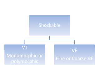
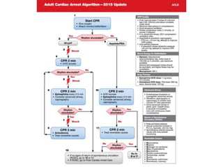
Adult Advanced Cardiovascular Life Support
Circulation. 2015;132[suppl 2]:S444–S464
• The 2015 American Heart Association(AHA)
Guidelines Update for Cardiopulmonary
Resuscitation (CPR) and Emergency
Cardiovascular Care (ECC) is based on the
International Liaison Committee on
Resuscitation(ILCOR) 2015 international
evidence review process.
• Advanced cardiac life support or advanced
cardiovascular life support (ACLS) refers to a set
of clinical interventions for the urgent treatment of
cardiac arrest, stroke and other life-threatening
medical emergencies, as well as the knowledge and
skills to deploy those interventions.
• ACLS is built heavily upon the foundation of
BLS.
• BLS is the foundation for saving lives after
cardiac arrest.
• Fundamental aspects of BLS includes
I. Immediate recognition of sudden cardiac
arrest.
II. Activation of emergency response system
III. Early CPR
IV. Rapid defibrillation with an AED.
Components of high quality CPR
• Scene safety:
1. Make sure the environment is safe for rescuers and
victim
• Recognition of cardiac arrest:
1. Check for responsiveness
2. No breathing or only gasping ( ie, no normal
breathing)
3. No definite pulse felt within 10 secs ( Carotid or
femoral pulse)
(Breathing and pulse check can be performed
simultaneously within 10 secs)
Activation of emergency response system:
• If alone with no mobile phone, leave the victim
to activate the emergency response system and
get the AED before beginning CPR
• Otherwise, send someone and begin CPR
immediately; use the AED as soon as it is
available
• In case of unwitnessed collapse of children or
infant give CPR for 2 mins before leaving the
victim and getting the AED then resume CPR
A Change From A-B-C to C-A-B
BLS Dos and Don’ts of Adult High-
Quality CPR
Rescuers Should
• Perform chest compressions at
a rate of 100-120/min.
• Compress to a depth of at least
2 inches (5 cm).
• Allow full recoil after each
compression
• Minimize pauses in
compressions
• Ventilate adequately (2 breaths
after 30 compressions, each
breath delivered over 1 second,
each causing chest rise)
Rescuers Should Not
• Compress at a rate slower than
100/min or faster than 120/min
• Compress to a depth of less
than 2 inches (5 cm) or greater
than 2.4 inches (6 cm)
• Lean on the chest between
compressions
• Interrupt compressions for
greater than 10 seconds
• Provide excessive ventilation(ie,
too many breaths or breaths
with excessive force)
Adjuncts to CPR
1.Adjuncts to CPR - Updated
• The immediate goals of CPR are to restore the
energy state of the heart so it can resume
mechanical work and to maintain the energy
state of the brain to minimize ischemic injury.
Adequate oxygen delivery is necessary to achieve
these goals. Oxygen delivery is dependent on
both blood flow and arterial oxygen content.
• . Because blood flow is typically the major
limiting factor to oxygen delivery during CPR, it is
theoretically important to maximize the oxygen
content of arterial blood by maximizing inspired
oxygen concentration. Maximal inspired oxygen
can be achieved with high-flow oxygen into a
resuscitation bag device attached to a mask or
an advanced airway.
• 1.1 2015 Recommendation - Updated
• When supplementary oxygen is available, it may
be reasonable to use the maximal feasible
inspired oxygen concentration during CPR
• 1.2 2015 Recommendation – Updated
Physiologic parameters
• Monitoring of pETCO2
• Coronary perfusion pressure (CPP)
• Central venous oxygen saturation (ScvO2)
• Intra-arterial pressure
• Abrupt increase in any of these parameters
is a sensitive indicator of ROSC that can be
monitored without interrupting chest
compressions
• Attempt to improve CPR quality if…
etco2 <10 mm Hg
diastolic pressure <20 mm Hg
ScvO2 is < 30%,
• Ultrasound may be applied to patients receiving CPR to
help assess myocardial contractility and to help identify
potentially treatable causes of cardiac arrest such as
hypovolemia, pneumothorax, pulmonary
thromboembolism, or pericardial tamponade.
• 1.3 2015 Recommendations - Updated
• Ultrasound (cardiac or noncardiac) may be considered
during the management of cardiac arrest, although its
usefulness has not been well established
• If a qualified sonographer is present and use of
ultrasound does not interfere with the standard cardiac
arrest treatment protocol, then ultrasound may be
considered as an adjunct to standard patient evaluation.
Adjuncts for
Airway Control
and Ventilation
2.Adjuncts for Airway Control and
Ventilation-Updated
• Both ventilation and chest compressions are thought to
be important for victims of prolonged ventricular
fibrillation (VF) cardiac arrest and for all victims with
other presenting rhythms.
• Because both systemic and pulmonary perfusion are
substantially reduced during CPR, normal ventilation-
perfusion relationships can be maintained with a
minute ventilation that is much lower than normal.
• During CPR with an advanced airway in place, a lower
rate of rescue breathing is needed to avoid
hyperventilation.
• During low blood flow states such as CPR, oxygen
delivery to the heart and brain is limited by blood
flow rather than by arterial oxygen content.
• Thus, during the first few minutes of witnessed
cardiac arrest a lone rescuer should not interrupt
chest compressions for ventilation. Advanced
airway placement in cardiac arrest should not
delay initial CPR and defibrillation for VF cardiac
arrest.
• routine use is not recommended.
• can be used in unconscious (unresponsive)
patients with no cough or gag reflex and should be
inserted only by persons trained in their use.
• can be used in patients with an obstructed airway.
• In the presence of known or suspected basal skull
fracture or severe coagulopathy, an oral airway is
preferred
• If advanced airway placement will interrupt chest
compressions, providers may consider deferring
insertion of the airway until the patient fails to
respond to initial CPR and defibrillation attempts or
demonstrates ROSC.
• 2015 Recommendations-Updated
• Either a bag-mask device or an advanced
airway(ETT/SGA Devices) may be used for
oxygenation and ventilation during CPR in both the
in-hospital and out-of-hospital setting.
• ETT/SGA Vs Bag Mask Ventillation-
Updated
• There is no high quality evidence favouring the use of
an ETT/SGA Devices compared to bag mask
ventillation in relation to overall survival or favorable
neurologic outcome.
• Continuous waveform capnography is the most
reliable method of confirming and monitoring
correct placement of an ETT.
• Deliver 1 breath every 6 seconds (10 breaths/min)
while continuous chest compressions are being
performed.
Management of
Cardiac Arrest
H’s T’s
• Hypoxia Toxins
• Hypovolemia Tamponade (cardiac)
• Hydrogen ion(acidosis) Tension pneumothorax
• Hypo-/hyperkalemia Thrombosis, pulmonary
• Hypothermia Thrombosis, coronary
I. ventricular fibrillation (VF)
II. pulseless ventricular tachycardia (pVT)
III. pulseless electric activity (PEA)
IV. asystole.
Shockable
VT
Monomorphic or
polymorphic
VF
Fine or Coarse VF
Unshockable
Asystole
PEA- pulseless electrical
activity or EMD-
electromechanical
dissociation
• In addition to high-quality CPR, the only
rhythm-specific therapy proven to increase
survival to hospital discharge is defibrillation
of VF/pulseless VT. Therefore, this intervention
is included as an integral part of the CPR cycle
when the rhythm check reveals VF/pulseless
VT.
4 pad positions
• anterolateral
• anteroposterior
• anterior-left infrascapular
• anterior-right infrascapular
• For adults, an electrode size of 8 to 12 cm is
reasonable.
• Any of the 4 pad positions is reasonable for
defibrillation.
3.1.Rhythm-Based Management of
Cardiac Arrest
 Defibrillation Strategies for Ventricular
Fibrillation or Pulseless Ventricular Tachycardia
• Currently manufactured manual and automated external
defibrillators use biphasic waveforms of 3 different designs:
biphasic truncated exponential (BTE), rectilinear biphasic
(RLB), and pulsed biphasic waveforms.
• These deliver different peak currents at the same
programmed energy setting and may adjust their energy
output in relation to patient impedance in differing ways.
• 2015 Recommendations – Update
I. Based on their greater success in arrhythmia
termination, defibrillators using biphasic
waveforms (BTE or RLB) are preferred to
monophasic defibrillators for treatment of both
atrial and ventricular arrhythmias.
II. If using a manual defibrillator capable of
escalating energies, higher energy for second and
subsequent shocks may be considered.
III. Defibrillation should be continued until VF is
terminated or maximal defibrillator energy output
is reached.
IV. A single-shock strategy (as opposed to stacked
shocks) is reasonable for defibrillation.
V. Current evidence indicates that the benefit of
using a multimodal defibrillator in manual instead
of automatic mode during cardiac arrest is
uncertain.
• When a rhythm check by an AED reveals a
nonshockable rhythm, CPR should be resumed
immediately,beginning with chest compressions,
and should continue for 2 minutes before the
rhythm check is repeated.
• When a rhythm check using a manual defibrillator
or cardiac monitor reveals an organized rhythm, a
pulse-check is performed. If a pulse is detected,
post–cardiac arrest care should be initiated
immediately.
• If the rhythm is asystole or the pulse is absent (eg,
PEA), CPR should be resumed immediately,
beginning with chest compressions, and should
continue for 2 minutes before the rhythm check is
repeated.
• During each 2-minute period of CPR the provider
should recall the H’s and T’s to identify factors that
may have caused the arrest or may be complicating
the resuscitative effort. For example--
-pulmonary embolism->start empirical fibrinolytic
therapy
-tension pneumothorax->do needle decompression
• Asystole is commonly the end-stage rhythm that
follows prolonged VF or PEA, and for this reason the
prognosis is generally much worse.
3.2. Medications for Arrest Rhythms
I. AMIODARONE may be considered for VF/pVT that is
unresponsive to CPR, defibrillation, and a vasopressor
therapy.
II. LIDOCAINE may be considered as an alternative to
amiodarone for VF/pVT that is unresponsive to
CPR,defibrillation, and vasopressor therapy.
III. The routine use of magnesium for VF/pVT is not
recommended in adult patients.
• Lidocaine
• 2015 (New): There is inadequate evidence to
support the routine use of lidocaine after
cardiac arrest. However, the initiation or
continuation of lidocaine may be considered
immediately after ROSC from cardiac arrest
due to VF/pVT to prevent recurrent VF/pVT.
• ß-Blockers
• 2015 (New): There is inadequate evidence to
support the routine use of a ß-blocker after
cardiac arrest. However, the initiation or
continuation of an oral or IV ß-blocker may be
considered early after hospitalization from
cardiac arrest due to VF/pVT.
WHY….
• In an observational study of patients who had
ROSC after VF/pVT cardiac arrest, ß-blocker
administration was associated with higher survival
rates. However, this finding is only an associative
relationship, and the routine use of ß-blockers
after cardiac arrest is potentially hazardous
because ß-blockers can cause or worsen
hemodynamic instability, exacerbate heart failure,
and cause bradyarrhythmias. Therefore, providers
should evaluate patients individually for their
suitability for ß-blockers.
• Vasopressin
• 2015 (Updated): Vasopressin in combination with
epinephrine offers no advantage as a substitute for
standard- dose epinephrine in cardiac arrest.
• 2010 (Old): One dose of vasopressin 40 units IV/
intraosseously may replace either the first or
second dose of epinephrine in the treatment of
cardiac arrest.
WHY….
• Both epinephrine and vasopressin
administration during cardiac arrest have been
shown to improve ROSC. Review of the
available evidence shows that efficacy of the 2
drugs is similar and that there is no
demonstrable benefit from administering both
epinephrine and vasopressin as compared
with epinephrine alone.
•Epinephrine
• 2015 (New): It may be reasonable to
administer epinephrine as soon as feasible
after the onset of cardiac arrest due to an
initial nonshockable rhythm.
WHY….
• A very large observational study of cardiac
arrest with nonshockable rhythm compared
epinephrine given at 1 to 3 minutes with
epinephrine given at 3 later time intervals (4
to 6, 7 to 9, and greater than 9 minutes). The
study found an association between early
administration of epinephrine and increased
ROSC, survival to hospital discharge, and
neurologically intact survival.
I. Standard-dose epinephrine (1 mg every 3 to 5 minutes)
may be reasonable for patients in cardiac arrest.
II. High-dose epinephrine is not recommended for routine
use in cardiac arrest.
III. Vasopressin offers no advantage as a substitute for
epinephrine in cardiac arrest.
IV. Vasopressin in combination with epinephrine offers no
advantage as a substitute for standard-dose epinephrine
in cardiac arrest.
V. It may be reasonable to administer epinephrine as soon
as feasible after the onset of cardiac arrest caused due
to an initial non- shockable rhythm.
• In IHCA, the combination of intra-arrest
vasopressin, epinephrine, and methylprednisolone
and postarrest hydrocortisone may be considered;
however, further studies are needed before
recommending the routine use of this therapeutic
strategy.
• For patients with OHCA, use of steroids during CPR
is of uncertain benefit.
3.5.Prognostication During CPR
• In intubated patients, failure to achieve an ETCO2
of greater than 10 mmHg by waveform
capnography after 20 minutes of CPR may be
considered as one component of a multimodal
approach to decide when to end resuscitative
efforts, but it should not be used in isolation.
• ECPR refers to venoarterial
extracorporeal membrane oxygenation
during cardiac arrest, including
extracorporeal membrane oxygenation
and cardiopulmonary bypass.
• There is insufficient evidence to recommend
the routine use of ECPR for patients with
cardiac arrest. In settings where it can be
rapidly implemented, ECPR may be considered
for select cardiac arrest patients for whom the
suspected etiology of the cardiac arrest is
potentially reversible during a limited period
of mechanical cardiorespiratory support.
4.Interventions Not Recommended
for Routine Use During Cardiac Arrest
I. Available evidence suggests that routine use of atropine
during PEA or asystole is unlikely to have a therapeutic
benefit.
II. Routine use of sodium bicarbonate is not
recommended for patients in cardiac arrest.(In some
special resuscitation situations, such as preexisting
metabolic acidosis, hyperkalemia, or tricyclic
antidepressant overdose, bicarbonate can be beneficial.)
III. Routine administration of calcium for treatment of in-
hospital and out-of-hospital cardiac arrest is not
recommended.
IV. Fibrinolytic therapy should not be routinely
used in cardiac arrest.
V. Electric pacing is not recommended for routine
use in cardiac arrest.
VI. The precordial thump(striking at the middle of a
person’s sternum with the ulnar aspect of the fist)
may be considered for termination of witnessed
monitored unstable ventricular tachyarrhythmias
when a defibrillator is not immediately ready for
use but should not delay CPR and shock delivery.
2015 acls
2015 acls
2015 acls
2015 acls

2015 acls

  • 1.
    Adult Advanced CardiovascularLife Support Circulation. 2015;132[suppl 2]:S444–S464
  • 2.
    • The 2015American Heart Association(AHA) Guidelines Update for Cardiopulmonary Resuscitation (CPR) and Emergency Cardiovascular Care (ECC) is based on the International Liaison Committee on Resuscitation(ILCOR) 2015 international evidence review process.
  • 3.
    • Advanced cardiaclife support or advanced cardiovascular life support (ACLS) refers to a set of clinical interventions for the urgent treatment of cardiac arrest, stroke and other life-threatening medical emergencies, as well as the knowledge and skills to deploy those interventions. • ACLS is built heavily upon the foundation of BLS.
  • 4.
    • BLS isthe foundation for saving lives after cardiac arrest. • Fundamental aspects of BLS includes I. Immediate recognition of sudden cardiac arrest. II. Activation of emergency response system III. Early CPR IV. Rapid defibrillation with an AED.
  • 5.
    Components of highquality CPR • Scene safety: 1. Make sure the environment is safe for rescuers and victim • Recognition of cardiac arrest: 1. Check for responsiveness 2. No breathing or only gasping ( ie, no normal breathing) 3. No definite pulse felt within 10 secs ( Carotid or femoral pulse) (Breathing and pulse check can be performed simultaneously within 10 secs)
  • 6.
    Activation of emergencyresponse system: • If alone with no mobile phone, leave the victim to activate the emergency response system and get the AED before beginning CPR • Otherwise, send someone and begin CPR immediately; use the AED as soon as it is available • In case of unwitnessed collapse of children or infant give CPR for 2 mins before leaving the victim and getting the AED then resume CPR
  • 9.
    A Change FromA-B-C to C-A-B
  • 10.
    BLS Dos andDon’ts of Adult High- Quality CPR Rescuers Should • Perform chest compressions at a rate of 100-120/min. • Compress to a depth of at least 2 inches (5 cm). • Allow full recoil after each compression • Minimize pauses in compressions • Ventilate adequately (2 breaths after 30 compressions, each breath delivered over 1 second, each causing chest rise) Rescuers Should Not • Compress at a rate slower than 100/min or faster than 120/min • Compress to a depth of less than 2 inches (5 cm) or greater than 2.4 inches (6 cm) • Lean on the chest between compressions • Interrupt compressions for greater than 10 seconds • Provide excessive ventilation(ie, too many breaths or breaths with excessive force)
  • 17.
  • 18.
    1.Adjuncts to CPR- Updated • The immediate goals of CPR are to restore the energy state of the heart so it can resume mechanical work and to maintain the energy state of the brain to minimize ischemic injury. Adequate oxygen delivery is necessary to achieve these goals. Oxygen delivery is dependent on both blood flow and arterial oxygen content.
  • 19.
    • . Becauseblood flow is typically the major limiting factor to oxygen delivery during CPR, it is theoretically important to maximize the oxygen content of arterial blood by maximizing inspired oxygen concentration. Maximal inspired oxygen can be achieved with high-flow oxygen into a resuscitation bag device attached to a mask or an advanced airway. • 1.1 2015 Recommendation - Updated • When supplementary oxygen is available, it may be reasonable to use the maximal feasible inspired oxygen concentration during CPR
  • 20.
    • 1.2 2015Recommendation – Updated Physiologic parameters • Monitoring of pETCO2 • Coronary perfusion pressure (CPP) • Central venous oxygen saturation (ScvO2) • Intra-arterial pressure • Abrupt increase in any of these parameters is a sensitive indicator of ROSC that can be monitored without interrupting chest compressions
  • 21.
    • Attempt toimprove CPR quality if… etco2 <10 mm Hg diastolic pressure <20 mm Hg ScvO2 is < 30%,
  • 22.
    • Ultrasound maybe applied to patients receiving CPR to help assess myocardial contractility and to help identify potentially treatable causes of cardiac arrest such as hypovolemia, pneumothorax, pulmonary thromboembolism, or pericardial tamponade. • 1.3 2015 Recommendations - Updated • Ultrasound (cardiac or noncardiac) may be considered during the management of cardiac arrest, although its usefulness has not been well established • If a qualified sonographer is present and use of ultrasound does not interfere with the standard cardiac arrest treatment protocol, then ultrasound may be considered as an adjunct to standard patient evaluation.
  • 23.
  • 24.
    2.Adjuncts for AirwayControl and Ventilation-Updated • Both ventilation and chest compressions are thought to be important for victims of prolonged ventricular fibrillation (VF) cardiac arrest and for all victims with other presenting rhythms. • Because both systemic and pulmonary perfusion are substantially reduced during CPR, normal ventilation- perfusion relationships can be maintained with a minute ventilation that is much lower than normal. • During CPR with an advanced airway in place, a lower rate of rescue breathing is needed to avoid hyperventilation.
  • 25.
    • During lowblood flow states such as CPR, oxygen delivery to the heart and brain is limited by blood flow rather than by arterial oxygen content. • Thus, during the first few minutes of witnessed cardiac arrest a lone rescuer should not interrupt chest compressions for ventilation. Advanced airway placement in cardiac arrest should not delay initial CPR and defibrillation for VF cardiac arrest.
  • 26.
    • routine useis not recommended. • can be used in unconscious (unresponsive) patients with no cough or gag reflex and should be inserted only by persons trained in their use. • can be used in patients with an obstructed airway. • In the presence of known or suspected basal skull fracture or severe coagulopathy, an oral airway is preferred
  • 27.
    • If advancedairway placement will interrupt chest compressions, providers may consider deferring insertion of the airway until the patient fails to respond to initial CPR and defibrillation attempts or demonstrates ROSC. • 2015 Recommendations-Updated • Either a bag-mask device or an advanced airway(ETT/SGA Devices) may be used for oxygenation and ventilation during CPR in both the in-hospital and out-of-hospital setting.
  • 28.
    • ETT/SGA VsBag Mask Ventillation- Updated • There is no high quality evidence favouring the use of an ETT/SGA Devices compared to bag mask ventillation in relation to overall survival or favorable neurologic outcome.
  • 29.
    • Continuous waveformcapnography is the most reliable method of confirming and monitoring correct placement of an ETT. • Deliver 1 breath every 6 seconds (10 breaths/min) while continuous chest compressions are being performed.
  • 30.
  • 31.
    H’s T’s • HypoxiaToxins • Hypovolemia Tamponade (cardiac) • Hydrogen ion(acidosis) Tension pneumothorax • Hypo-/hyperkalemia Thrombosis, pulmonary • Hypothermia Thrombosis, coronary
  • 32.
    I. ventricular fibrillation(VF) II. pulseless ventricular tachycardia (pVT) III. pulseless electric activity (PEA) IV. asystole.
  • 33.
  • 34.
    Unshockable Asystole PEA- pulseless electrical activityor EMD- electromechanical dissociation
  • 35.
    • In additionto high-quality CPR, the only rhythm-specific therapy proven to increase survival to hospital discharge is defibrillation of VF/pulseless VT. Therefore, this intervention is included as an integral part of the CPR cycle when the rhythm check reveals VF/pulseless VT.
  • 36.
    4 pad positions •anterolateral • anteroposterior • anterior-left infrascapular • anterior-right infrascapular • For adults, an electrode size of 8 to 12 cm is reasonable. • Any of the 4 pad positions is reasonable for defibrillation.
  • 37.
    3.1.Rhythm-Based Management of CardiacArrest  Defibrillation Strategies for Ventricular Fibrillation or Pulseless Ventricular Tachycardia • Currently manufactured manual and automated external defibrillators use biphasic waveforms of 3 different designs: biphasic truncated exponential (BTE), rectilinear biphasic (RLB), and pulsed biphasic waveforms. • These deliver different peak currents at the same programmed energy setting and may adjust their energy output in relation to patient impedance in differing ways.
  • 38.
    • 2015 Recommendations– Update I. Based on their greater success in arrhythmia termination, defibrillators using biphasic waveforms (BTE or RLB) are preferred to monophasic defibrillators for treatment of both atrial and ventricular arrhythmias. II. If using a manual defibrillator capable of escalating energies, higher energy for second and subsequent shocks may be considered.
  • 39.
    III. Defibrillation shouldbe continued until VF is terminated or maximal defibrillator energy output is reached. IV. A single-shock strategy (as opposed to stacked shocks) is reasonable for defibrillation. V. Current evidence indicates that the benefit of using a multimodal defibrillator in manual instead of automatic mode during cardiac arrest is uncertain.
  • 40.
    • When arhythm check by an AED reveals a nonshockable rhythm, CPR should be resumed immediately,beginning with chest compressions, and should continue for 2 minutes before the rhythm check is repeated. • When a rhythm check using a manual defibrillator or cardiac monitor reveals an organized rhythm, a pulse-check is performed. If a pulse is detected, post–cardiac arrest care should be initiated immediately. • If the rhythm is asystole or the pulse is absent (eg, PEA), CPR should be resumed immediately, beginning with chest compressions, and should continue for 2 minutes before the rhythm check is repeated.
  • 41.
    • During each2-minute period of CPR the provider should recall the H’s and T’s to identify factors that may have caused the arrest or may be complicating the resuscitative effort. For example-- -pulmonary embolism->start empirical fibrinolytic therapy -tension pneumothorax->do needle decompression • Asystole is commonly the end-stage rhythm that follows prolonged VF or PEA, and for this reason the prognosis is generally much worse.
  • 42.
    3.2. Medications forArrest Rhythms I. AMIODARONE may be considered for VF/pVT that is unresponsive to CPR, defibrillation, and a vasopressor therapy. II. LIDOCAINE may be considered as an alternative to amiodarone for VF/pVT that is unresponsive to CPR,defibrillation, and vasopressor therapy. III. The routine use of magnesium for VF/pVT is not recommended in adult patients.
  • 43.
    • Lidocaine • 2015(New): There is inadequate evidence to support the routine use of lidocaine after cardiac arrest. However, the initiation or continuation of lidocaine may be considered immediately after ROSC from cardiac arrest due to VF/pVT to prevent recurrent VF/pVT.
  • 44.
    • ß-Blockers • 2015(New): There is inadequate evidence to support the routine use of a ß-blocker after cardiac arrest. However, the initiation or continuation of an oral or IV ß-blocker may be considered early after hospitalization from cardiac arrest due to VF/pVT. WHY….
  • 45.
    • In anobservational study of patients who had ROSC after VF/pVT cardiac arrest, ß-blocker administration was associated with higher survival rates. However, this finding is only an associative relationship, and the routine use of ß-blockers after cardiac arrest is potentially hazardous because ß-blockers can cause or worsen hemodynamic instability, exacerbate heart failure, and cause bradyarrhythmias. Therefore, providers should evaluate patients individually for their suitability for ß-blockers.
  • 46.
    • Vasopressin • 2015(Updated): Vasopressin in combination with epinephrine offers no advantage as a substitute for standard- dose epinephrine in cardiac arrest. • 2010 (Old): One dose of vasopressin 40 units IV/ intraosseously may replace either the first or second dose of epinephrine in the treatment of cardiac arrest. WHY….
  • 47.
    • Both epinephrineand vasopressin administration during cardiac arrest have been shown to improve ROSC. Review of the available evidence shows that efficacy of the 2 drugs is similar and that there is no demonstrable benefit from administering both epinephrine and vasopressin as compared with epinephrine alone.
  • 48.
    •Epinephrine • 2015 (New):It may be reasonable to administer epinephrine as soon as feasible after the onset of cardiac arrest due to an initial nonshockable rhythm. WHY….
  • 49.
    • A verylarge observational study of cardiac arrest with nonshockable rhythm compared epinephrine given at 1 to 3 minutes with epinephrine given at 3 later time intervals (4 to 6, 7 to 9, and greater than 9 minutes). The study found an association between early administration of epinephrine and increased ROSC, survival to hospital discharge, and neurologically intact survival.
  • 50.
    I. Standard-dose epinephrine(1 mg every 3 to 5 minutes) may be reasonable for patients in cardiac arrest. II. High-dose epinephrine is not recommended for routine use in cardiac arrest. III. Vasopressin offers no advantage as a substitute for epinephrine in cardiac arrest. IV. Vasopressin in combination with epinephrine offers no advantage as a substitute for standard-dose epinephrine in cardiac arrest. V. It may be reasonable to administer epinephrine as soon as feasible after the onset of cardiac arrest caused due to an initial non- shockable rhythm.
  • 51.
    • In IHCA,the combination of intra-arrest vasopressin, epinephrine, and methylprednisolone and postarrest hydrocortisone may be considered; however, further studies are needed before recommending the routine use of this therapeutic strategy. • For patients with OHCA, use of steroids during CPR is of uncertain benefit.
  • 52.
    3.5.Prognostication During CPR •In intubated patients, failure to achieve an ETCO2 of greater than 10 mmHg by waveform capnography after 20 minutes of CPR may be considered as one component of a multimodal approach to decide when to end resuscitative efforts, but it should not be used in isolation.
  • 53.
    • ECPR refersto venoarterial extracorporeal membrane oxygenation during cardiac arrest, including extracorporeal membrane oxygenation and cardiopulmonary bypass.
  • 55.
    • There isinsufficient evidence to recommend the routine use of ECPR for patients with cardiac arrest. In settings where it can be rapidly implemented, ECPR may be considered for select cardiac arrest patients for whom the suspected etiology of the cardiac arrest is potentially reversible during a limited period of mechanical cardiorespiratory support.
  • 56.
    4.Interventions Not Recommended forRoutine Use During Cardiac Arrest I. Available evidence suggests that routine use of atropine during PEA or asystole is unlikely to have a therapeutic benefit. II. Routine use of sodium bicarbonate is not recommended for patients in cardiac arrest.(In some special resuscitation situations, such as preexisting metabolic acidosis, hyperkalemia, or tricyclic antidepressant overdose, bicarbonate can be beneficial.) III. Routine administration of calcium for treatment of in- hospital and out-of-hospital cardiac arrest is not recommended.
  • 57.
    IV. Fibrinolytic therapyshould not be routinely used in cardiac arrest. V. Electric pacing is not recommended for routine use in cardiac arrest. VI. The precordial thump(striking at the middle of a person’s sternum with the ulnar aspect of the fist) may be considered for termination of witnessed monitored unstable ventricular tachyarrhythmias when a defibrillator is not immediately ready for use but should not delay CPR and shock delivery.