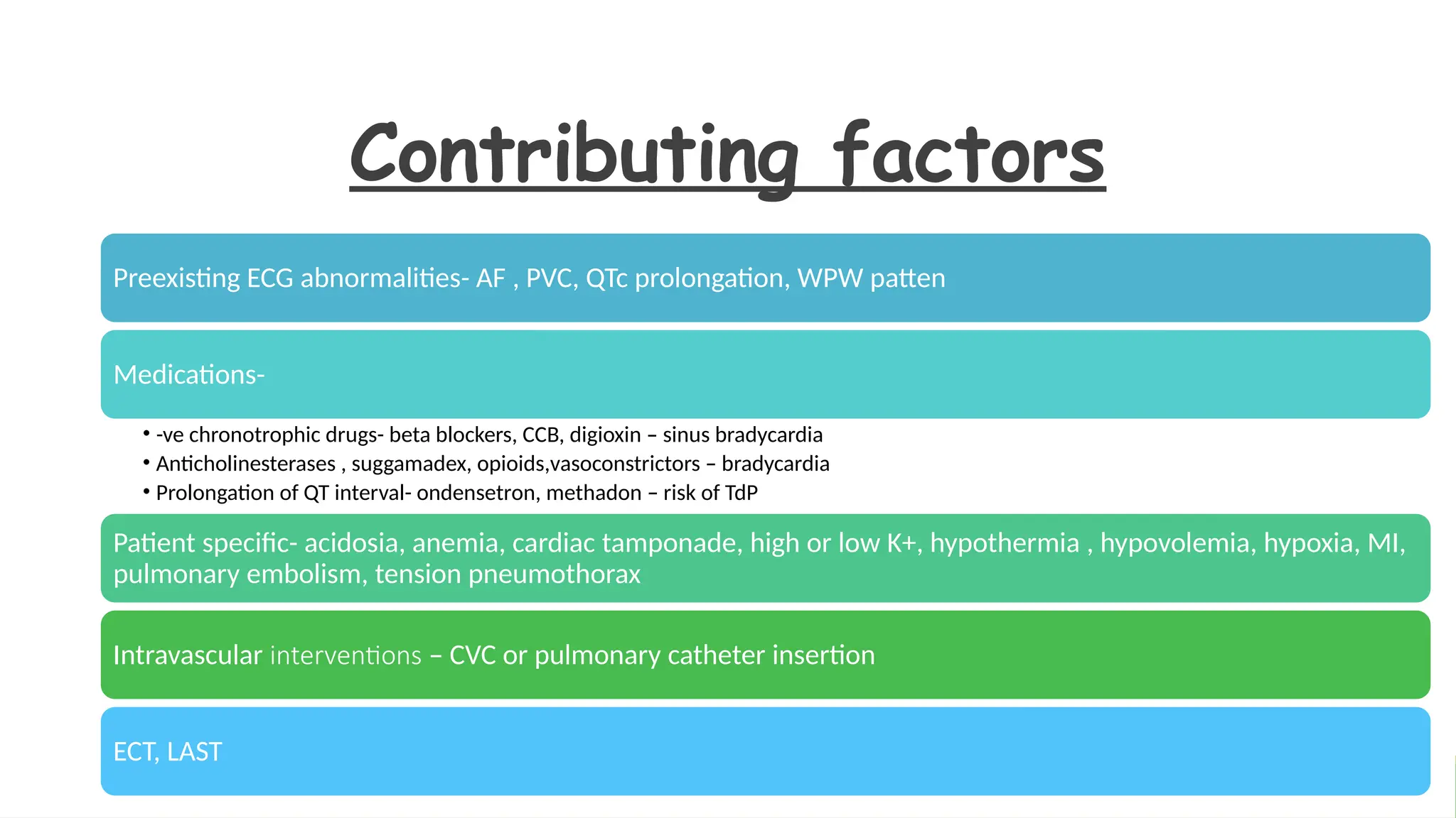
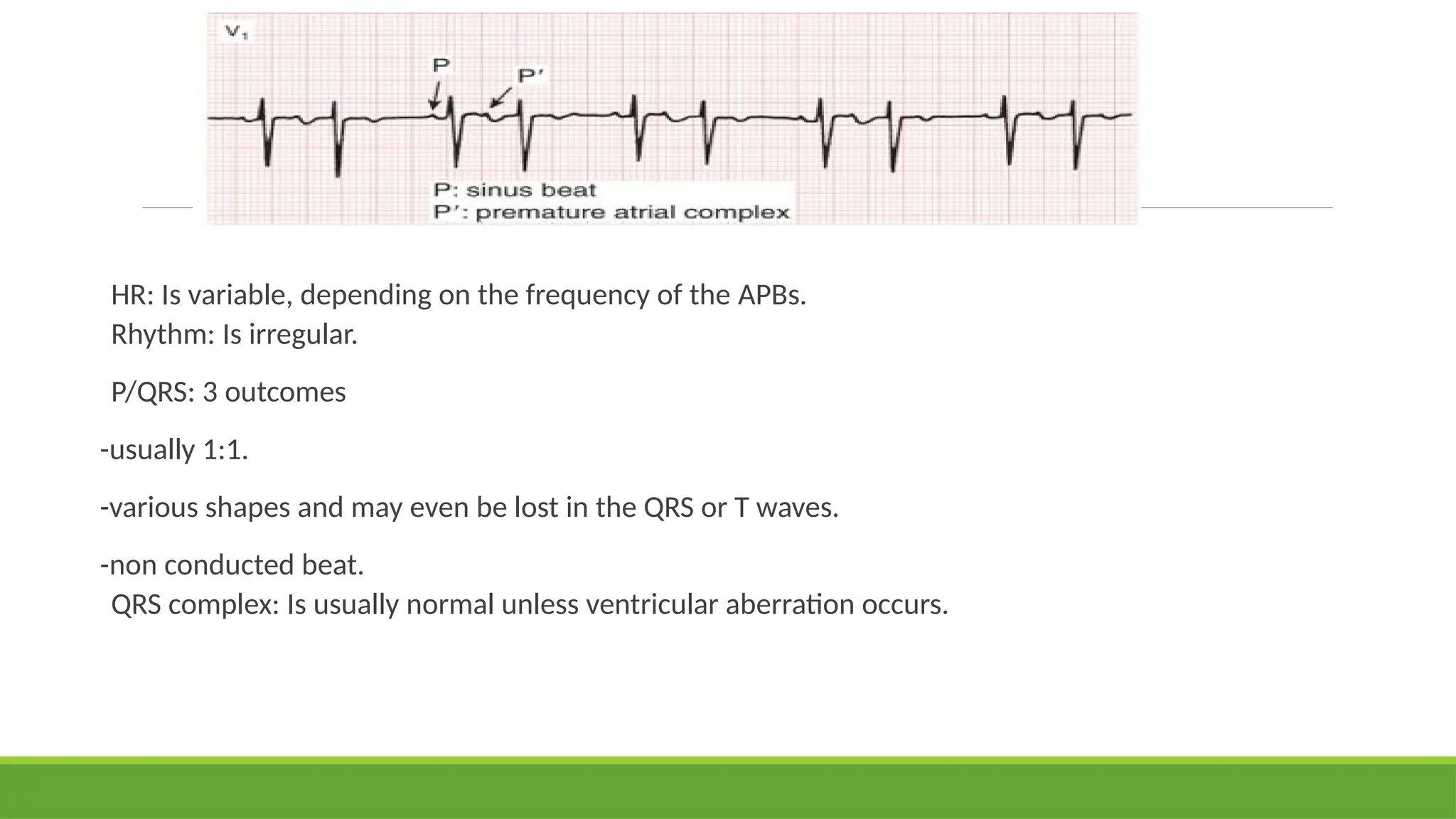
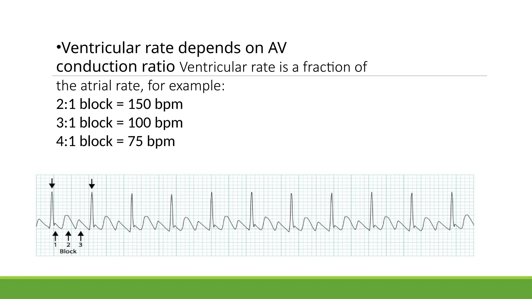
Perioperative
cardiac
arrhythmias
and their
management
Moderator – DrAvinash
Assistant professor
Presenter – Dr Shraya D S
Junior resident
This Photo by Unknown author is licensed under CC BY-SA.
Content
Contributing factors
mechanisms
Bradyarrhythmias and their management
Tachyarrhthmias and their management
Contributing factors
Preexisting ECG abnormalities- AF , PVC, QTc prolongation, WPW patten
Medications-
• -ve chronotrophic drugs- beta blockers, CCB, digioxin – sinus bradycardia
• Anticholinesterases , suggamadex, opioids,vasoconstrictors – bradycardia
• Prolongation of QT interval- ondensetron, methadon – risk of TdP
Patient specific- acidosia, anemia, cardiac tamponade, high or low K+, hypothermia , hypovolemia, hypoxia, MI,
pulmonary embolism, tension pneumothorax
Intravascular interventions – CVC or pulmonary catheter insertion
ECT, LAST
Mechanisms of arrythmias
Increased automaticity- repetitive firing focus other than
the sinus node
Reentry pathways- mechanism that may precipitate a wide
variety of supraventricular and ventricular arrhythmias.
Triggering by afterdepolarisation- early or delayed
after depolarisation .
Intraoperative
diagnosis
If tachy or bradyarrhythmias develop that cannot be
readily diagnosed , all available leads are displayed on the
monitor and 12 lead ECG id obtained as soon as possible
This Photo by Unknown author is licensed under CC BY-NC.
Interpreting
Perioperative Arrhythmias
1. What is the HR?
2. Is the rhythm regular?
3. Is one P wave present for each QRS complex?
4. Is the QRS complex normal?
5. Is the rhythm dangerous?
6. Does the rhythm require treatment?
Check RR interval in mm
SAN disaese
Above bundle of
His(supraventric
ular origin )
➢ 25mm
Bradyarrhythmia
< 15 mm
Tachyarrhythmia
AV BLOCK
Below bundle of
His( ventricuilar
origin)
-sinus
bradycardia
-Sinus
arrest( junction
al rhythm)
First degree
Second degree
Third degree
Narrow complex
tachyarrhythmia
Broad complex
tachyarrhythmia
Sinus bradycardia
• APPROX. 11% OF INTRAOPERATIVE ARRHYTHMIAS
• HR: < 60 BPM
• RHYTHM: REGULAR
• P/QRS: RATIO IS 1:1
• QRS : NORMAL MORPHOLOGY
Pacemaker site in Sinus Node but rate is lower
than Normal.
Causes
1)vagal reflexes- oculocardiac, peritoneal
stretching during laparoscopic surgery
2) neuraxial anesthesia- T1-T4 ( treated with beta
agonists)
3) drugs- CCB, beta blockers, digoxin, amiodarone,
anticholinesterases, sugammadex, opioids
Significance:
Treatment is recommended if
- Hypotension
- Ventricular Arrhythmia
- Signs of Poor Peripheral Perfusion
- May be a part of Sick Sinus
Syndrome
Management
Atropine ​
(0.5 to 1.0 mg by intravenous (IV) bolus, repeated every 3 to 5 minutes, up to 0.04 mg/kg or approximately a
3.0-mg total dose for the average 75-kg male patient​
Ephedrine, 5 to 25 mg by IV bolus​( bradycardia with hypotension)
Dopamine or Dobutamine (if blood pressure is adequate), 5 to 20 μg/kg/min by IV​
infusion​
Epinephrine, 2 to 10 μg/min IV infusion;​
​
Isoproterenol, 2 to 10 μg/min by IV infusion​
( in heart transplantation patients)
​
Temporary transcutaneous or trans venous pacing may be necessary for severe, recurrent, drug-refractory sinus
bradycardia.​
Junctional
rhythm
Pacemaker- tissue surrounding AVN
Classification:
1)Junctional bradycardia: < 40 bpm
2)Accelerated Junctional Rhythm: 60-100
bpm
3)Junctional Tachycardia: > 100 bpm
Causes- halogenated volatile anesthetics,
digitalis toxicity, MI
Atropine if hemodinamically significant
slow rhythm.
Heart
blocks
3 types
1) First degree heart block
2) Second degree heart block
- Mobitz type 1
- Mobitz type 2
3) third degree heart block
This Photo by Unknown author is licensed under CC BY-SA.
Causes
Diseases- acute MI,
myocarditis, Rheumatic
fever, mononucleosis,
Lymes diseases,
infiltrative diseases,
Iatrogenic- traumatic
injury from monitoring,
ablation catheters,
cardiac surgery
Drugs- digitalis
toxicity, beta blockers ,
CCB
First degree heart block
Delay in passage through AVN
PR interval > 200ms(>1 big square)
Each P wave followed by QRS complex pf normal duration
Asymptomatic
Management – avoid drugs/ situations increasing vagal tone
- treat underlying condition
Second degree heart block
–Mobitz type 1(Wenckebach)
Characterized by progressive lengthening of the PR interval until an impulse is not conducted and the beat
is dropped​
Relatively benign , often reversible​
rarely progresses to third degree block
Digitalis toxicity or MI and is usually transient.​
Mobitz type I block reflects disease of the AV node​
Mobitz type 2
•Dropped beats occur without any progressive lengthening of the PR interval.​
•This type of block has a serious prognosis because it frequently progresses to complete heart
block and may require pacemaker insertion​
•Conduction interruption below AVN
•Symptomatic – palpitations, near syncope
•Management- pacemaker placement
- Atropine ineffective
Third degree heart block
Complete heart block, complete interruption of AV conduction
Ventricular activity d/t ectopic pacemaker distal to site of block
If block near AVN – HR 45 – 55 bpm, narrow QRS complex
Infranodal block – HR 30 – 40 bpm , wide QRS complex
Symptoms – vertigo , syncope (stokes adams attack)
M/C/C - fibrotic degeneration d/t aging ( Lenegre disease)
In anesthetised pts – cardiac ischemia, metabolic or
electrolyte abnormalities, infection, reperfusion injury, stunned myocardium after cardiac surgery
Management – transcutaneous or transvenous cardiac pacemeker
- IV Isoproterenol infusion ( chemical pacemaker)
Caution- antiarrythmics suppress functioning ectopic pacemaker responsible for maintaing HR.
Preoperative placement of transvenous pacemake or availability of transcutaneous pacemaker
before administration of anesthesia for permanent cardiac pacemaker placement
Bradycardia
algorithm
Tachyarrhythmias : classification
Narrow complex tachycardia
- regular rhythm
- irregular rhythm
Broad complex tachycardia
- regular rhythm
- irregular rhythm
Maximum HR for a person=
220 - age in yrs
Narrow complex tachycardia : QRS < 0.12 sec
Regular (supraventricular tachycardia [SVT])
 Sinus tachycardia
Physiological response to insult. Impulse originates from sino-atrial (SA) node.
 Atrial tachycardia
Aberrant atrial focus producing impulse independent of SA node
 Atrioventricular nodal re-entry tachycardia (AVNRT)
Re-entry circuit within or near AV node
 AV re-entry tachycardia (AVRT)
Re-entry circuit conducted from atria to ventricles via abnormal accessory pathway; usually due to
Wolff-Parkinson-White (WPW) syndrome
 Atrial flutter with regular AV block(e.g., 2:1, 3:1)
Re-entry circuit within the atria
 IrregularAtrial fibrillation (AF)
Atria twitch instead of beating in a coordinated manner
Broad complex tachycardia : QRS > 0.12 sec
- Regular
- Ventricular tachycardia (VT)
- Generated by a single ventricular focus
- SVT with bundle branch block (BBB)
- This is rare. Any broad complex tachycardia should be treated as VT unless there the patient has an old ECG
with clear previous bundle branch block of unchanged morphology.
- Irregular
- Polymorphic VT (Torsades de pointes)
- Sinusoidal morphology usually due to abnormal ventricular repolarisation (long QT)
- AF with bundle branch block
Sinus tachycardia
Most common arrhythmia in the perioperative Period
The pacemaker site is in the sinus node, and the rate is faster than normal
Causes –
-Anaemia because of blood loss
-Pain
-Inadequate anaesthesia
-Hypovolaemia
-Fever
-Hypercarbia, hypoxia
-Thyrotoxicosis/ thyroid crisis, MI ,
-Cardiac failure with compensatory sinus tachycardia
-Caffine, atropine, sympathetic drugs
•HR: > 100 bpm​
•Rhythm: Is Regular ​
•P/QRS: Ratio is 1:1​
•QRS : normal, but ST-segment depression with severe increases in HR and resulting
myocardial ischemia may be associated​
•Significance: even short episodes of tachycardia may be associated with significant
myocardial ischemia in susceptible patients due to shortening of diastolic time period for
coronary blood flow to the LV and increased myocardial oxygen demand.
Management
Treat the cause
Boluses of Esmolol
20 – 50 mg every 2-3
min.
Avoid beta blockers
in hypovolemia,
decompensated
heart failure
Deepen the plane of
anesthesia, opioids,
dexmeditomidine
Sinus Arrhythmia
The impulses arise from the SA node
Variable HR characterizes the rhythm.
Normal PR interval and QRS complex.
Rate increases with inspiration and decreases with expiration.
Occurs more often in children than in adults
Paroxysmal Supraventricular Tachycardia(PSVT)
commonest cause of palpitations in patients with structurally normal hearts
Abrupt Onset and termination
Multiple variants based on where re-entry occurs
AVNRT( AVN reentry tachycardia) is m/c, typically paroxysmal and may occur spontaneously or
upon provocation with exertion, caffeine, alcohol, beta-agonists (salbutamol) or
sympathomimetics
more common in women than men
generally well tolerated and rarely life threatening
Regular tachycardia ~140-280 bpm
Narrow QRS complexes (< 120ms) unless there is co-existing bundle branch block,
accessory pathway,
P waves if visible exhibit retrograde conduction with P-wave inversion in leads II, III, aVF. They may
be buried within, visible after, or very rarely visible before the QRS complex
Mechanism
Functional pathways within the AV
node
There are two pathways within the AV
node:
-The slow pathway (alpha): a slowly-
conducting pathway with a short
refractory period.
-The fast pathway (beta): a rapidly-
conducting pathway with a long refractory
period.
1) A premature atrial contraction
(PAC) arrives while the fast
pathway is still refractory, and is
directed down the slow pathway
2) The ERP in the fast pathway
ends, and the PAC impulse
travels retrogradely up the fast
pathway
3) The impulse continually cycles
around the two pathways
Patients will typically complain of the sudden onset of rapid, regular palpitations.
Other associated symptoms may include:
- Presyncope or syncope due to a transient fall in blood pressure
- Chest pain, especially in the context of underlying coronary artery disease
- Dyspnoea
- Anxiety
- Rarely, polyuria due to elevated atrial pressures causing release of atrial
natriuretic peptide
Management of SVT
Vagal manoeuvres such as carotid sinus massage​
Adenosine (drug of choice) - 6-mg rapid (2 seconds) IV bolus​
If no response, second and third doses of 12 to 18 mg
of adenosine may be administered by rapid IV bolus.​
Verapamil (2.5 to 10 mg given IV) successfully terminates AV
nodal re-entry in approximately 90% of patients. ​
Amiodarone (150-mg infusion over 10 minutes for the loading
dose)​
Esmolol (1 mg/kg by bolus and 50 to 200 mg/kg/ min by
infusion) ​
In unstable patients- Synchronized cardioversion may be
performed with incremental doses of energy of 100, 200, 300,
and 360 J, preferably after light sedative premedication.​
​
Electrode catheter ablation using radiofrequency energy has evolved as
the definitive, long-term treatment for most persistent AV re entrant or
focal atrial SVTs​
​
​
​
Premature
atrial beat
•An ectopic pacemaker site - left or the right atrium ​
•Shape of the P wave is different from the usual SA node
P wave and may be inverted​
•APBs represent 10% of all intraoperative arrhythmias. ​
•They have little clinical significance, but frequent APBs
may lead to other, more serious supraventricular
arrhythmias or may be a sign of digitalis intoxication.​
•Treatment: Is rarely necessary​
, avoid sympathetic
stimulation
• Beta blockers ,CCB can be used
HR: Is variable, depending on the frequency of the APBs.
Rhythm: Is irregular.
P/QRS: 3 outcomes
-usually 1:1.
-various shapes and may even be lost in the QRS or T waves.
-non conducted beat.
QRS complex: Is usually normal unless ventricular aberration occurs.
Multifocal
Atrial
tachycardia
A rapid, irregular atrial rhythm arising from
multiple ectopic foci within the atria.
Most commonly seen in patients with
severe COPD or congestive heart failure.
It is typically a transitional rhythm between
frequent premature atrial complexes (PACs)
and atrial flutter / fibrillation.
Electrocardiographic Features
Heart rate > 100 bpm (usually 100-150 bpm; may be as high as 250 bpm).
Irregularly irregular rhythm with varying PP, PR and RR intervals.
At least 3 distinct P-wave morphologies in the same lead.
Isoelectric baseline between P-waves (i.e. no flutter waves).
Absence of a single dominant atrial pacemaker (i.e. not just sinus rhythm
with frequent PACs).
Some P waves may be nonconducted; others may be aberrantly
conducted to the ventricles.
Management
Avoid drugs or procedures that worsen pulmonary status
Avoid hypoxemia
Treatment – MgSO4 2gm IV over 1 hr
- verapamil , beta blockers
Theophylline exacerbates the condition
Cardioversion has no effect ( since multiple ectopic sites)
Atrial Flutter
a macro-reentrant arrhythmia that circulates in a specific manner in the right atrium ​
Usually accompanied by AV block because of very fast heart Rates.​
Classic sawtooth flutter waves (F waves) are usually present​
Causes- acute exacerbation of COPD, acute MI , thyrotoxicosis, after cardiac surgery
•Ventricular rate depends on AV
conduction ratio Ventricular rate is a fraction of
the atrial rate, for example:
2:1 block = 150 bpm
3:1 block = 100 bpm
4:1 block = 75 bpm
Treatment
Hemodynamically unstable – synchronised cardioversion , often 50 J monophasic adequate
Hemodynamically stable – initial goal is to control ventricular rate
a. β-Adrenergic blockers such as IV Esmolol (1 mg/kg by IV bolus) or propranolol
b. Calcium channel blockers such as verapamil (5 to 10 mg given IV) or diltiazem
The class III antiarrhythmic agent IBUTILIDE (1 mg in 10 mL saline, or 5% dextrose in water,(infused IV
slowly over 10 minutes)
Procainamide (5 to 10 mg/kg for the IV loading dose, infused no faster than 0.5 mg/kg/min) may rarely
be used in an attempt to restore sinus rhythm after the ventricular response has been adequately
Controlled
Atrial Fibrillation
Most common postoperative arrhythmia
associated with increased morbidity; mortality; longer hospital stays.
Excessively rapid and irregular atrial focus with no P waves appearing
on the ECG; instead, a fine fibrillatory activity is seen.
➢ Signs and symptoms- generalised weakness, palpitations,
syncope, hypotension, angina, shortness of breath,
orthopnea.
Unstable features - chest pain, dyspnoea, heart failure,
hypotension
HR: The atrial rate is 350 to 500 bpm, and the ventricular rate is 60 to 170 bpm​
Rhythm: Is irregularly irregular.​
P/QRS: The P wave is absent and replaced by F waves or no obvious atrial activity.​
QRS complex: Is normal​
​
Causes
1)Catecholamine excess or increased sensitivity
Exogenous (eg. adrenaline infusion, hydrocarbon exposure)
Endogenous- Subarachnoid haemorrhage, Stress, Phaeochromocytoma, Thyrotoxicosis
2) Valvular disease (e.g. mitral stenosis), including infective endocarditis
3) Ischemic heart disease, hypertension (m/c/c)
4) Increased atrial automaticity / irritation
Alcohol (“holiday heart”)
Caffeine
Electrolyte derangement (hypokalaemia, hypomagnesaemia)
Myocarditis
Complications
Adverse effects on hemodynamics:
-loss of atrial systole (aka “atrial kick”) (normally responsible for about 20% of
ventricular filling)
-Decreased diastolic filling time due to tachycardia
-Rate-related cardiomyopathy (can occur over weeks)
Atrial thrombus formation
-Systemic embolism, especially stroke
-Pulmonary embolism
Management
address life threats, determine if stable or unstable
Unstable features -Electrical cardioversion
- start at 100J (synhronised), then increased to 150J provide procedural
sedation
rate control versus rhythm control
Anticoagulation- direct acting oral anticoagulants preferred
Seek and treat underlying cause and complications
Ablation therapy is indicated if:
-permanent AF where ventricular rate difficult to control and persistent
symptoms/LV dysfunction
-rate-related cardiomyopathy due to refractory AF
-refractory symptomatic paroxysmal AF
Rate control with anticoagulation is the strategy of choice, rather than rhythm control, for most
patients
Rate control strategies
- β-blocker or calcium channel blocker for paroxysmal AF
◦ can be given IV if hemodynamically stable (CCBs preferred in COPD patients)
- IV amiodarone for critically ill patients
- AV nodal blockade (e.g. BB, CCB, amiodarone) is not recommended in patients with pre-excitation (e.g.
Wolff-Parkinson-White)
- target a rate of 80 as the endpoint, however, higher rates (e.g. ~110/min) are acceptable in stable patients
with preserved LV function
Rhythm control strategies
1)DC cardioversion
◦ essential in unstable patients
◦ effective strategy when performed within 48 hours of AF onset or later
following adequate anticoagulation and TOE
2) Pharmacological options for cardioversion can be used in hemodynamically
stable patients
◦ Amiodarone ( in OR- good choice for chemical cardioversion and rate control)
◦ flecanide (only an option in structurally normal hearts, sudden cardiac death in
structurally abnormal hearts)
◦ propafenone
◦ vernakalant
Patients on anticoagulation
Pts for urgent or emergent surgery – reverse warfarin effect by FFP, Prothrombin complex,
vit K
Elective surgery – goal- normal coagulation on the day of surgery
- stop anticoagulants 3-7 days prior , depending on pharmacokinetics of drug
- bridging with heparin or LMWH
If pt is undergoing cardiac surgery for other indications, surgical maze procedure and left
atrial appendage exclusion to eliminate site of blood stasis.
Arrhythmias with
wide QRS complex
Premature ventricular ectopic
-Ectopic pacemaker activity below the AV junction.
Causes
Anxiety, Excess caffeine
Sympathomimetics
Beta-agonists
Hypokalaemia, Hypomagnesaemia, Digoxin toxicity
Myocardial ischemia
ECG features
•Broad QRS complex (≥ 120 ms) with
abnormal morphology
•Premature — i.e. occurs earlier than would
be expected for the next sinus impulse
•Discordant ST segment and T wave changes.
•Usually followed by a full compensatory
pause
PVCs often occur in repeating patterns:
Bigeminy — every other beat is a PVC
Trigeminy — every third beat is a PVC
Quadrigeminy — every fourth beat is a PVC
Couplet — two consecutive PVCs
Clinical significance:
PVCs are a normal electrophysiological phenomenon not usually
requiring investigation or treatment
Frequent PVCs may cause palpitations and a sense of the heart “skipping
a beat”
In patients with underlying predispositions (e.g. ischaemic heart disease,
WPW), a PVC may trigger the onset of a re-entrant tachydysrhythmia — e.g.
VT, AVNRT, AVRT
During admnistration of anesthetic , VPC -More likely to lead to fibrillation
if they are multiple, multifocal, or bigeminal
No treatment is generally required for isolated VPB in asymptomatic and
healthy patients.
Correct any underlying abnormalities such as decreased serum potassium or
low arterial oxygen tension
Lignocaine – Drug of Choice - initial bolus dose of 1.5 mg/kg can be followed
by Infusion of 1 to 4 mg/min
Additional therapies: Esmolol, propranolol, procainamide, quinidine, Disopyramide
Treatment :
Ventricular
Tachycardia
three or more sequential VPBs defines VT​
HR: Is 100 to 200 bpm.​
Rhythm: regular( irregular if the VT is paroxysmal.)
P/QRS: No fixed relationship (VT - form of AV dissociation
in which the P waves can be observed marching through
the QRS complex.)
QRS complex: Is wide
Significance: Acute onset is life threatening and requires
immediate treatment​
If > 30 seconds = sustained
can be monomorphic or polymorphic
TYPES
1)Monomorphic
most common
associated with MI
2)Polymorphic
QRS at 200 beats/min or more which change amplitude and axis so they appear to twist around the
baseline
treatment is the same for both
causes
Electrolytes- hypokalaemia, hyperkalaemia, hypomagnesaemia,
hypocalcaemia
Hypothermia
Structural heart disease- LV dysfunction, coronary artery disease,
MI, HOCM
Iatrogenic- mechanical ventilation, drug therapy(digoxin), CVC
insretion
Ventricular Tachycardia Classification is based on:
1. Clinical Presentation
Hemodynamically stable
Hemodynamically unstable — e.g hypotension, chest pain, cardiac failure, decreased conscious level
This is the most important classification clinically and influences immediate management.
2. Duration
Sustained = Duration > 30 seconds, or requiring intervention due to hemodynamic compromise
Non-sustained = terminating spontaneously in < 30 seconds
Clinical Significance
VT may impair cardiac output with consequent hypotension, collapse, and acute cardiac
failure. This is due to extreme heart rates and loss of coordinated atrial contraction (“atrial
kick”)
The presence of pre-existing poor ventricular function is strongly associated with
cardiovascular compromise
Decreased cardiac output - decreased myocardial perfusion with degeneration into VF
Prompt recognition and initiation of treatment (e.g. electrical cardioversion) is required in
all cases of VT
Management
hemodynamically stable – Amiodarone 150 mg IV in 100 mL saline or D5W over 10
minutes, followed by an IV infusion of 1 mg/min for 6 hours and 0.5 mg/min
thereafter (maximum IV dose 2.2 g/24 hr).​
Lidocaine and Procainamide have been used in the past with varying degrees of success to
treat VT​
Pts with symptoms, unstable VT – cardioversion
Pulseless VT – defibrillation
Polymorphic VT with prolonged QT interval is a more serious rhythm Disturbance - IV
infusion of 1 g of magnesium over 2 to 3 minutes​
Ventricular Fibrillation
Irregular rhythm that results from a rapid discharge of impulses from one or more ventricular
foci or from multiple wandering re-entrant circuit in the ventricles
Major causes – Myocardial ischemia, hypoxia, hypothermia, electric shock, electrolyte
imbalance, and drug effects
M/C/C of sudden cardiac death
ECG
HR: Is rapid and grossly disorganized.​
Rhythm: Is totally irregular.​
P/QRS: No relationship is seen​
QRS complex: Is not present​
Significance: ​
No effective cardiac output exists​
, Incompatible with life
Treatment: ​
- CPR immediately initiated, and then defibrillation must be performed as rapidly as
possible. ​
- Asynchronous external defibrillation should be performed with a DC defibrillator, using
incremental energies in the range of 200 to 360 J.​
​
Defibrillation - delivery of high energy electric current throughout heart to depolarise all
myocardial cells at once . ideally single intrinsic pacemaker focus then restores myocardial
synchrony .
Prolonged QT syndrome
normal QT = < 440ms (two large squares) – prolonged QT > 460ms
produces prolonged ventricular repolarisation -> predisposes to malignant ventricular
arrhythmias
2 types- congenital and acquired
CAUSES
drugs: amiodarone, TCA’s, many antibiotics, fluconazole,
erythromycin, metoclopramide, quinidine, haloperidol,
droperidol, methadone, ondansetron, SSRI’s
genetic: cardiac ion channel mutation (Na+, K+)
myocardial disease: MI, RF, 3rd degree HB, cardiomyopathy
electrolytes: low Ca2+, low K+, low Mg2+
Polymorphic ventricular tachycardia (PVT)- multiple ventricular foci with the resultant QRS
complex varying in amplitude, axis, and duration.
Torsades de pointes (TdP) is a specific form of PVT occurring in the context of QT prolongation
— it has a characteristic morphology in which the QRS complexes “twist” around the isoelectric
line.
Antiarrhythmics
Class 0 – HCN channel blockers – Ivabradine
Class I – voltage gated sodium channel blockers
Class II- Autonomic inhibitors /activators
Class III – K + channel blockers / openers
Class IV- Ca2+ handling modulators
Class V- mechanosensitive channel blockers
Class VI- gap junction channel blockers
Class VII- molecules underlying longer term signalling processes affecting structural remodelling
Thank you

Arrhythmias vascular and hemorrhage (2) (1).pptx

  • 1.
    Perioperative cardiac arrhythmias and their management Moderator –DrAvinash Assistant professor Presenter – Dr Shraya D S Junior resident This Photo by Unknown author is licensed under CC BY-SA.
  • 2.
    Content Contributing factors mechanisms Bradyarrhythmias andtheir management Tachyarrhthmias and their management
  • 3.
    Contributing factors Preexisting ECGabnormalities- AF , PVC, QTc prolongation, WPW patten Medications- • -ve chronotrophic drugs- beta blockers, CCB, digioxin – sinus bradycardia • Anticholinesterases , suggamadex, opioids,vasoconstrictors – bradycardia • Prolongation of QT interval- ondensetron, methadon – risk of TdP Patient specific- acidosia, anemia, cardiac tamponade, high or low K+, hypothermia , hypovolemia, hypoxia, MI, pulmonary embolism, tension pneumothorax Intravascular interventions – CVC or pulmonary catheter insertion ECT, LAST
  • 4.
    Mechanisms of arrythmias Increasedautomaticity- repetitive firing focus other than the sinus node Reentry pathways- mechanism that may precipitate a wide variety of supraventricular and ventricular arrhythmias. Triggering by afterdepolarisation- early or delayed after depolarisation .
  • 5.
    Intraoperative diagnosis If tachy orbradyarrhythmias develop that cannot be readily diagnosed , all available leads are displayed on the monitor and 12 lead ECG id obtained as soon as possible This Photo by Unknown author is licensed under CC BY-NC.
  • 7.
    Interpreting Perioperative Arrhythmias 1. Whatis the HR? 2. Is the rhythm regular? 3. Is one P wave present for each QRS complex? 4. Is the QRS complex normal? 5. Is the rhythm dangerous? 6. Does the rhythm require treatment?
  • 8.
    Check RR intervalin mm SAN disaese Above bundle of His(supraventric ular origin ) ➢ 25mm Bradyarrhythmia < 15 mm Tachyarrhythmia AV BLOCK Below bundle of His( ventricuilar origin) -sinus bradycardia -Sinus arrest( junction al rhythm) First degree Second degree Third degree Narrow complex tachyarrhythmia Broad complex tachyarrhythmia
  • 9.
    Sinus bradycardia • APPROX.11% OF INTRAOPERATIVE ARRHYTHMIAS • HR: < 60 BPM • RHYTHM: REGULAR • P/QRS: RATIO IS 1:1 • QRS : NORMAL MORPHOLOGY
  • 10.
    Pacemaker site inSinus Node but rate is lower than Normal. Causes 1)vagal reflexes- oculocardiac, peritoneal stretching during laparoscopic surgery 2) neuraxial anesthesia- T1-T4 ( treated with beta agonists) 3) drugs- CCB, beta blockers, digoxin, amiodarone, anticholinesterases, sugammadex, opioids
  • 11.
    Significance: Treatment is recommendedif - Hypotension - Ventricular Arrhythmia - Signs of Poor Peripheral Perfusion - May be a part of Sick Sinus Syndrome
  • 12.
    Management Atropine ​ (0.5 to1.0 mg by intravenous (IV) bolus, repeated every 3 to 5 minutes, up to 0.04 mg/kg or approximately a 3.0-mg total dose for the average 75-kg male patient​ Ephedrine, 5 to 25 mg by IV bolus​( bradycardia with hypotension) Dopamine or Dobutamine (if blood pressure is adequate), 5 to 20 μg/kg/min by IV​ infusion​ Epinephrine, 2 to 10 μg/min IV infusion;​ ​ Isoproterenol, 2 to 10 μg/min by IV infusion​ ( in heart transplantation patients) ​ Temporary transcutaneous or trans venous pacing may be necessary for severe, recurrent, drug-refractory sinus bradycardia.​
  • 13.
    Junctional rhythm Pacemaker- tissue surroundingAVN Classification: 1)Junctional bradycardia: < 40 bpm 2)Accelerated Junctional Rhythm: 60-100 bpm 3)Junctional Tachycardia: > 100 bpm Causes- halogenated volatile anesthetics, digitalis toxicity, MI Atropine if hemodinamically significant slow rhythm.
  • 14.
    Heart blocks 3 types 1) Firstdegree heart block 2) Second degree heart block - Mobitz type 1 - Mobitz type 2 3) third degree heart block This Photo by Unknown author is licensed under CC BY-SA.
  • 15.
    Causes Diseases- acute MI, myocarditis,Rheumatic fever, mononucleosis, Lymes diseases, infiltrative diseases, Iatrogenic- traumatic injury from monitoring, ablation catheters, cardiac surgery Drugs- digitalis toxicity, beta blockers , CCB
  • 16.
    First degree heartblock Delay in passage through AVN PR interval > 200ms(>1 big square) Each P wave followed by QRS complex pf normal duration Asymptomatic Management – avoid drugs/ situations increasing vagal tone - treat underlying condition
  • 17.
    Second degree heartblock –Mobitz type 1(Wenckebach) Characterized by progressive lengthening of the PR interval until an impulse is not conducted and the beat is dropped​ Relatively benign , often reversible​ rarely progresses to third degree block Digitalis toxicity or MI and is usually transient.​ Mobitz type I block reflects disease of the AV node​
  • 18.
    Mobitz type 2 •Droppedbeats occur without any progressive lengthening of the PR interval.​ •This type of block has a serious prognosis because it frequently progresses to complete heart block and may require pacemaker insertion​ •Conduction interruption below AVN •Symptomatic – palpitations, near syncope •Management- pacemaker placement - Atropine ineffective
  • 19.
    Third degree heartblock Complete heart block, complete interruption of AV conduction Ventricular activity d/t ectopic pacemaker distal to site of block If block near AVN – HR 45 – 55 bpm, narrow QRS complex Infranodal block – HR 30 – 40 bpm , wide QRS complex Symptoms – vertigo , syncope (stokes adams attack)
  • 20.
    M/C/C - fibroticdegeneration d/t aging ( Lenegre disease) In anesthetised pts – cardiac ischemia, metabolic or electrolyte abnormalities, infection, reperfusion injury, stunned myocardium after cardiac surgery Management – transcutaneous or transvenous cardiac pacemeker - IV Isoproterenol infusion ( chemical pacemaker) Caution- antiarrythmics suppress functioning ectopic pacemaker responsible for maintaing HR. Preoperative placement of transvenous pacemake or availability of transcutaneous pacemaker before administration of anesthesia for permanent cardiac pacemaker placement
  • 21.
  • 22.
    Tachyarrhythmias : classification Narrowcomplex tachycardia - regular rhythm - irregular rhythm Broad complex tachycardia - regular rhythm - irregular rhythm Maximum HR for a person= 220 - age in yrs
  • 23.
    Narrow complex tachycardia: QRS < 0.12 sec Regular (supraventricular tachycardia [SVT])  Sinus tachycardia Physiological response to insult. Impulse originates from sino-atrial (SA) node.  Atrial tachycardia Aberrant atrial focus producing impulse independent of SA node  Atrioventricular nodal re-entry tachycardia (AVNRT) Re-entry circuit within or near AV node  AV re-entry tachycardia (AVRT) Re-entry circuit conducted from atria to ventricles via abnormal accessory pathway; usually due to Wolff-Parkinson-White (WPW) syndrome  Atrial flutter with regular AV block(e.g., 2:1, 3:1) Re-entry circuit within the atria  IrregularAtrial fibrillation (AF) Atria twitch instead of beating in a coordinated manner
  • 24.
    Broad complex tachycardia: QRS > 0.12 sec - Regular - Ventricular tachycardia (VT) - Generated by a single ventricular focus - SVT with bundle branch block (BBB) - This is rare. Any broad complex tachycardia should be treated as VT unless there the patient has an old ECG with clear previous bundle branch block of unchanged morphology. - Irregular - Polymorphic VT (Torsades de pointes) - Sinusoidal morphology usually due to abnormal ventricular repolarisation (long QT) - AF with bundle branch block
  • 25.
    Sinus tachycardia Most commonarrhythmia in the perioperative Period The pacemaker site is in the sinus node, and the rate is faster than normal Causes – -Anaemia because of blood loss -Pain -Inadequate anaesthesia -Hypovolaemia -Fever -Hypercarbia, hypoxia -Thyrotoxicosis/ thyroid crisis, MI , -Cardiac failure with compensatory sinus tachycardia -Caffine, atropine, sympathetic drugs
  • 26.
    •HR: > 100bpm​ •Rhythm: Is Regular ​ •P/QRS: Ratio is 1:1​ •QRS : normal, but ST-segment depression with severe increases in HR and resulting myocardial ischemia may be associated​ •Significance: even short episodes of tachycardia may be associated with significant myocardial ischemia in susceptible patients due to shortening of diastolic time period for coronary blood flow to the LV and increased myocardial oxygen demand.
  • 27.
    Management Treat the cause Bolusesof Esmolol 20 – 50 mg every 2-3 min. Avoid beta blockers in hypovolemia, decompensated heart failure Deepen the plane of anesthesia, opioids, dexmeditomidine
  • 28.
    Sinus Arrhythmia The impulsesarise from the SA node Variable HR characterizes the rhythm. Normal PR interval and QRS complex. Rate increases with inspiration and decreases with expiration. Occurs more often in children than in adults
  • 29.
    Paroxysmal Supraventricular Tachycardia(PSVT) commonestcause of palpitations in patients with structurally normal hearts Abrupt Onset and termination Multiple variants based on where re-entry occurs AVNRT( AVN reentry tachycardia) is m/c, typically paroxysmal and may occur spontaneously or upon provocation with exertion, caffeine, alcohol, beta-agonists (salbutamol) or sympathomimetics more common in women than men generally well tolerated and rarely life threatening
  • 30.
    Regular tachycardia ~140-280bpm Narrow QRS complexes (< 120ms) unless there is co-existing bundle branch block, accessory pathway, P waves if visible exhibit retrograde conduction with P-wave inversion in leads II, III, aVF. They may be buried within, visible after, or very rarely visible before the QRS complex
  • 31.
    Mechanism Functional pathways withinthe AV node There are two pathways within the AV node: -The slow pathway (alpha): a slowly- conducting pathway with a short refractory period. -The fast pathway (beta): a rapidly- conducting pathway with a long refractory period.
  • 32.
    1) A prematureatrial contraction (PAC) arrives while the fast pathway is still refractory, and is directed down the slow pathway 2) The ERP in the fast pathway ends, and the PAC impulse travels retrogradely up the fast pathway 3) The impulse continually cycles around the two pathways
  • 33.
    Patients will typicallycomplain of the sudden onset of rapid, regular palpitations. Other associated symptoms may include: - Presyncope or syncope due to a transient fall in blood pressure - Chest pain, especially in the context of underlying coronary artery disease - Dyspnoea - Anxiety - Rarely, polyuria due to elevated atrial pressures causing release of atrial natriuretic peptide
  • 34.
    Management of SVT Vagalmanoeuvres such as carotid sinus massage​ Adenosine (drug of choice) - 6-mg rapid (2 seconds) IV bolus​ If no response, second and third doses of 12 to 18 mg of adenosine may be administered by rapid IV bolus.​ Verapamil (2.5 to 10 mg given IV) successfully terminates AV nodal re-entry in approximately 90% of patients. ​ Amiodarone (150-mg infusion over 10 minutes for the loading dose)​ Esmolol (1 mg/kg by bolus and 50 to 200 mg/kg/ min by infusion) ​
  • 35.
    In unstable patients-Synchronized cardioversion may be performed with incremental doses of energy of 100, 200, 300, and 360 J, preferably after light sedative premedication.​ ​ Electrode catheter ablation using radiofrequency energy has evolved as the definitive, long-term treatment for most persistent AV re entrant or focal atrial SVTs​ ​ ​ ​
  • 36.
    Premature atrial beat •An ectopicpacemaker site - left or the right atrium ​ •Shape of the P wave is different from the usual SA node P wave and may be inverted​ •APBs represent 10% of all intraoperative arrhythmias. ​ •They have little clinical significance, but frequent APBs may lead to other, more serious supraventricular arrhythmias or may be a sign of digitalis intoxication.​ •Treatment: Is rarely necessary​ , avoid sympathetic stimulation • Beta blockers ,CCB can be used
  • 37.
    HR: Is variable,depending on the frequency of the APBs. Rhythm: Is irregular. P/QRS: 3 outcomes -usually 1:1. -various shapes and may even be lost in the QRS or T waves. -non conducted beat. QRS complex: Is usually normal unless ventricular aberration occurs.
  • 38.
    Multifocal Atrial tachycardia A rapid, irregularatrial rhythm arising from multiple ectopic foci within the atria. Most commonly seen in patients with severe COPD or congestive heart failure. It is typically a transitional rhythm between frequent premature atrial complexes (PACs) and atrial flutter / fibrillation.
  • 39.
    Electrocardiographic Features Heart rate> 100 bpm (usually 100-150 bpm; may be as high as 250 bpm). Irregularly irregular rhythm with varying PP, PR and RR intervals. At least 3 distinct P-wave morphologies in the same lead. Isoelectric baseline between P-waves (i.e. no flutter waves). Absence of a single dominant atrial pacemaker (i.e. not just sinus rhythm with frequent PACs). Some P waves may be nonconducted; others may be aberrantly conducted to the ventricles.
  • 40.
    Management Avoid drugs orprocedures that worsen pulmonary status Avoid hypoxemia Treatment – MgSO4 2gm IV over 1 hr - verapamil , beta blockers Theophylline exacerbates the condition Cardioversion has no effect ( since multiple ectopic sites)
  • 41.
    Atrial Flutter a macro-reentrantarrhythmia that circulates in a specific manner in the right atrium ​ Usually accompanied by AV block because of very fast heart Rates.​ Classic sawtooth flutter waves (F waves) are usually present​ Causes- acute exacerbation of COPD, acute MI , thyrotoxicosis, after cardiac surgery
  • 42.
    •Ventricular rate dependson AV conduction ratio Ventricular rate is a fraction of the atrial rate, for example: 2:1 block = 150 bpm 3:1 block = 100 bpm 4:1 block = 75 bpm
  • 43.
    Treatment Hemodynamically unstable –synchronised cardioversion , often 50 J monophasic adequate Hemodynamically stable – initial goal is to control ventricular rate a. β-Adrenergic blockers such as IV Esmolol (1 mg/kg by IV bolus) or propranolol b. Calcium channel blockers such as verapamil (5 to 10 mg given IV) or diltiazem The class III antiarrhythmic agent IBUTILIDE (1 mg in 10 mL saline, or 5% dextrose in water,(infused IV slowly over 10 minutes) Procainamide (5 to 10 mg/kg for the IV loading dose, infused no faster than 0.5 mg/kg/min) may rarely be used in an attempt to restore sinus rhythm after the ventricular response has been adequately Controlled
  • 44.
    Atrial Fibrillation Most commonpostoperative arrhythmia associated with increased morbidity; mortality; longer hospital stays. Excessively rapid and irregular atrial focus with no P waves appearing on the ECG; instead, a fine fibrillatory activity is seen.
  • 45.
    ➢ Signs andsymptoms- generalised weakness, palpitations, syncope, hypotension, angina, shortness of breath, orthopnea. Unstable features - chest pain, dyspnoea, heart failure, hypotension HR: The atrial rate is 350 to 500 bpm, and the ventricular rate is 60 to 170 bpm​ Rhythm: Is irregularly irregular.​ P/QRS: The P wave is absent and replaced by F waves or no obvious atrial activity.​ QRS complex: Is normal​ ​
  • 46.
    Causes 1)Catecholamine excess orincreased sensitivity Exogenous (eg. adrenaline infusion, hydrocarbon exposure) Endogenous- Subarachnoid haemorrhage, Stress, Phaeochromocytoma, Thyrotoxicosis 2) Valvular disease (e.g. mitral stenosis), including infective endocarditis 3) Ischemic heart disease, hypertension (m/c/c) 4) Increased atrial automaticity / irritation Alcohol (“holiday heart”) Caffeine Electrolyte derangement (hypokalaemia, hypomagnesaemia) Myocarditis
  • 47.
    Complications Adverse effects onhemodynamics: -loss of atrial systole (aka “atrial kick”) (normally responsible for about 20% of ventricular filling) -Decreased diastolic filling time due to tachycardia -Rate-related cardiomyopathy (can occur over weeks) Atrial thrombus formation -Systemic embolism, especially stroke -Pulmonary embolism
  • 49.
    Management address life threats,determine if stable or unstable Unstable features -Electrical cardioversion - start at 100J (synhronised), then increased to 150J provide procedural sedation rate control versus rhythm control Anticoagulation- direct acting oral anticoagulants preferred Seek and treat underlying cause and complications Ablation therapy is indicated if: -permanent AF where ventricular rate difficult to control and persistent symptoms/LV dysfunction -rate-related cardiomyopathy due to refractory AF -refractory symptomatic paroxysmal AF
  • 50.
    Rate control withanticoagulation is the strategy of choice, rather than rhythm control, for most patients Rate control strategies - β-blocker or calcium channel blocker for paroxysmal AF ◦ can be given IV if hemodynamically stable (CCBs preferred in COPD patients) - IV amiodarone for critically ill patients - AV nodal blockade (e.g. BB, CCB, amiodarone) is not recommended in patients with pre-excitation (e.g. Wolff-Parkinson-White) - target a rate of 80 as the endpoint, however, higher rates (e.g. ~110/min) are acceptable in stable patients with preserved LV function
  • 51.
    Rhythm control strategies 1)DCcardioversion ◦ essential in unstable patients ◦ effective strategy when performed within 48 hours of AF onset or later following adequate anticoagulation and TOE 2) Pharmacological options for cardioversion can be used in hemodynamically stable patients ◦ Amiodarone ( in OR- good choice for chemical cardioversion and rate control) ◦ flecanide (only an option in structurally normal hearts, sudden cardiac death in structurally abnormal hearts) ◦ propafenone ◦ vernakalant
  • 52.
    Patients on anticoagulation Ptsfor urgent or emergent surgery – reverse warfarin effect by FFP, Prothrombin complex, vit K Elective surgery – goal- normal coagulation on the day of surgery - stop anticoagulants 3-7 days prior , depending on pharmacokinetics of drug - bridging with heparin or LMWH If pt is undergoing cardiac surgery for other indications, surgical maze procedure and left atrial appendage exclusion to eliminate site of blood stasis.
  • 53.
    Arrhythmias with wide QRScomplex Premature ventricular ectopic -Ectopic pacemaker activity below the AV junction. Causes Anxiety, Excess caffeine Sympathomimetics Beta-agonists Hypokalaemia, Hypomagnesaemia, Digoxin toxicity Myocardial ischemia
  • 54.
    ECG features •Broad QRScomplex (≥ 120 ms) with abnormal morphology •Premature — i.e. occurs earlier than would be expected for the next sinus impulse •Discordant ST segment and T wave changes. •Usually followed by a full compensatory pause
  • 55.
    PVCs often occurin repeating patterns: Bigeminy — every other beat is a PVC Trigeminy — every third beat is a PVC Quadrigeminy — every fourth beat is a PVC Couplet — two consecutive PVCs
  • 56.
    Clinical significance: PVCs area normal electrophysiological phenomenon not usually requiring investigation or treatment Frequent PVCs may cause palpitations and a sense of the heart “skipping a beat” In patients with underlying predispositions (e.g. ischaemic heart disease, WPW), a PVC may trigger the onset of a re-entrant tachydysrhythmia — e.g. VT, AVNRT, AVRT During admnistration of anesthetic , VPC -More likely to lead to fibrillation if they are multiple, multifocal, or bigeminal
  • 57.
    No treatment isgenerally required for isolated VPB in asymptomatic and healthy patients. Correct any underlying abnormalities such as decreased serum potassium or low arterial oxygen tension Lignocaine – Drug of Choice - initial bolus dose of 1.5 mg/kg can be followed by Infusion of 1 to 4 mg/min Additional therapies: Esmolol, propranolol, procainamide, quinidine, Disopyramide Treatment :
  • 58.
    Ventricular Tachycardia three or moresequential VPBs defines VT​ HR: Is 100 to 200 bpm.​ Rhythm: regular( irregular if the VT is paroxysmal.) P/QRS: No fixed relationship (VT - form of AV dissociation in which the P waves can be observed marching through the QRS complex.) QRS complex: Is wide Significance: Acute onset is life threatening and requires immediate treatment​
  • 59.
    If > 30seconds = sustained can be monomorphic or polymorphic TYPES 1)Monomorphic most common associated with MI 2)Polymorphic QRS at 200 beats/min or more which change amplitude and axis so they appear to twist around the baseline treatment is the same for both
  • 60.
    causes Electrolytes- hypokalaemia, hyperkalaemia,hypomagnesaemia, hypocalcaemia Hypothermia Structural heart disease- LV dysfunction, coronary artery disease, MI, HOCM Iatrogenic- mechanical ventilation, drug therapy(digoxin), CVC insretion
  • 61.
    Ventricular Tachycardia Classificationis based on: 1. Clinical Presentation Hemodynamically stable Hemodynamically unstable — e.g hypotension, chest pain, cardiac failure, decreased conscious level This is the most important classification clinically and influences immediate management. 2. Duration Sustained = Duration > 30 seconds, or requiring intervention due to hemodynamic compromise Non-sustained = terminating spontaneously in < 30 seconds
  • 62.
    Clinical Significance VT mayimpair cardiac output with consequent hypotension, collapse, and acute cardiac failure. This is due to extreme heart rates and loss of coordinated atrial contraction (“atrial kick”) The presence of pre-existing poor ventricular function is strongly associated with cardiovascular compromise Decreased cardiac output - decreased myocardial perfusion with degeneration into VF Prompt recognition and initiation of treatment (e.g. electrical cardioversion) is required in all cases of VT
  • 63.
    Management hemodynamically stable –Amiodarone 150 mg IV in 100 mL saline or D5W over 10 minutes, followed by an IV infusion of 1 mg/min for 6 hours and 0.5 mg/min thereafter (maximum IV dose 2.2 g/24 hr).​ Lidocaine and Procainamide have been used in the past with varying degrees of success to treat VT​ Pts with symptoms, unstable VT – cardioversion Pulseless VT – defibrillation Polymorphic VT with prolonged QT interval is a more serious rhythm Disturbance - IV infusion of 1 g of magnesium over 2 to 3 minutes​
  • 64.
    Ventricular Fibrillation Irregular rhythmthat results from a rapid discharge of impulses from one or more ventricular foci or from multiple wandering re-entrant circuit in the ventricles Major causes – Myocardial ischemia, hypoxia, hypothermia, electric shock, electrolyte imbalance, and drug effects M/C/C of sudden cardiac death
  • 65.
    ECG HR: Is rapidand grossly disorganized.​ Rhythm: Is totally irregular.​ P/QRS: No relationship is seen​ QRS complex: Is not present​
  • 66.
    Significance: ​ No effectivecardiac output exists​ , Incompatible with life Treatment: ​ - CPR immediately initiated, and then defibrillation must be performed as rapidly as possible. ​ - Asynchronous external defibrillation should be performed with a DC defibrillator, using incremental energies in the range of 200 to 360 J.​ ​ Defibrillation - delivery of high energy electric current throughout heart to depolarise all myocardial cells at once . ideally single intrinsic pacemaker focus then restores myocardial synchrony .
  • 67.
    Prolonged QT syndrome normalQT = < 440ms (two large squares) – prolonged QT > 460ms produces prolonged ventricular repolarisation -> predisposes to malignant ventricular arrhythmias 2 types- congenital and acquired
  • 68.
    CAUSES drugs: amiodarone, TCA’s,many antibiotics, fluconazole, erythromycin, metoclopramide, quinidine, haloperidol, droperidol, methadone, ondansetron, SSRI’s genetic: cardiac ion channel mutation (Na+, K+) myocardial disease: MI, RF, 3rd degree HB, cardiomyopathy electrolytes: low Ca2+, low K+, low Mg2+
  • 69.
    Polymorphic ventricular tachycardia(PVT)- multiple ventricular foci with the resultant QRS complex varying in amplitude, axis, and duration. Torsades de pointes (TdP) is a specific form of PVT occurring in the context of QT prolongation — it has a characteristic morphology in which the QRS complexes “twist” around the isoelectric line.
  • 70.
    Antiarrhythmics Class 0 –HCN channel blockers – Ivabradine Class I – voltage gated sodium channel blockers Class II- Autonomic inhibitors /activators Class III – K + channel blockers / openers Class IV- Ca2+ handling modulators Class V- mechanosensitive channel blockers Class VI- gap junction channel blockers Class VII- molecules underlying longer term signalling processes affecting structural remodelling
  • 71.