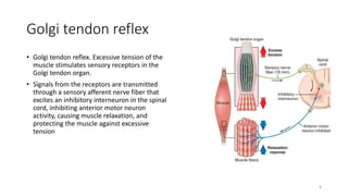
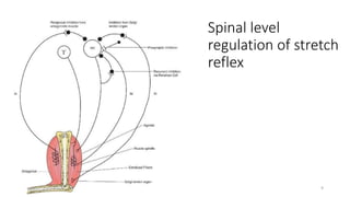
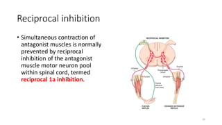
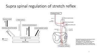
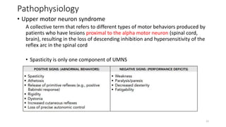
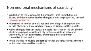
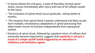
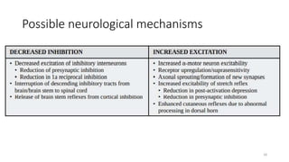
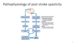
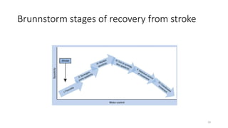
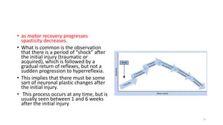
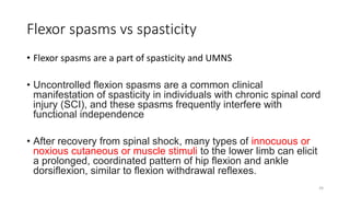
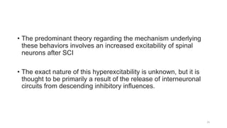
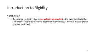
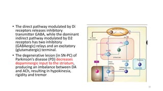
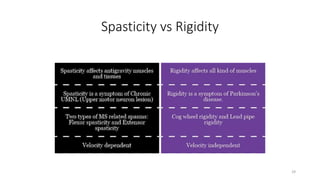
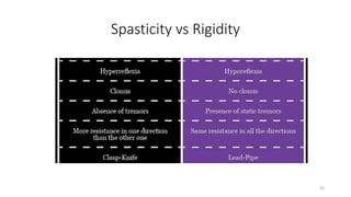
Spasticity is a common motor control disorder frequently encountered in the
spectrum of the upper motor neuron (UMN) syndrome. It can result in pain,
fatigue, joint restrictions, functional impairments, and skin breakdown that may
negatively affect many domains of life by causing social avoidance and
diminished life satisfaction . Spasticity was originally defined as a velocity dependent increase in tonic stretch reflexes or muscle tone with exaggerated
tendon jerks resulting from increased excitability of the stretch reflex . This
definition has been criticized for being too narrow and inadequately depicting
the clinical sequelae. In 2005, a European Thematic Network to Develop
Standardized Measures of Spasticity (the SPASM consortium) suggested
broadening the definition to reflect a more clinical entity . They defined
spasticity as “disordered sensory-motor control, resulting from an upper motor
neuron lesion, presenting as intermittent or sustained involuntary activation of
muscles.
1. Pathophysiology of spasticity and differences
with rigidity and flexor spasms
• Presenter
Dr Joe Antony
Junior resident
PMR
Kgmu ,Lucknow
• Moderator
Prof A K Gupta
Professor and HOD
PMR
Kgmu ,Lucknow
1
2. Contents
• Definitions
• Conditions causing spasticity
• Normal physiology
• Pathophysiology of spasticity
• Differences with flexor spasms
• Introduction to rigidity
2
3. Definitions
• Tone is defined as resistance to passive stretch of muscle fiber.
• Spasticity is defined as a velocity-dependent increase in muscle tone
• Clonus is a rhythmic pattern of muscle contraction provoked by a
sudden stretch, which activates muscle spindles reflexes.
• Spastic co-contraction is the inappropriate co-activation of
antagonistic muscle groups during voluntary activity resulting from
loss of reciprocal inhibition.
3
4. Epidemiology
• Spasticity is a component of the UMNS and can result from numerous
conditions, such as
• Stroke
• Cerebral palsy,
• Anoxic brain injury
• Traumatic brain injury (TBI)
• Spinal cord injury (SCI)
• Multiple sclerosis (MS)
• other CNS neurodegenerative diseases.
4
5. Normal Physiology
• Muscle sensory receptors
• Muscle spindle
• Muscle length
• Rate of change of length
• Golgi tendon organ
• Tendon tension
• Rate of change of tension
5
6. Muscle spindle
• Each muscle spindle has three essential
elements:
1. Specialized intrafusal muscle fibers
• Nuclear bag fiber - Dynamic
• Nuclear chain fiber- Static
2. Large diameter myelinated afferent nerves
(types Ia and II) originating in the central
portion of the intrafusal fibers.
3. Small diameter myelinated efferent nerves
(gamma) supplying the polar contractile
regions of the intrafusal fibers .
Guytons and hall physiology 6
7. Stretch reflex
• Whenever a muscle is stretched suddenly,
excitation of the spindles causes reflex
contraction of the large skeletal muscle fibers
of the stretched muscle and also of closely
allied synergistic muscles.
• Mono synaptic pathway
Muscle
spindle
stretch
1a nerve
fiber
Anterior
horn neuron
( alpha
motor
neuron)
Same
muscle
Contraction
7
8. Golgi tendon reflex
• Golgi tendon reflex. Excessive tension of the
muscle stimulates sensory receptors in the
Golgi tendon organ.
• Signals from the receptors are transmitted
through a sensory afferent nerve fiber that
excites an inhibitory interneuron in the spinal
cord, inhibiting anterior motor neuron
activity, causing muscle relaxation, and
protecting the muscle against excessive
tension
8
10. Reciprocal inhibition
• Simultaneous contraction of
antagonist muscles is normally
prevented by reciprocal
inhibition of the antagonist
muscle motor neuron pool
within spinal cord, termed
reciprocal 1a inhibition.
10
12. Pathophysiology
• Upper motor neuron syndrome
A collective term that refers to different types of motor behaviors produced by
patients who have lesions proximal to the alpha motor neuron (spinal cord,
brain), resulting in the loss of descending inhibition and hypersensitivity of the
reflex arc in the spinal cord
• Spasticity is only one component of UMNS
12
13. • Spasticity is characterized by an exaggeration of the stretch reflex
• mechanism of which remains poorly understood
• likely due to abnormal intraspinal processing of afferent impulses,
• loss of descending inhibitory regulation of segmental reflexes,
• increased excitability of the motor neurons
13
14. • exaggeration of stretch reflex in patients with spasticity is due to an
abnormal processing of sensory inputs in the spinal cord with
increased segmental spinal excitation and loss of presynaptic
inhibition.
• This loss of inhibition is due, in part, to both reduced descending
supraspinal inhibition and reciprocal inhibition
14
16. Non neuronal mechanisms of spasticity
• In addition to these neuronal disturbances, limb immobilization,
disuse, and denervation lead to changes in muscle properties, termed
rheologic properties.
• Alterations in tendon compliance and physiological changes in the
muscle fiber cause increased mechanical resistance of the muscle.
• Other changes that can increase muscle resistance in the absence of
electromyographic muscle activity include muscle atrophy and
shortening, loss of sarcomeres, and muscle infiltration with
connective tissue and fat.
• These changes in muscle properties further exacerbate impairment in
motor control caused by spasticity.
16
17. • In lesions below the mid-pons, a state of flaccidity, termed spinal
shock, ensues immediately after injury with loss of all reflexes caudal
to the injury.
• The resolution of spinal shock occurs gradually , taking weeks to
months.
• The recovery from spinal shock is poorly understood and likely results
from multiple, simultaneous adaptations in spinal processing that
allow motor neuron to function independently from supraspinal
control.
• Existence of spinal shock, followed by a gradual return of reflexes that
eventually become hyperactive, suggests that spasticity is not just a
result of a simple on/off switch triggered by an alteration in
inhibitory and facilitative signals
17
21. • as motor recovery progresses
spasticity decreases.
• What is common is the observation
that there is a period of “shock” after
the initial injury (traumatic or
acquired), which is followed by a
gradual return of reflexes, but not a
sudden progression to hyperreflexia.
• This implies that there must be some
sort of neuronal plastic changes after
the initial injury.
• This process occurs at any time, but is
usually seen between 1 and 6 weeks
after the initial injury
21
22. • Plastic rearrangement occurs within the brain and spinal cord and is
regarded as an attempt at restoration of function through emergence
of novel neuronal circuitry.
• This process of plastic rearrangement often results in muscle
overactivity and hyperreflexia, and thus spasticity
22
23. Benefits
• patients may not realize that they benefit from spasticity until it decreases or resolves.
• Facilitates transfer via stand-pivot (and patients may falsely equate this ability to
preserved muscle strength).
• Psychologic benefits to patients when they still experience movement in a paretic limb.
• Facilitation of circulation,
• Orthostatic blood pressure support,
• Maintenance of muscle mass.
• May assist in preventing osteoporosis
• Decrease of pressure ulcer formation over bony prominences
• Thus it is of paramount importance to obtain a thorough yet focused history to guide the
examination and formulate mutually agreed upon treatment goals and plans.
23
24. Flexor spasms vs spasticity
• Flexor spasms are a part of spasticity and UMNS
• Uncontrolled flexion spasms are a common clinical
manifestation of spasticity in individuals with chronic spinal cord
injury (SCI), and these spasms frequently interfere with
functional independence
• After recovery from spinal shock, many types of innocuous or
noxious cutaneous or muscle stimuli to the lower limb can elicit
a prolonged, coordinated pattern of hip flexion and ankle
dorsiflexion, similar to flexion withdrawal reflexes.
24
25. • The predominant theory regarding the mechanism underlying
these behaviors involves an increased excitability of spinal
neurons after SCI
• The exact nature of this hyperexcitability is unknown, but it is
thought to be primarily a result of the release of interneuronal
circuits from descending inhibitory influences.
25
26. Introduction to Rigidity
• Definition
• Resistance to stretch that is not velocity dependent—the examiner feels the
same resistance to stretch irrespective of the velocity at which a muscle group
is being stretched.
26
27. • The striatal GABAergic neurones
receive side-loop excitatory
glutamatergic (Glu) input from the
motor cortex and modulatory
dopaminergic (DA) projections
from the substantia nigra pars
compacta (SN-PC).
• There are also balancing
cholinergic (ACh) interneurones.
The striatal neurones express both
excitatory D1 and inhibitory D2
receptors.
• The output from the striatum to
substania nigra pars reticulata (SN-
PR) and internal globus pallidus
(GP) follows a direct and an
indirect pathway.
27
28. • The direct pathway modulated by Di
receptors releases inhibitory
transmitter GABA, while the dominant
indirect pathway modulated by D2
receptors has two inhibitory
(GABAergic) relays and an excitatory
(glutamatergic) terminal.
• The degenerative lesion (in SN-PC) of
Parkinson's disease (PD) decreases
dopaminergic input to the striatum,
producing an imbalance between DA
and ACh, resulting in hypokinesia,
rigidity and tremor
28
Inhibitory inputs
Recurrent inhibition by Renshaw cell, presynaptic inhibition by interneuron from golgitendon rogan, reciprocal inhibition by antagonistic muscles
Crossed extensor reflex
Athetosis- slow involuntary writhing movements
Motor recovery commences almost immediately after the onset of a central nervous lesion, when reversible changes resolve. following a stroke regardless of type (hemorrhagic or not) or location (cortical or subcortical), a relatively predictable pattern of recovery sets in. this empirically describes the stereotypical stages of motor recovery from flaccidity to full recovery of motor function. As a stroke survivor improves, progression from one recovery stage to the next toward recovery of normal movement occurs in an orderly manner, but this evolution may be arrested at any stage.