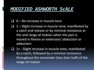
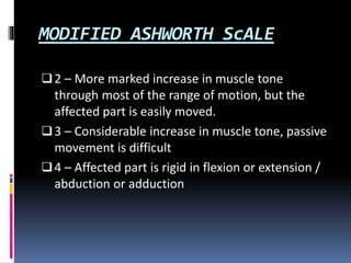
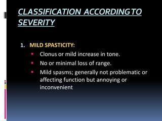
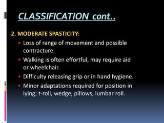
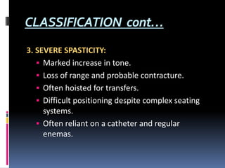
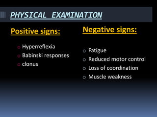
SPASTICITY
PRATIGYA DEUJA
DEFINITION
Spasticity is defined as velocity dependent
increase in muscle tone with exaggerated tendon
jerks, resulting from hyperexcitabilty of the
stretch reflex i.e, the faster the passive
movements of the limb through it’s range, the
greater the increase in muscle tone.
Increased resistance to passive movements in
one direction.
It is an upper motor lesion.
It is noted in cases like; SCI, multiple sclerosis,,
traumatic brain injury, amyotrophic lateral
sclerosis, cp, spinal cord tumors.
CLASSIFICATION ACCORDINGTO
SEVERITY
1. MILD SPASTICITY:
 Clonus or mild increase in tone.
 No or minimal loss of range.
 Mild spasms; generally not problematic or
affecting function but annoying or
inconvenient
CLASSIFICATION cont..
2. MODERATE SPASTICITY:
 Loss of range of movement and possible
contracture.
 Walking is often effortful, may require aid
or wheelchair.
 Difficulty releasing grip or in hand hygiene.
 Minor adaptations required for position in
lying; t-roll, wedge, pillows, lumbar roll.
CLASSIFICATION cont…
3. SEVERE SPASTICITY:
 Marked increase in tone.
 Loss of range and probable contracture.
 Often hoisted for transfers.
 Difficult positioning despite complex seating
systems.
 Often reliant on a catheter and regular
enemas.
CLINICAL FEATURES
 Increasing tightness
 Worsening spasm
 Physical activities like; walking, transferring are
affected
 Functional activities like picking, grasping are
affected
PHYSICAL EXAMINATION
Positive signs: Negative signs:
o Hyperreflexia
o Babinski responses
o clonus
o Fatigue
o Reduced motor control
o Loss of coordination
o Muscle weakness
PATHOPHYSIOLOGY
 Spasticity resulted from a loss of descending,
facilitatory inhibitory influences that act on Ia
interneuron inhibition.
 Reciprocal inhibition mediated through Ia
interneuron requires facilitation from higher
centers. With injury to CNS the interneuron is
unable to shut off antagonist muscle firing with
resultant increased velocity dependent
resistance to movement.
PATHOPHYSIOLOGY
 The combination of both decrease inhibition,
increased depolarization state of cell membrane,
decrease action potential threshold for nerve
signal conduction thus increase activity of
structures innervated by the affected nerves.
 Recent studies suggest that intrinsic changes in
the motor neurone develop over time following a
lesion. These result in abnormally long plateau
like potentials that prolong motor neurone
discharge and thus muscle contraction in
response to synaptic inputs.
ASSESSMENT
 Patient history
 DTR
 Range of motion
 Test for clonus
 Functional observation
 Aggravating factors
 Modified Ashworth scale
MODIFIED ASHWORTH ScALE
 0 – No increase in muscle tone
 1 – Slight increase in muscle tone, manifested by
a catch and release or by minimal resistance at
the end range of motion when the part is
moved in flexion or extension/ abduction or
adduction
 1+ - Slight increase in muscle tone, manifested
by a catch, followed by a minimal resistance
throughout the remainder (less than half) of the
range of motion
MODIFIED ASHWORTH ScALE
2 – More marked increase in muscle tone
through most of the range of motion, but the
affected part is easily moved.
3 – Considerable increase in muscle tone, passive
movement is difficult
4 – Affected part is rigid in flexion or extension /
abduction or adduction
PHARMACOLOGICAL MANAGEMENT
 Baclofen
 Tizanidine
 Dentrolene
 Phenol block
 Botulinum toxin
SURGICAL MANAGEMENT
 Baclofen pump
 Tendon lengthening
 Tendon transfer
 Osteotomy
 Arthodesis
PHYSIOTHERAPY MANAGEMENT
 Positioning
 Ultrasound: 10 minutes/session
: Continuous mode for 5 weeks
: frequency : 1 mhz
: intensity : 1.5 w/cm2
 Cryotherapay : 20 minutes
 Thermotherapy : 20 minutes, prior to stretching
 Hydrotherapy
 Proprioceptive neuromuscular facilitation
PHYSIOTHERAPY MANAGEMENT
 Massage
 Passive movements
 Weight bearing through affected extremity
 Rood’s technique
 Yoga
 Relaxation techniques

Spasticity

  • 1.
  • 2.
    DEFINITION Spasticity is definedas velocity dependent increase in muscle tone with exaggerated tendon jerks, resulting from hyperexcitabilty of the stretch reflex i.e, the faster the passive movements of the limb through it’s range, the greater the increase in muscle tone. Increased resistance to passive movements in one direction. It is an upper motor lesion. It is noted in cases like; SCI, multiple sclerosis,, traumatic brain injury, amyotrophic lateral sclerosis, cp, spinal cord tumors.
  • 3.
    CLASSIFICATION ACCORDINGTO SEVERITY 1. MILDSPASTICITY:  Clonus or mild increase in tone.  No or minimal loss of range.  Mild spasms; generally not problematic or affecting function but annoying or inconvenient
  • 4.
    CLASSIFICATION cont.. 2. MODERATESPASTICITY:  Loss of range of movement and possible contracture.  Walking is often effortful, may require aid or wheelchair.  Difficulty releasing grip or in hand hygiene.  Minor adaptations required for position in lying; t-roll, wedge, pillows, lumbar roll.
  • 5.
    CLASSIFICATION cont… 3. SEVERESPASTICITY:  Marked increase in tone.  Loss of range and probable contracture.  Often hoisted for transfers.  Difficult positioning despite complex seating systems.  Often reliant on a catheter and regular enemas.
  • 6.
    CLINICAL FEATURES  Increasingtightness  Worsening spasm  Physical activities like; walking, transferring are affected  Functional activities like picking, grasping are affected
  • 7.
    PHYSICAL EXAMINATION Positive signs:Negative signs: o Hyperreflexia o Babinski responses o clonus o Fatigue o Reduced motor control o Loss of coordination o Muscle weakness
  • 8.
    PATHOPHYSIOLOGY  Spasticity resultedfrom a loss of descending, facilitatory inhibitory influences that act on Ia interneuron inhibition.  Reciprocal inhibition mediated through Ia interneuron requires facilitation from higher centers. With injury to CNS the interneuron is unable to shut off antagonist muscle firing with resultant increased velocity dependent resistance to movement.
  • 9.
    PATHOPHYSIOLOGY  The combinationof both decrease inhibition, increased depolarization state of cell membrane, decrease action potential threshold for nerve signal conduction thus increase activity of structures innervated by the affected nerves.  Recent studies suggest that intrinsic changes in the motor neurone develop over time following a lesion. These result in abnormally long plateau like potentials that prolong motor neurone discharge and thus muscle contraction in response to synaptic inputs.
  • 10.
    ASSESSMENT  Patient history DTR  Range of motion  Test for clonus  Functional observation  Aggravating factors  Modified Ashworth scale
  • 11.
    MODIFIED ASHWORTH ScALE 0 – No increase in muscle tone  1 – Slight increase in muscle tone, manifested by a catch and release or by minimal resistance at the end range of motion when the part is moved in flexion or extension/ abduction or adduction  1+ - Slight increase in muscle tone, manifested by a catch, followed by a minimal resistance throughout the remainder (less than half) of the range of motion
  • 12.
    MODIFIED ASHWORTH ScALE 2– More marked increase in muscle tone through most of the range of motion, but the affected part is easily moved. 3 – Considerable increase in muscle tone, passive movement is difficult 4 – Affected part is rigid in flexion or extension / abduction or adduction
  • 13.
    PHARMACOLOGICAL MANAGEMENT  Baclofen Tizanidine  Dentrolene  Phenol block  Botulinum toxin
  • 14.
    SURGICAL MANAGEMENT  Baclofenpump  Tendon lengthening  Tendon transfer  Osteotomy  Arthodesis
  • 15.
    PHYSIOTHERAPY MANAGEMENT  Positioning Ultrasound: 10 minutes/session : Continuous mode for 5 weeks : frequency : 1 mhz : intensity : 1.5 w/cm2  Cryotherapay : 20 minutes  Thermotherapy : 20 minutes, prior to stretching  Hydrotherapy  Proprioceptive neuromuscular facilitation
  • 16.
    PHYSIOTHERAPY MANAGEMENT  Massage Passive movements  Weight bearing through affected extremity  Rood’s technique  Yoga  Relaxation techniques