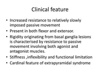
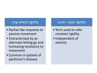
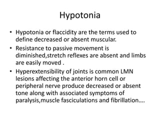
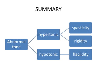
Muscle tone
Dr.Satish K Pimpale PT
MPTh in Neurosciences
Definition
• Tone is the resistance offered by muscles to
continuous passive stretch (Brooks1986)
• Muscle tone can be defined clinically as the
resistance that is encountered when the joint
of a relaxed patient is moved passively (Maria
Strokes)
• Muscle tone degree of residual contraction in
normally innervated ,resting muscles, or
steady contraction.
• Resistance is due to number of factors.
1. Physical inertia
2. Intrinsic mechanical elastic stiffness of
muscle and connective tissue.
3. Reflex muscle contraction caused by muscle
stretch
• In a relaxed human subject with no
neurological deficit, the resistance to passive
movement is due to the mechanical factors
such as the :
• Compliance of muscle
• Tendons
• Ligaments
• Joints rather than neural mechanism
• Clinically muscle tone may be abnormally
increased or decreased
• Hypertonia
• Hypotonia
• Dystonia
Hypertonia
• Spasticity and rigidity
There are two rare types of hypertonia
1. Gegenhaltan
2. Alpha rigidity
Spasticity
• Spasticity may be defined as a velocity
dependent increase in resistance to passive
stretch of a muscle with exagggerated tendon
reflex resulting from hyperexcitability of the
stretch reflex as one component of the Upper
motor neuron
• Spasticity -arises from injury to the
corticofugal pathways (pyramidal tracts)a
• Occurs as a part of UMN syndrome.
• Loss of inhibitory control on LMN results in
disordered spinal segmental reflexes
1. Increase alpha motor neuron excitability
2. Increase spindle Ia
3. Flexor reflex afferent excitability
4. Altered synaptic activity
5. Decrease presynaptic Ia inhibition rate
Additional sign and symtoms
• Brisk tendon reflex
• Involuntary flexor and extensor spasms
• Clonus
• Babinski’s sign
• Exaggerated cutaneous reflexes
• Loss of precise autonomic control
• Dyssynergic movement patterns
• Abnormal timing
• Paresis
• Loss of dexterity and fatigability
Chronic spasticity is associated with
• Contracture
• Abnormal posture
• Deformity
• Functional limitation
Clinical features
Spasticity is recognised clinically by
1. The characteristic pattern of involvement of
certain muscle groups.
2. The increase responsiveness of muscle to
strecth
3. Markedly increase tendon reflex
Spasticity predominately affects the antigravity
muscles i.e
• The flexors of the arms and
• The extensors of the legs.
As a result the arms tend to assume a flexed and
a pronated posture while the legs are usually
held in extended and adducted
In spasticy resistance increase with increase in
amplitude and velocity of strecth .thus the
larger and quicker the stretch ,the stronger
the resistance of the spastic muscle.
• Initially stretch produces high resistance
followed by a sudden inhibition or letting go
of resistance , followed by a sudden inhibition
or letting go of resistance ,termed the clasp
knife response.
Clonus
• It is characterized by spasmodic alternation of
muscular contraction and relaxation in
response to sustained stretch of a spastic
muscle.
• Clonus is most commonly seen in the ankle
and characterised by excess or inappropriate
motor activity.
Pathophysiology of spasticity
Neural
component
• Abnormal enhancement of spinal stretch reflex
• Increased muscle spindle sensitivity
• Increased excitability of central synapse
• Inhibititory pathway interrupted or increased activity in
facilitatory pathway
Non neural
component
• Weakness/disuse of muscle=>shortening=>reduction of no. of
sarcomere=>increase in collagen content
• Decrease proportion of type II ms fibre=>passive
stiffness=>contracture
Rigidity
• Clinically defined as increased resistance to
stretch and the inability to achieve complete
muscle relaxation
• The stiffness or involuntary muscle
contraction is maintained throughout the
ROM ,relatively independent of velocity of
stretch and for as long as the stretch is
maintained.
Factors contribute to rigidity
• Inability of the patient to relax and completely
eliminate activity in the muscles
• Increase stiffness due to altered viscoelastic
properties of the muscles.
• Abnormal co-activition of agonist and
antagonist muscle group.
• Increase stretch reflex.
• Decerebrate and decorticate rigidity are
abnormal posture associated with coma.
• The decerebrate and decorticate posturing
would be more appropriate
Decorticate rigidity
• Refers to sustained
contraction and
posturing of the trunk
and lower limb in
extension and the upper
limb in flexion.
• Indicate corticospinal
tract lesion at the level
of diencephlon
Decerebrate rigidity
• Refers to sustained
contraction and
posturing of the trunk
and limbs in a position
of full extension.
• Brainstem lesion
Opisthamus
• Strong and sustained contraction of the
extensor muscles of the neck and trunk
• The patient assumes a rigid hyperextended
posture.
Gegenhalten rigidity
• Elderly patient
• Unable to relax limb during examination
• Appears to Resist voluntarily but unable to
prevent such movement and therefore not
voluntory resistance
• Caused by damage to frontal lobe and also
associated with CVA and Neurodegenerative
condition
Alpha rigidity
• Increase in tone of both flexors and extensors
• Tendon reflex absent or reduced
• Increase motor unit excitability
Clinical feature
• Increased resistance to relatively slowly
imposed passive movement
• Present in both flexor and extensor.
• Rigidity originating from basal ganglia lesions
is characterised by resistance to passive
movement involving both agonist and
antagonist muscles.
• Stiffness ,inflexibility and functional limitation
• Cardinal feature of extrapyramidal syndrome
Cog-wheel rigidity
• Rachet like response to
passive movement
• Characterized by an
alternate letting go and
increasing resistance to
movement
• Common in patient of
parkinson’s disease
Lead – pipe rigidity
• Term used to refer
constant rigidity.
• Independent of
velocity
Hypotonia
• Hypotonia or flaccidity are the terms used to
define decreased or absent muscular.
• Resistance to passive movement is
diminished,stretch reflexes are absent and limbs
are easily moved .
• Hyperextensibility of joints is common LMN
lesions affecting the anterior horn cell or
peripheral nerve produce decreased or absent
tone along with associated symptoms of
paralysis,muscle fasciculations and fibrillation….
… with denervation and neurogenic atrophy
,decreased tone is also associated with UMN
lesion affecting the cerebellum or pyramidal
tracts .
These may be temporary states ,termed spinal
shock or cerebral shock,depending on the
location of the lesion.
The duration of the CNS depression that occurs
with shock is highly variable lasting days or
weeks.
SUMMARY
Abnormal
tone
hypertonic
spasticity
rigidity
hypotonic flaciidity
Thank you

abnormal muscle tone

  • 1.
    Muscle tone Dr.Satish KPimpale PT MPTh in Neurosciences
  • 2.
    Definition • Tone isthe resistance offered by muscles to continuous passive stretch (Brooks1986) • Muscle tone can be defined clinically as the resistance that is encountered when the joint of a relaxed patient is moved passively (Maria Strokes)
  • 3.
    • Muscle tonedegree of residual contraction in normally innervated ,resting muscles, or steady contraction. • Resistance is due to number of factors. 1. Physical inertia 2. Intrinsic mechanical elastic stiffness of muscle and connective tissue. 3. Reflex muscle contraction caused by muscle stretch
  • 4.
    • In arelaxed human subject with no neurological deficit, the resistance to passive movement is due to the mechanical factors such as the : • Compliance of muscle • Tendons • Ligaments • Joints rather than neural mechanism
  • 5.
    • Clinically muscletone may be abnormally increased or decreased • Hypertonia • Hypotonia • Dystonia
  • 6.
    Hypertonia • Spasticity andrigidity There are two rare types of hypertonia 1. Gegenhaltan 2. Alpha rigidity
  • 7.
    Spasticity • Spasticity maybe defined as a velocity dependent increase in resistance to passive stretch of a muscle with exagggerated tendon reflex resulting from hyperexcitability of the stretch reflex as one component of the Upper motor neuron
  • 8.
    • Spasticity -arisesfrom injury to the corticofugal pathways (pyramidal tracts)a • Occurs as a part of UMN syndrome. • Loss of inhibitory control on LMN results in disordered spinal segmental reflexes 1. Increase alpha motor neuron excitability 2. Increase spindle Ia 3. Flexor reflex afferent excitability 4. Altered synaptic activity 5. Decrease presynaptic Ia inhibition rate
  • 9.
    Additional sign andsymtoms • Brisk tendon reflex • Involuntary flexor and extensor spasms • Clonus • Babinski’s sign • Exaggerated cutaneous reflexes • Loss of precise autonomic control • Dyssynergic movement patterns • Abnormal timing • Paresis • Loss of dexterity and fatigability
  • 10.
    Chronic spasticity isassociated with • Contracture • Abnormal posture • Deformity • Functional limitation
  • 11.
    Clinical features Spasticity isrecognised clinically by 1. The characteristic pattern of involvement of certain muscle groups. 2. The increase responsiveness of muscle to strecth 3. Markedly increase tendon reflex
  • 12.
    Spasticity predominately affectsthe antigravity muscles i.e • The flexors of the arms and • The extensors of the legs. As a result the arms tend to assume a flexed and a pronated posture while the legs are usually held in extended and adducted
  • 13.
    In spasticy resistanceincrease with increase in amplitude and velocity of strecth .thus the larger and quicker the stretch ,the stronger the resistance of the spastic muscle. • Initially stretch produces high resistance followed by a sudden inhibition or letting go of resistance , followed by a sudden inhibition or letting go of resistance ,termed the clasp knife response.
  • 14.
    Clonus • It ischaracterized by spasmodic alternation of muscular contraction and relaxation in response to sustained stretch of a spastic muscle. • Clonus is most commonly seen in the ankle and characterised by excess or inappropriate motor activity.
  • 15.
    Pathophysiology of spasticity Neural component •Abnormal enhancement of spinal stretch reflex • Increased muscle spindle sensitivity • Increased excitability of central synapse • Inhibititory pathway interrupted or increased activity in facilitatory pathway Non neural component • Weakness/disuse of muscle=>shortening=>reduction of no. of sarcomere=>increase in collagen content • Decrease proportion of type II ms fibre=>passive stiffness=>contracture
  • 16.
    Rigidity • Clinically definedas increased resistance to stretch and the inability to achieve complete muscle relaxation • The stiffness or involuntary muscle contraction is maintained throughout the ROM ,relatively independent of velocity of stretch and for as long as the stretch is maintained.
  • 17.
    Factors contribute torigidity • Inability of the patient to relax and completely eliminate activity in the muscles • Increase stiffness due to altered viscoelastic properties of the muscles. • Abnormal co-activition of agonist and antagonist muscle group. • Increase stretch reflex.
  • 18.
    • Decerebrate anddecorticate rigidity are abnormal posture associated with coma. • The decerebrate and decorticate posturing would be more appropriate
  • 19.
    Decorticate rigidity • Refersto sustained contraction and posturing of the trunk and lower limb in extension and the upper limb in flexion. • Indicate corticospinal tract lesion at the level of diencephlon Decerebrate rigidity • Refers to sustained contraction and posturing of the trunk and limbs in a position of full extension. • Brainstem lesion
  • 20.
    Opisthamus • Strong andsustained contraction of the extensor muscles of the neck and trunk • The patient assumes a rigid hyperextended posture.
  • 21.
    Gegenhalten rigidity • Elderlypatient • Unable to relax limb during examination • Appears to Resist voluntarily but unable to prevent such movement and therefore not voluntory resistance • Caused by damage to frontal lobe and also associated with CVA and Neurodegenerative condition
  • 22.
    Alpha rigidity • Increasein tone of both flexors and extensors • Tendon reflex absent or reduced • Increase motor unit excitability
  • 23.
    Clinical feature • Increasedresistance to relatively slowly imposed passive movement • Present in both flexor and extensor. • Rigidity originating from basal ganglia lesions is characterised by resistance to passive movement involving both agonist and antagonist muscles. • Stiffness ,inflexibility and functional limitation • Cardinal feature of extrapyramidal syndrome
  • 24.
    Cog-wheel rigidity • Rachetlike response to passive movement • Characterized by an alternate letting go and increasing resistance to movement • Common in patient of parkinson’s disease Lead – pipe rigidity • Term used to refer constant rigidity. • Independent of velocity
  • 25.
    Hypotonia • Hypotonia orflaccidity are the terms used to define decreased or absent muscular. • Resistance to passive movement is diminished,stretch reflexes are absent and limbs are easily moved . • Hyperextensibility of joints is common LMN lesions affecting the anterior horn cell or peripheral nerve produce decreased or absent tone along with associated symptoms of paralysis,muscle fasciculations and fibrillation….
  • 26.
    … with denervationand neurogenic atrophy ,decreased tone is also associated with UMN lesion affecting the cerebellum or pyramidal tracts . These may be temporary states ,termed spinal shock or cerebral shock,depending on the location of the lesion. The duration of the CNS depression that occurs with shock is highly variable lasting days or weeks.
  • 27.
  • 28.