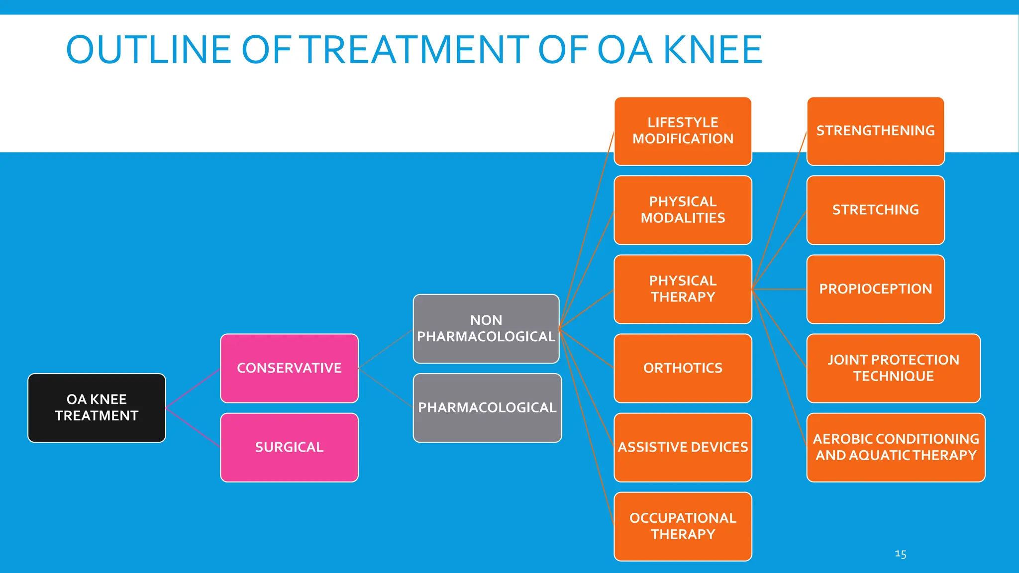
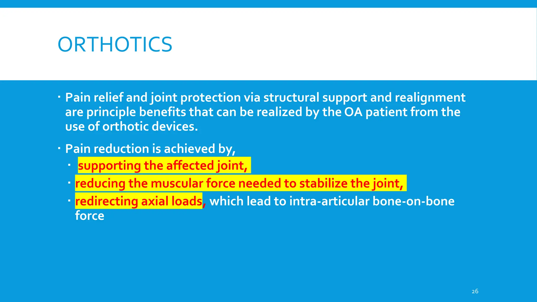
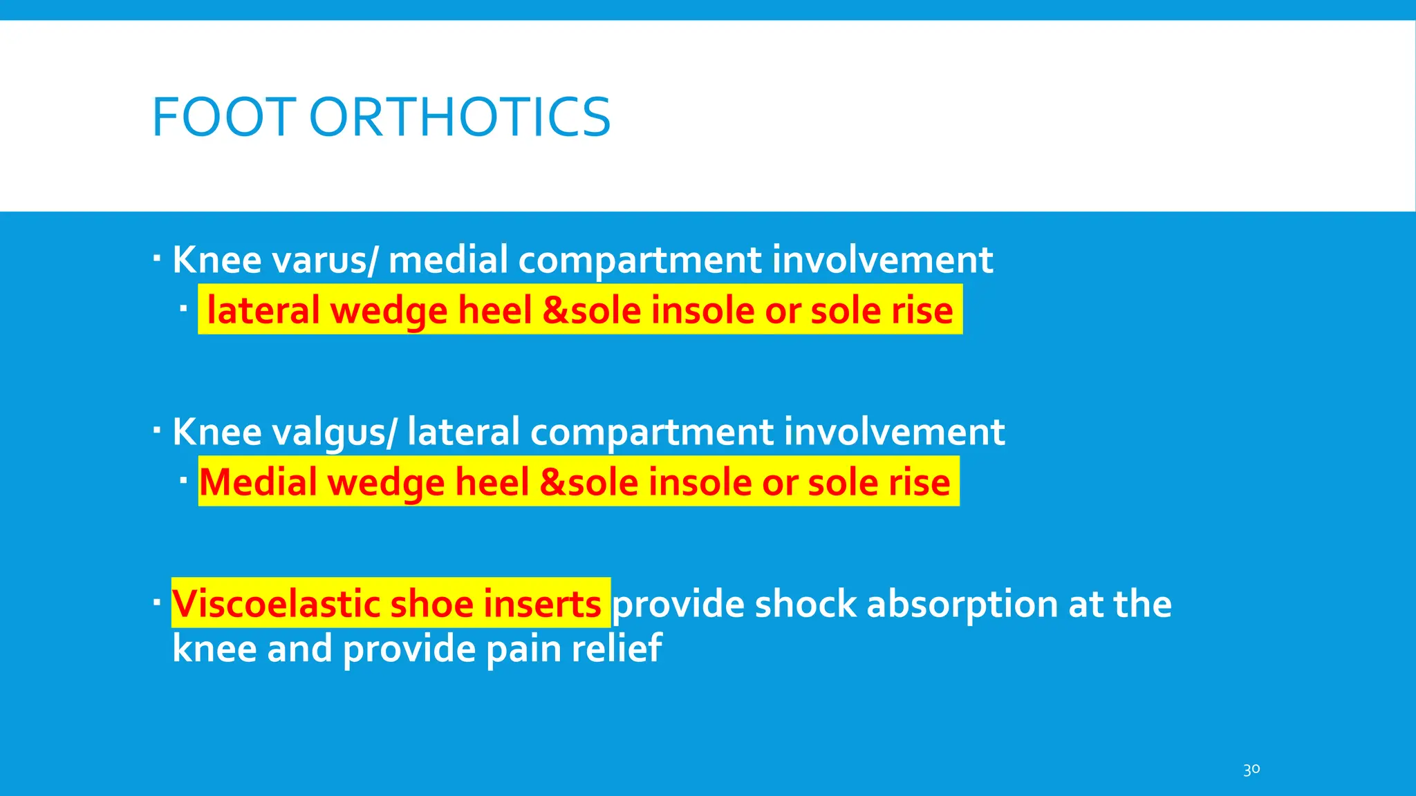
Osteoarthritis (OA) of the knee is a degenerative joint disorder characterized by structural changes like cartilage loss, synovial inflammation, and bone remodeling . Knee OA commonly affects daily activities due to symptoms like joint pain and stiffness, impacting functional abilities . Various factors contribute to knee OA development, including mechanical, enzymatic, and biological factors . In knee OA patients, proinflammatory cytokines like IL-6 and TNF-α have been found to correlate with functional impairment assessed by WOMAC scores, indicating a potential impact on knee joint function . Understanding the interplay between aging and knee OA is crucial, as aging processes can exacerbate the degenerative changes in the knee joint, leading to functional limitations . Evaluating patients' perspectives on knee OA management through instruments like the Knee Outcome Survey Activity Daily Living Scale (KOS-ADLS) is essential for assessing the success of interventions
3. INTRODUCTION
Definition- nonerosive, noninflammatory progressive disorder of the joints leading
to deterioration of the articular cartilage and new bone formation at the joint
surfaces and margins.
Among the chronic rheumatic diseases, hip and knee osteoarthritis (OA) is the
most prevalent and is a leading cause of pain and disability in most countries
worldwide.
Overall prevalence of knee OA was found to be 28.7% in India
Pal CP, Singh P, Chaturvedi S, Pruthi KK, Vij A. Epidemiology of knee osteoarthritis in India and related factors. Indian journal of
orthopaedics. 2016 Oct;50(5):518-22.
3
4. TYPES OF OSTEOARTHRITIS
1. Primary Osteoarthritis
Occurs in a joint de novo.
It occurs in old age, mainly in the weight bearing joints (knee and hip).
In a generalised variety, the trapezio-metacarpal joint of the thumb and the
distal inter-phalangeal joints of the fingers are also affected.
Primary OA is commoner than secondary OA
2. Secondary Osteoarthritis
There is an underlying primary disease of the joint which leads to
degeneration of the joint
4
5. CAUSES OF SECONDARY OSTEOARTHRITIS KNEE
Congenital maldevelopment
Irregularity of the joint surfaces from previous trauma
Previous disease producing a damaged articular surface
Internal derangement , such as a loose body
Mal-alignment
Obesity and excessive weight.
5
6. PATHOLOGY
6
Increase in
water content
and depletion
of
proteoglycans
Repeated
weight
bearing on
such
cartilage
Cartilage
gets abraded
on points of
contact
Subchondral
bone
become
eburnated
(Hard and
glossy)
Bone at the
margins
hypertrophie
s to form
osteophytes
8. SYMPTOMS
Dull aching pain increased with activity, relieved by rest.
Later pain occurs at rest.
Joint stiffness for <30 minutes; becomes worse as the day goes on.
Joint giving away.
Articular gelling- stiffness after immobility lasting short periods and
dissipating after brief period of movement.
Crepitus on ROM.
Deformities in later stages
8
9. SIGNS
Mono- or pauciarticular; shows no obvious joint
pattern.
Localized tenderness of joints.
Pain and crepitus of involved joints.
Enlargement of the joint - changes in the cartilage
and bone secondary to proliferation of synovial fluid
and synovitis.
9
10. RADIOLOGICAL APPROACHTO OA
Melnic CM, Gordon J, Courtney PM, Sheth NP. A Systematic Approach to Evaluating Knee Radiographs with a
Focus on Osteoarthritis. J Orthopedics Rheumatol. 2014;2(1): 6.
10
11. RECOMMENDEDVIEWS
1) weight bearing anteroposterior
2) weight bearing 45 degree
posteroanterior (PA) (Rosenberg view)
Melnic CM, Gordon J, Courtney PM, Sheth NP. A Systematic Approach to Evaluating Knee
Radiographs with a Focus on Osteoarthritis. J Orthopedics Rheumatol. 2014;2(1): 6.
11
15. OUTLINE OFTREATMENT OF OA KNEE
OA KNEE
TREATMENT
CONSERVATIVE
NON
PHARMACOLOGICAL
LIFESTYLE
MODIFICATION
PHYSICAL
MODALITIES
PHYSICAL
THERAPY
STRENGTHENING
STRETCHING
PROPIOCEPTION
JOINT PROTECTION
TECHNIQUE
AEROBIC CONDITIONING
AND AQUATICTHERAPY
ORTHOTICS
ASSISTIVE DEVICES
OCCUPATIONAL
THERAPY
PHARMACOLOGICAL
SURGICAL
15
16. NON PHARMACOLOGICAL MANAGEMENT
Initial counseling should include a discussion of the etiology,
natural history, and prognosis of OA
In the home,
Raised toilet seats,
Grab rails,
Walk-in showers,
Higher seating surfaces
Ramps instead of steps
16
17. WEIGHT LOSS
Weight loss is recommended for all individuals who are overweight or obese,
Those with a healthy body weight should be encouraged to maintain their
weight.
Weight loss has been shown to improve pain and disability related to OA.
Some may require referral to a nutritionist or weight loss clinic for assistance.
Most effective nonpharmacologic weight loss interventions combine
Fat and caloric restriction,
Increased physical activity,
Behavioral reinforcement,
Extended weight maintenance program, with support from the physician and weight-loss support groups
17
18. PHYSICALTHERAPY
An individualized program is important
for adherence to and maintenance of an
exercise program.
Low-impact exercises are often better
tolerated as well as shorter bouts of
exercise.
18
PHYSICALTHERAPY
STRENGTHENING
STRETCHING
PROPIOCEPTION
JOINT PROTECTION
TECHNIQUE
AEROBIC
CONDITIONINGAND
AQUATICTHERAPY
19. STRETCHING EXERCISES
Knee OA classically involves extension lag, but flexion may also be limited.
The pathophysiology of ROM deficits is probably multifactorial, including articular
changes within the joint as well as shortening of myotendinous structures in areas of
pain and/ or weakness.
Decreased ROM is often found not only at the OA joint but also at other joints within
the same lower limb and even in the contralateral lower limb.
When muscles are shorter than their ideal length, they are at a biomechanical
disadvantage when they are required to generate force.
Thus, a stretching program to address inflexibilities should probably be incorporated
early in an exercise program for OA patients.
19
20. STRETCHING PROGRAM- FLEXIBILITY
PROGRAM
GENTLE ROM
EXERCISES
STRETCINGTO
REVERSE
SOME OF LOST
ROM
Slow, gentle, and sustained stretching.
Sustained stretching generally involves
holding the stretch for at least 20 to 40
seconds, and perhaps longer, before
relaxing and then repeating the stretch.
Sudden, jerky or ballistic stretching should
be avoided since it may cause exacerbation
of the OA
20
21. STRENGTHENING EXERCISES
Quadriceps weakness is a better indicator of functional
limitation than pain
Preferred strengthening technique in OA knee
Closed chain kinetic exercises
Initially- Isometric
Benefits limited
Goal- isotonic exercise in pain free ROM of mixed program of
closed and open chain exercises 21
22. BALANCE AND PROPRIOCEPTIVETRAINING
If lower extremity proprioception is suboptimal, the
force of impact transmitted up to the hip and knee
will be increased during weight-bearing activities
Repetitively, such forces may promote progression of
OA and the associated symptoms.
Hence , propioception and balance training must be
included in OA knee Physical therapy program
22
23. JOINT PROTECTION
Joint protection is one goal of exercise in a patient with OA.
Flexibility, strength, and proprioception are optimized in hopes of reducing
joint stresses, decreasing shock impacts to the joint, and maximizing joint
movement and alignment.
23
24. AEROBIC CONDITIONING AND AQUATIC
THERAPY
Aerobic exercise for OA patients commonly includes a daily walking
program since physical activity levels are often reduced.
Using the guideline of 30 minutes of accumulated of moderate activity on
most days is an excellent goal for those patients whose lifestyles are
sedentary
Aquatic therapy
increased sensory input, relaxation from warm water, and decreased
joint compression often allow individuals to move with less significant
pain.
24
25. MODALITIES
Heat – hydrotherapy , hydrocollator packs, Pulse diathermy
Improve myotendinous flexibility, help in stretching and flexibility, there by improve pain also
Cold- Ice packs/massage
Improved muscle strength and ROM or decreased swelling, good analgesia
Electrical- high-intensity burst modes and acupuncture-like transcutaneous
electrical nerve stimulation (TENS)-
Especially in acute exacerbation
No evidence for use of routine use of electrical stimulation, iontophoresis, or
ultrasound in OA
25
26. ORTHOTICS
Pain relief and joint protection via structural support and realignment
are principle benefits that can be realized by the OA patient from the
use of orthotic devices.
Pain reduction is achieved by,
supporting the affected joint,
reducing the muscular force needed to stabilize the joint,
redirecting axial loads, which lead to intra-articular bone-on-bone
force
26
27. Canadian Arthritis Research Symposium—
University of British Columbia knee orthosis
(CARS UBC)- 1975
comprised of plastic thigh and calf shells,
utilized universal hinges
a telescoping tube assembly,
a waistband for suspension.
27
28. UNLOADING BRACES
Bracing for medial compartment gonarthrosis
currently involves application of a three-point
force system across the coronal plane of the
knee joint
Valgus bracing that “unloads” the medial
compartment
28
29. A simple knee sleeve for those patients who present
with mild oa-related knee pain but without any
significant angular deformity
No structural advantage
Improvement in proprioception- pain relief
29
30. FOOT ORTHOTICS
Knee varus/ medial compartment involvement
lateral wedge heel &sole insole or sole rise
Knee valgus/ lateral compartment involvement
Medial wedge heel &sole insole or sole rise
Viscoelastic shoe inserts provide shock absorption at the
knee and provide pain relief
30
31. ORTHOTICS IN PATELLO FEMORAL OA
neoprene-sleeve patellar stabilizing brace consists of a patellar cutout
and force inducing buttress pads around the inferior and lateral aspects
of the patella.
The brace has two circumferentially wrapped rubber straps that apply
dynamic tension to a crescent-shaped lateral patellar pad
31
32. ASSISTIVE DEVICES
Utilizing a cane in the contralateral hand can reduce pain and improve
function in hip and knee OA.
For those concerned about the appearance of a cane, a good alternative
is a walking stick.
If there is not sufficient joint offloading with a cane or in the presence of
bilateral OA, a walker can be used.
32
33. THANKYOU
References
Braddoms 6th edition
Delisas physical medicine and rehabilitaion
Essentials of orthopedics- Maheswari
European Society for Clinical and EconomicAspects of Osteoporosis and Osteoarthritis
(ESCEO) algorithm for the management of knee osteoarthritis
A Systematic Approach to Evaluating Knee Radiographs with a Focus on Osteoarthritis-
Christopher M Melnic
33
Editor's Notes
Quality ap- fibular head is approximately one centimeter below the tibial plateau and one fourth of the head will overlap the tibia.
Morphology of articular surface of tf joints, medial and lateral tf joint space- > 5mm, osteophytes (pain), SC cycsts and sclerosis
Rosenberg view- the most frequently involved zones of articular cartilage were the contact areas of the knees that were between 30 and 60 degrees of flexion.ntercondylar notch -Specific pathologies include osteochondritis dissecans, osteonecrosis, presence of osteophytes, and loose bodies
overlap of the medial and lateral femoral condyles indicating a properly rotated radiograph (left). The sulcus terminalis identifies the lateral femoral condyle (Yellow Arrow). (A) The medial plateau is concave, while (B) the lateral plateau is convex. The patella height is within normal limits. On the right is a lateral radiograph depicting osteoarthritis of the knee. Subchondral sclerosis, patellar osteophytes, and tibiofemoral joint space narrowing can be seen.
Merchant view allows for excellent visualization of the patellofemoral joint and analysis of the joint space for osteophytes, subchondral cysts, and sclerosis. sulcus; this measurement is normally approximately 138 degrees. The congruence angle is measured as the angle of intersection of a line drawn from the deepest portion of the sulcus to the apex of the patella (anterior-most point), and a line from the deepest portion of the sulcus to the posterior-most aspect of the articular surface of the patella. This typically measures -6 degrees +/- 11 degrees
Isotonic and closed chain is most beneficial
Squats, lunges, wall slides