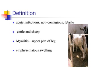
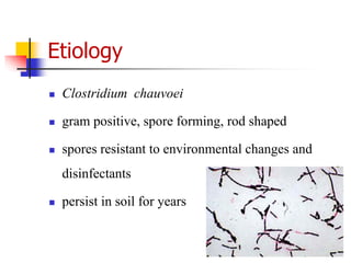
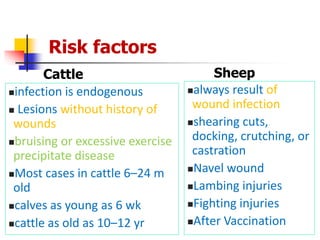
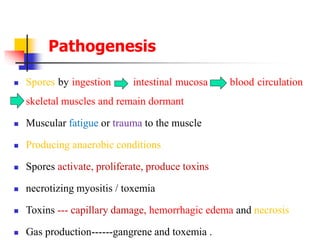
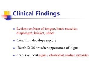
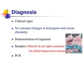
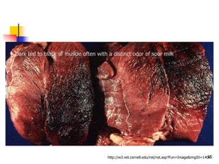
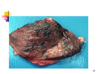
Blackleg Disease
(Black quarter)
Chorray Maar
Definition
 acute, infectious, non-contagious, febrile
 cattle and sheep
 Myositis—upper part of leg
 emphysematous swelling
Etiology
 Clostridium chauvoei
 gram positive, spore forming, rod shaped
 spores resistant to environmental changes and
disinfectants
 persist in soil for years
Route of Infection
 Ingested spores
 pass wall of GI tract/erupting teeth
 access to bloodstream
 deposited in muscle and other tissues (spleen, liver,
and alimentary tract)
 remain dormant indefinitely
Occurrence
• Worldwide
• When disease occurs number of animals affected
• Enzootic in particular areas
• Group of farms to individual in field
• case fatality rate 100%
• seasonal—warm months
• Excavation of soil
Risk factors
Cattle
infection is endogenous
 Lesions without history of
wounds
bruising or excessive exercise
precipitate disease
Most cases in cattle 6–24 m
old
calves as young as 6 wk
cattle as old as 10–12 yr
Sheep
always result of
wound infection
shearing cuts,
docking, crutching, or
castration
Navel wound
Lambing injuries
Fighting injuries
After Vaccination
Pathogenesis
 Spores by ingestion intestinal mucosa blood circulation
skeletal muscles and remain dormant
 Muscular fatigue or trauma to the muscle
 Producing anaerobic conditions
 Spores activate, proliferate, produce toxins
 necrotizing myositis / toxemia
 Toxins --- capillary damage, hemorrhagic edema and necrosis
 Gas production------gangrene and toxemia .
Clinical Findings
 Sever lameness
 Pronounced swelling of upper part of leg
 Depression
 Complete anorexia
 Ruminal stasis
 High temperature and pulse rate
Clinical Findings
 Early stages --- swelling hot and painful
 Later--- cold and painless
 Edematous and emphysematous
 Crepitation on palpation
 Discolored, dry and cracked skin
Clinical Findings
 Lesions on base of tongue, heart muscles,
diaphragm, brisket, udder
 Condition develops rapidly
 Death12-36 hrs after appearance of signs
 deaths without signs / clostridial cardiac myositis
Diagnosis
 Clinical signs
 No constant changes in hemogram and serum
chemistry
 Demonstration of organism
 Samples: Muscle in air tight container
Air dried impression smears
 PCR
Necropsy findings
 Lying on the side with the affected hind limb stuck
out stiffly---characteristic position
 Bloating and putrefaction occurs quickly
 Blood stained froth from nostrils and anus
 Incision ---- dark red to black swollen tissue with
rancid odor---gas bubbles
Black leg. Dark-red skeletal
muscle of a heifer showing
hemorrhage, necrosis, edema
and emphysema.
: Black leg disease. Heart muscle
showing dark red hemorrhagic and
emphysematous
15http://w3.vet.cornell.edu/nst/nst.asp?Fun=Image&imgID=1404
Dark red to black of muscle often with a distinct odor of sour milk
16
False blackleg
 C. septicum and C. novyi
 Mixed infection C. chouvoei and C. septicum
Differential diagnosis
 Malignant edema
 Anthrax
 Lightning stroke
 Bacillary hemoglobinuria
Treatment
 Penicillin--- Large doses (40,000IU/kg BW)
 Surgical debridement of the lesion including
fasciotomy
 Recovery rate is low
 Blackleg antiserum
Control
 Vaccination in enzootic areas
 Booster vaccination
 Prior to anticipated danger period
Control
 Maternal immunity --- 3 months
 In outbreak ---vaccinate unaffected animals
 Penicillin at 10000IU/kg BW IM
 Carcasses destroyed by burning or deep burial

Blackleg (black quarter)

  • 1.
  • 2.
    Definition  acute, infectious,non-contagious, febrile  cattle and sheep  Myositis—upper part of leg  emphysematous swelling
  • 3.
    Etiology  Clostridium chauvoei gram positive, spore forming, rod shaped  spores resistant to environmental changes and disinfectants  persist in soil for years
  • 4.
    Route of Infection Ingested spores  pass wall of GI tract/erupting teeth  access to bloodstream  deposited in muscle and other tissues (spleen, liver, and alimentary tract)  remain dormant indefinitely
  • 5.
    Occurrence • Worldwide • Whendisease occurs number of animals affected • Enzootic in particular areas • Group of farms to individual in field • case fatality rate 100% • seasonal—warm months • Excavation of soil
  • 6.
    Risk factors Cattle infection isendogenous  Lesions without history of wounds bruising or excessive exercise precipitate disease Most cases in cattle 6–24 m old calves as young as 6 wk cattle as old as 10–12 yr Sheep always result of wound infection shearing cuts, docking, crutching, or castration Navel wound Lambing injuries Fighting injuries After Vaccination
  • 7.
    Pathogenesis  Spores byingestion intestinal mucosa blood circulation skeletal muscles and remain dormant  Muscular fatigue or trauma to the muscle  Producing anaerobic conditions  Spores activate, proliferate, produce toxins  necrotizing myositis / toxemia  Toxins --- capillary damage, hemorrhagic edema and necrosis  Gas production------gangrene and toxemia .
  • 8.
    Clinical Findings  Severlameness  Pronounced swelling of upper part of leg  Depression  Complete anorexia  Ruminal stasis  High temperature and pulse rate
  • 9.
    Clinical Findings  Earlystages --- swelling hot and painful  Later--- cold and painless  Edematous and emphysematous  Crepitation on palpation  Discolored, dry and cracked skin
  • 10.
    Clinical Findings  Lesionson base of tongue, heart muscles, diaphragm, brisket, udder  Condition develops rapidly  Death12-36 hrs after appearance of signs  deaths without signs / clostridial cardiac myositis
  • 11.
    Diagnosis  Clinical signs No constant changes in hemogram and serum chemistry  Demonstration of organism  Samples: Muscle in air tight container Air dried impression smears  PCR
  • 12.
    Necropsy findings  Lyingon the side with the affected hind limb stuck out stiffly---characteristic position  Bloating and putrefaction occurs quickly  Blood stained froth from nostrils and anus  Incision ---- dark red to black swollen tissue with rancid odor---gas bubbles
  • 14.
    Black leg. Dark-redskeletal muscle of a heifer showing hemorrhage, necrosis, edema and emphysema. : Black leg disease. Heart muscle showing dark red hemorrhagic and emphysematous
  • 15.
    15http://w3.vet.cornell.edu/nst/nst.asp?Fun=Image&imgID=1404 Dark red toblack of muscle often with a distinct odor of sour milk
  • 16.
  • 17.
    False blackleg  C.septicum and C. novyi  Mixed infection C. chouvoei and C. septicum
  • 18.
    Differential diagnosis  Malignantedema  Anthrax  Lightning stroke  Bacillary hemoglobinuria
  • 19.
    Treatment  Penicillin--- Largedoses (40,000IU/kg BW)  Surgical debridement of the lesion including fasciotomy  Recovery rate is low  Blackleg antiserum
  • 20.
    Control  Vaccination inenzootic areas  Booster vaccination  Prior to anticipated danger period
  • 21.
    Control  Maternal immunity--- 3 months  In outbreak ---vaccinate unaffected animals  Penicillin at 10000IU/kg BW IM  Carcasses destroyed by burning or deep burial