Types of cortex and localization of brain functions
•Download as PPT, PDF•
7 likes•2,686 views

![The surface of the cortex is divided into four lobes which feature key localized functions , but functions are usually elaborated in circuits that involve at least several cortical and subcortical areas Frontal Temporal Parietal Occipital ,[object Object],[object Object],[object Object],[object Object],[object Object],[object Object],[object Object],[object Object],[object Object],[object Object],[object Object],[object Object],[object Object],[object Object],[object Object],[object Object],[object Object],[object Object],[object Object],[object Object],[object Object],… ..from Cortex II lecture slides](data:image/gif;base64,R0lGODlhAQABAIAAAAAAAP///yH5BAEAAAAALAAAAAABAAEAAAIBRAA7)
Recommended
More Related Content
What's hot
What's hot (20)
Similar to Types of cortex and localization of brain functions
Similar to Types of cortex and localization of brain functions (20)
Recently uploaded
Recently uploaded (20)
Types of cortex and localization of brain functions
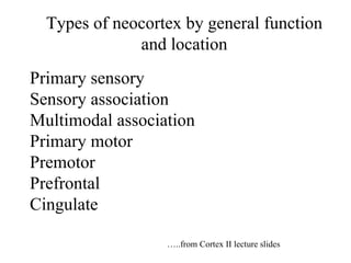
- 1. Types of neocortex by general function and location Primary sensory Sensory association Multimodal association Primary motor Premotor Prefrontal Cingulate … ..from Cortex II lecture slides
- 3. Agnosias Symptoms of association cortex dysfunction See Blumenfeld text Chapter 19 for excellent coverage of higher cerebral functions
- 5. Agnosias-examples for posterior association cortex in parietal and temporal lobes Acalculia -impaired arithmetic calculation Finger agnosia -can’t name individual fingers Right-left disorientation Paracusis -hear a sound, keep hearing it over and over Echolalia -repeating sounds heard over and over Allesthesia -report localization of stimuli on wrong side of the body Hemi-neglect -sensory, motor, combined, conceptual types Anosognosia -lack of awareness of the illness Anosodiaphoria -aware of severe deficits but don’t care (Chicago Cubs fan syndrome) Hemiasomatognosia -Denial that the left half of body belongs to them
- 6. Visual Agnosias, illusions-examples Simultagnosia -cannot see visual scene as a whole, items jump around randomly.Dorsal type stops movement intermittently Micropsia, macropsia -visual objects wrong size Metamorphosia -distorted shape and size, Alice in Wonderland syndrome Palinopsia -previously viewed object reappears sporadically Polyopia -see two or more images of the same object Erythropsia -unnatural coloring of the visual field Prosopagnosia -can’t recognize people by face, but can by voice, touch
- 7. Agnosias Gerstmann’s syndrome-lower 7/39 on left side-dysgraphia, dyscalcula, right left confusion finger agnosia, dyslexia Prosopagnosia-20/21 (inferior temporal): Inability to recognize faces Dyslexia Auditory agnosia
- 8. Agnosias Sensory neglect-; superior parietal Especially on non dom hem- get a right para-sylvian area syndrome-dressing apraxia, constructional apraxia, anosognosia (inability to recognize contra body, disease denial) Unilateral neglect Non-dominant hem superior parietal strokes can cause neglect, even of visual memories. Patients will describe only the right side of their home town square, mall. Asked to imagine the same scene when turned 180 o , they describe the other half, still neglecting their left visual field
- 9. Language A broadly distributed network of nearly half of cortex Aoccdrnig to a rscheearch at Cmabrigde Uinervtisy, it deosn't mttaer in waht oredr the ltteers in a wrod are, the olny iprmoatnt tihng is that the frist and lsat ltteer be in the rghit pclae. The rset can be a taotl mses and you can sitll raed it wouthit a porbelm. Tihs is bcuseae the huamn mnid deos not raed ervey lteter by istlef, but the wrod as a wlohe.
- 10. Thompson 2001 Is the capacity for language predominantly genetically or environmentally determined? - twin study Areas in red show high genetic impact- note dominant hemisphere Wernicke and Broca areas
- 11. Ideational speech area Wernicke’s speech area Broca’s area Semantic speech area
- 12. Blumenfeld 2002 Primary Language Areas
- 13. 40 39 22 43 47 45 44 Visual Somatosensory Auditory Ideational speech area of inferior parietal lobule influenced by sensory association areas-impact on reading/writing/speech
- 15. Language related cortices- for dominant hemisphere 40 39 22 43 47 45 44 8 38 Ideational speech area-39, 40 Wernicke’s speech area- 22+39+40 Broca’s area-44+45 Semantic speech area -Speech apraxia or Broca’s aphasia-44/45 -Pure word deafness-22 -Pure word blindness- 39/37 boundary -Fluent aphasia or Wernicke’s aphasia-22/ 39/40 -Conduction aphasia-43/underlying arcuate bundle -Agraphia-39/40 -Anomia-nouns-38 37
- 16. Left side is dominant in 95% of righties, 60% of lefties Basic distribution of cortical speech pathology “ Anterior aphasia” “ Frontal” “ Expressive” “ Motor” “ Non-fluent” (phrase length, content vs function words) “ Posterior” “ Temporo- Parietal” “ Receptive” “ Sensory” “ Fluent” Broca’s Aphasia Wernicke’s Aphasia Dominant side (usually left) Non-dominant side (usually right) Syntax Grammar Prosody Emotional expression
- 17. Blumenfeld Blood supply of language cortices
- 18. Blumenfeld Types of aphasia and their primary localization Aphasia Fluent? Comprehends? Repeats?
- 19. Broca’s aphasia- Damage to Broca’s Area (BA 44,45) in dominant (left) hemisphere. Decreased premotor and motor aspects of speech. Fluency is diminished, with phrase length less than 5 or 6 words, increased ratio of content words (nouns) to function words (articles, prepositions, syntax modifiers); agrammatism, telegraphic speech) Poorer naming of items, and repetition of words is impaired. Understanding/comprehension is intact, except for syntactically dependent structures (Dog bites man or man bites dog??) Writing and reading is also slow, laborious (Broca’s Patient gets frustrated when being examined )
- 20. Wernicke’s Aphasia Damage to posterior superior temporal gyrus (BA 22) and adjacent inferior parietal (BA39) and temporo-occipital cortex (BA 37) in dominant hemisphere. Impaired comprehension of speech Fluent aphasia Full of nonsense words, empty speech, neologisms, paraphasia (jumbled, unintelligible speech) Impaired repetiton Anoganosia-unaware of their deficit Angry, paranoid Can have contralateral field loss, esp upper right quadrant Writing and reading also severely impaired ( Examiner gets frustrated with Wernicke’s patients) *
- 21. Fluent aphasia lesion-CT R L
- 22. Language/Speech Agnosias-examples Alexia- loss of reading ability Agraphia- loss of writing ability Anomia- can’t name objects, various lesions Paraphasia- inappropriate word substitution. Semantic type substitute “water” for “jacuzzi”. Phonemic type substitutes “trap” for “flap” Global- impaired fluency, comprehension, repetition (full MCA infarct) Conduction- impaired repetition, normal fluency, comprehension. Lesion of arcuate fasciculus, overlying cx Transcortical- impaired fluency, comprehension, repetition is spared. Follows a watershed lesion or basal ganglia/thalamus lesion Aprosodosia- Flat speech. Lesion of nondominant inf frontal
- 23. Alexia without Agraphia lesion (PCA infarct of dominant occipital cortex and corpus callosum) Blumenfeld 2002 Can write normally but can’t read, even their own writing Alexia with agraphia with lesion of dominant angular gyrus
- 24. Alexia without Agraphia lesion Blumenfeld 2002 Can write normally but can’t read, even their own writing Left visual field Can have an emotional response to what is seen, read (Right Wernicke connection OK) Can’t understand what is read (Motor cortex connection OK)
- 25. Alexia without agraphia lesion-MRI T2 R L
- 26. Amnesia But what’s worse, not remembering, or never being able to forget?
- 28. Memory Acquisition of new, non-emotional memories-declarative Hippocampal area Precuneus-31 episodic Dorsal lateral prefrontal cortex-46 Executive/short term memory Amygdala- emotional memory organizer Striatum- procedural Cerebellum- Motor/procedural Long term-everywhere Genetic-orbital/ant temp Faces
- 29. Convergence onto the final common pathway on the PFC pyramidal neuron..and its loops Fallon et al 2003 Optional slide
- 34. Blumenfeld 2002 Neuroanatomy through Clinical Cases Sectors of the Frontal Lobe
- 35. Blumenfeld 2002 Neuroanatomy through Clinical Cases Sectors of the Frontal Lobe
- 37. Functions of the Prefrontal Cortex: “Memory of the Future” Or “How would I look back on what I’m about to do” “ maybe I shouldn’t do that” Risk/Reward Decisions Behavioral Inhibition Ethics and Morality Extraversion/Introversion
- 38. Functions of the Prefrontal Cortex: “Memory of the Future” Or “How would I look back on what I’m about to do” D.T. L.H.
- 39. Progression of Myelination: Axons For all the cortical layers, myelination starts in primary cortices, then spreads to their association cortices, proximally to distally. The last area to myelinate is the frontal pole.
- 40. Progression of dopamine innervation of the prefrontal cortex: The last system in the brain to mature The lamination of DA input to the prefrontal cortex is not complete until about 18 years of age. This is typically when the “dopamine” disorders like schizophrenia really become obvious
- 41. Thompson 2004 Optional slide
- 42. Onset of adaptive behaviors in infants and children-Pt 2 Onset of 1st phase: 10-12 mo Narcissism, grandiosity, self, hyperactivity,exploratory, learning to deal with mother Hedonic dopamine phase in right orbital cortex , Onset of 2nd phase: 16-18 mo Development of “shame” as parents give child disapproving looks, hyper, elated behavior, becomes inhibited, urine and feces provoke disgust response, learning to deal with father Inhibition of dopamine loops, norepinephrine increases, cortisol up, endorphins down as right orbital cortex matures- Optional slide
- 43. Gene-environment-lesion interactions: A quadrillion unique humans are possible And Examples from psychopathology
- 44. Genetic impact on brain and behavior: A quadrillion unique humans are possible based on genetics alone
- 45. Monoamines Catecholamines Serotonin COMT (frontal cortex) MAO-A MAO-A , MAO-B
- 46. ‘ vv ’ - high COMT activity LOW synaptic dopamine Arnsten and Goldman-Rakic, 1986 Arnsten et al., 1994 Murphy et al., 1994, 1996 a,b, 1997 Williams and Goldman-Rakic, 1995 Verma and Moghaddam, 1996 ‘ vm ’ – intermediate ‘ mm ’ – low activity HIGH synaptic dopamine Predicted relative effects of COMT genotype on prefrontal cortical function ______ Optimal________ Optional slide
- 47. D1 Receptors: Inverted U-Shaped Curve Low DA High DA Greatest dopamine effects in Frontal Lobe Normal behavior Schiz, Mania OCD Schiz ADHD Depression Low, Noisy output High Output of PFC But uncoupled from ext. (Cf Yerkes-Dodson law) Fallon Optional slide
- 48. = SNP (Single Nucleotide Polymorphism) ... A C T T T G A ... ... A T T T T G A ... general population genomic sequences An extremely large number of SNPs--millions-- have been detected within the general population and can change quickly in human evolution The presence of millions of SNPs leads to multiple trillions of individual human beings based on genetic variability alone
- 49. Person A Person B Person C High risk Allele/SNP Low risk Allele/SNP High risk Allele/SNP Low risk Allele/SNP High risk Allele/SNP Low risk Allele/SNP GENE LOADS
- 50. Person A Person B Person C High risk allele Low risk allele High risk allele Low risk allele High risk allele Low risk allele Serotonin Transporter Long Serotonin Transporter Short Serotonin Transporter Long Serotonin Transporter Short Serotonin Transporter Long Serotonin Transporter Short MAO-A High active MAO-A Low active MAO-A High active MAO-A Low active MAO-A High active MAO-A Low active COMT High active COMT Low active COMT High active COMT Low active COMT High active COMT Low active Low Hostility Average Hostility High Hostility
- 51. Person A Person B Person C High risk allele Low risk allele High risk allele Low risk allele High risk allele Low risk allele Serotonin Transporter Long Serotonin Transporter Short Serotonin Transporter Long Serotonin Transporter Short Serotonin Transporter Long Serotonin Transporter Short MAO-A High active MAO-A Low active MAO-A High active MAO-A Low active MAO-A High active MAO-A Low active COMT High active COMT Low active COMT High active COMT Low active COMT High active COMT Low active Low Hostility Average Hostility High Hostility These are variants of genes in the normal population. These are not mutants. So what makes one a “ reasonable person”?
- 52. Gene-environment-lesion interactions: A quadrillion unique humans are possible And Examples from psychopathology
- 53. David Berkowitz Aileen Wuornos Ted Bundy Albert Fish John Gacy Charles Manson Beltway Snipers
- 54. Palpatine: Darth Sidious aka His Imperial Majesty Emperor Palpatine of the Galactic Empire Movie characters inspiring real killers
- 55. Issei Sagawa Hannibal Lecter Albert Fish HAL 9000 Ed Gein* *Ed Gein inspired Psycho, The Texas Chainsaw Massacre, The Silence of the Lambs Real killers inspiring movie characters
- 56. Tommy (Joe Pesci) In “ Goodfellas” Excellent portrayal of a psychopathic killer Frank Booth (Dennis Hopper) in ‘ Blue Velvet’
- 57. The sociopath-born or made? Low cortisol in boys-correlations with cruelty to animals, violence, forcing sexual acts, weapon use
- 59. Anderson/ Damasio 1999 Damage at 16 months age Adult lack of moral reasoning
- 60. Psychopathic murderer Amen 2004
- 61. Normal 40 yo Normal 80 yo Adult murderer Teenaged murderer AT AT ORB AT AT ORB ORB AT ORB Orbital cortex (ORB) and anterior temporal cortex (AT)
- 62. Brain areas most likely to be damaged or altered in psychopaths Orbital cortex Anterior cingulate Frontal pole Ventromedial Anterior medial temporal and amygdala
- 63. There is one major group of single nucleotide polymorphisms (SNPs) in the MAO-A gene ; A combination in one group results is low MAO-A activity and very high serotonin levels, and is associated with High Risk for violence and aggression In humans, a Dutch kindred with a missense mutation in the MAO-A gene was described : hemizygous males, representing functional gene knockouts, exhibited a pattern of impulsively violent criminal behaviour for generations (Brunner 1993).
- 64. Monoamines Catecholamines Serotonin COMT (frontal cortex) MAO-A MAO-A , MAO-B
- 65. Structural data: MAOA Low vs High MAO-A expression Meyer-Lindenberg et al 2006 PNAS Low MAO-A activity, high serotonin levels, hyper-reactive amygdala High Risk, Females show no effect of genotype Male at high risk have Increased orbital volumes Optional slide
- 66. Meyer-Lindenberg et al 2006 PNAS Emotional memory scores- response in left amygdala in Males is higher in high risk (MAO-AL) individuals Hyper-responsive left amygdala in High Risk males Optional slide
- 67. Caspi and colleagues (2002) also found that the high risk genotype (ie low levels of MAO-A; high levels of serotonin) affects responses to harsh environmental stressors. The genetically high risk adolescents and adults who were also maltreated as children were much more likely to develop conduct disorder, antisocial personality disorder, and to be convicted for violent crimes. Gene-Environment Interactions
- 68. Dorsal PFC Orbital Cortex Amygdala Nucleus Accumbens DA DA _ _ Competition among prefrontal and limbic cortices for control of DA influence in nucleus accumbens Dorsal Anterior Cingulate Ventral Anterior Cingulate CRH VTA Dopamine Net Motor Output, i.e., Behavior: Do it or Don’t Do it? GO DON’T GO
Editor's Notes
- Catechol-O-methyl transferase is involved in the breakdown of the catecholamine neurotransmitters, dopamine, epinephrine and norepinephrine. The enzyme introduces a methyl group to the catecholamine which is donated by S-adenosyl methionine (SAM). A functional polymorphism (a common normal variant) of the gene for catechol-O-methyl transferase has been shown to affect cognitive tasks broadly related to executive function, such as set shifting, response inhibition, abstract thought and the acquisition of rules or task structure. The link between impairments in these sorts of cognitive tasks and the COMT gene is thought to be mediated by an effect on dopamine signalling in the frontal lobes. Arguably, the clearest link between genetic variation and aggression exists for Monoamine Oxidase A (MAO-A, MIM 309850), a key enzyme in the catabolism of monoamines, especially serotonin. The serotonergic system has been implicated in impulsivity and manifest violent behaviour in animals and both auto- and heteroaggression in humans . MAO A and B genes, likely derived from the same ancestral gene, are both located on the X-chromosome (Xp11.23), comprising 15 exons with identical intron-exon organization . MAO-A provides the major enzymatic clearing step for serotonin and norepinephrine during brain development, while MAO-B activity increases dramatically after birth . Mouse knockouts for MAO-A , but not MAO-B , have elevated brain levels of serotonin, norepinephrine, and dopamine. They show enhanced amygdala-dependent emotional, but not motor, learning , and males exhibit dramatically increased aggressive behaviour . In humans, a Dutch kindred with a missense mutation in the MAO-A gene was described : hemizygous males, representing functional gene knockouts, exhibited a pattern of impulsively violent criminal behaviour for generations.
- Catechol-O-methyl transferase is involved in the breakdown of the catecholamine neurotransmitters, dopamine, epinephrine and norepinephrine. The enzyme introduces a methyl group to the catecholamine which is donated by S-adenosyl methionine (SAM). A functional polymorphism (a common normal variant) of the gene for catechol-O-methyl transferase has been shown to affect cognitive tasks broadly related to executive function, such as set shifting, response inhibition, abstract thought and the acquisition of rules or task structure. The link between impairments in these sorts of cognitive tasks and the COMT gene is thought to be mediated by an effect on dopamine signalling in the frontal lobes. Arguably, the clearest link between genetic variation and aggression exists for Monoamine Oxidase A (MAO-A, MIM 309850), a key enzyme in the catabolism of monoamines, especially serotonin. The serotonergic system has been implicated in impulsivity and manifest violent behaviour in animals and both auto- and heteroaggression in humans . MAO A and B genes, likely derived from the same ancestral gene, are both located on the X-chromosome (Xp11.23), comprising 15 exons with identical intron-exon organization . MAO-A provides the major enzymatic clearing step for serotonin and norepinephrine during brain development, while MAO-B activity increases dramatically after birth . Mouse knockouts for MAO-A , but not MAO-B , have elevated brain levels of serotonin, norepinephrine, and dopamine. They show enhanced amygdala-dependent emotional, but not motor, learning , and males exhibit dramatically increased aggressive behaviour . In humans, a Dutch kindred with a missense mutation in the MAO-A gene was described : hemizygous males, representing functional gene knockouts, exhibited a pattern of impulsively violent criminal behaviour for generations.
- Fig. 1: Structural data demonstrates limbic and paralimbic regional volume changes in MAOA-L subjects (n = 97). Plots represent the summed volumes of voxels in pre-defined regions of interest, normalized to volume measures relative to the MAOA-H group mean. (a) Compared with MAOA-H subjects, MAOA-L individuals exhibit significant volume reductions in bilateral amygdala, supragenual anterior cingulate and subgenual anterior cingulate cortex. Male and female subjects were combined. (b) male MAOA-L individuals show increased lateral orbitofrontal volume, bilaterally, relative to MAOA-H subjects. Females show no effect of genotype, resulting in a highly significant sex by genotype interaction. Summary Biological mechanisms of violence in humans remain poorly understood. A genetic approach to this issue is the X-linked MAO-A gene, which is associated with impulsive aggression in animals and humans. Here, we identify underlying neural mechanisms by studying the impact of a common functional polymorphism in MAO-A on brain structure and function in a large sample of healthy human volunteers. We show that the risk (low expression) variant is associated with pronounced limbic volume reductions and hyperresponsive amygdala during emotional arousal, while reactivity in regulatory prefrontal regions is reduced. In men, this allele also is associated with changes in orbitofrontal volume, hyperreactivity of amygdala and hippocampus during aversive recall, and impaired cingulate activation during cognitive inhibition.. Our data identify dysfunctional limbic circuitry for emotional and cognitive regulation mediating association of MAO-A with impulsive aggression, demonstrate X-inactivation in human brain, and isolate potential targets for an interventional biological approach towards violence. Introduction Violent and criminal behaviour are likely related to complex environmental and social circumstances, but heritable factors also have been implicated . The specific neural mechanisms leading to delinquency and impulsive aggression are poorly understood, though they have been the subject of spirited speculation and debate for literally centuries . Arguably, the clearest link between genetic variation and aggression exists for Monoamine Oxidase A (MAO-A, MIM 309850), a key enzyme in the catabolism of monoamines, especially serotonin. The serotonergic system has been implicated in impulsivity and manifest violent behaviour in animals and both auto- and heteroaggression in humans . MAO A and B genes, likely derived from the same ancestral gene, are both located on the X-chromosome (Xp11.23), comprising 15 exons with identical intron-exon organization . MAO-A provides the major enzymatic clearing step for serotonin and norepinephrine during brain development, while MAO-B activity increases dramatically after birth . Mouse knockouts for MAO-A , but not MAO-B , have elevated brain levels of serotonin, norepinephrine, and dopamine. They show enhanced amygdala-dependent emotional, but not motor, learning , and males exhibit dramatically increased aggressive behaviour . In humans, a Dutch kindred with a missense mutation in the MAO-A gene was described : hemizygous males, representing functional gene knockouts, exhibited a pattern of impulsively violent criminal behaviour for generations. While functionally disabling variants of the gene are rarities outside the laboratory setting, a common variable number of tandem repeats polymorphism of the MAO-A gene has been described that strongly impacts transcriptional efficiency: enzyme expression is relatively high for carriers of 3.5 or 4 repeats (MAOA-H) and lower for carriers of 2,3, or 5 repeats ( MAOA-L ) . While conflicting evidence exists for the association of genotype with trait impulsivity in human cross sectional studies, a clear and pronounced gene by environment interaction was found in a large longitudinal study of children followed for 25 years in which MAOA-L predicted violent offences in males with adverse early experience (maltreatment) . This finding, replicated in the majority but not all further studies, suggests a deficiency in the neural mechanisms for emotional regulation and memory as a possible substrate for the observed gene-environment interaction. This agrees with current proposals linking brain structures involved in emotional control, such as amygdala and medial prefrontal and orbitofrontal cortices, to the emergence of violent behaviour . However, while two previous fMRI studies suggested an effect of MAO-A genotype during a cognitive task in small samples , no data related to emotional processing or brain structure are available.
- Fig. 3: Limbic activation during the retrieval of aversive memories varies according to MAO-A genotype (n=90). (a) Left amygdala response during emotional memory is higher for male MAOA-L subjects, compared with male MAOA-H individuals. (b) Left hippocampal engagement during emotional memory is more pronounced for male, but not female, MAOA-L subjects, relative to MAOA-H individuals (n = 90).