Download as PDF, PPTX

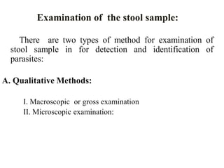
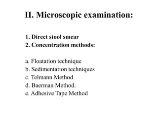


This document provides information about parasitology practice and laboratory diagnosis of parasites. It discusses different types of specimens that can be examined for parasites, including stool, blood, skin, sputum, vaginal discharge, and tissue biopsies. For stool samples, it describes proper collection and preservation methods. It also lists the common parasites that may be observed in different specimen types. Methods for microscopic examination of stool samples include direct smears and concentration techniques like floatation, sedimentation, and others.