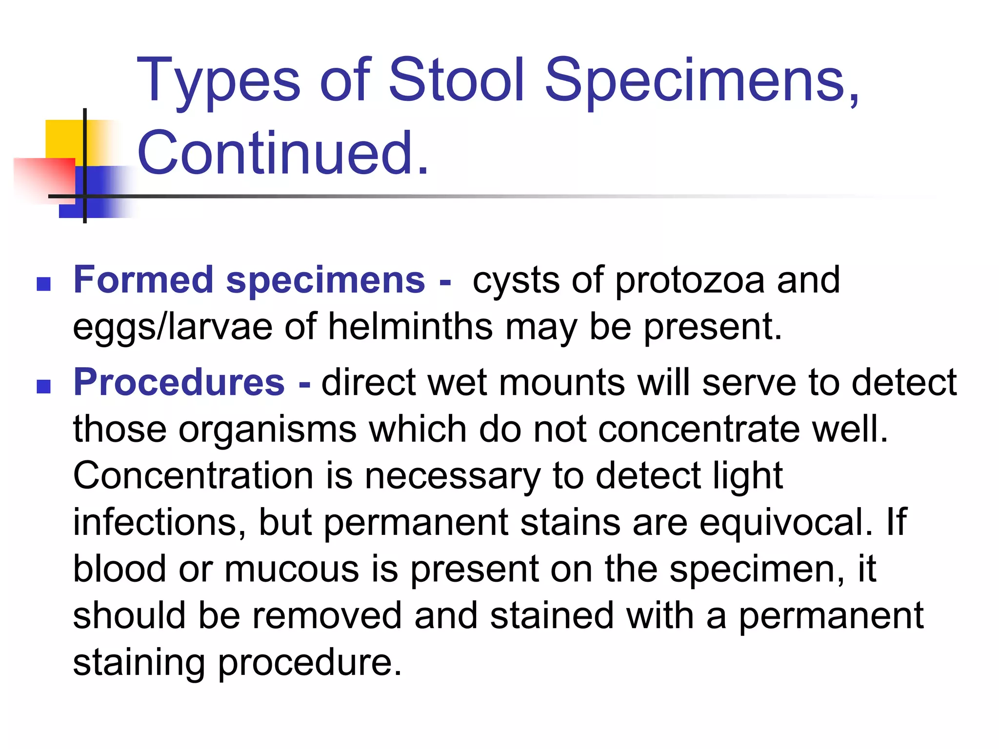
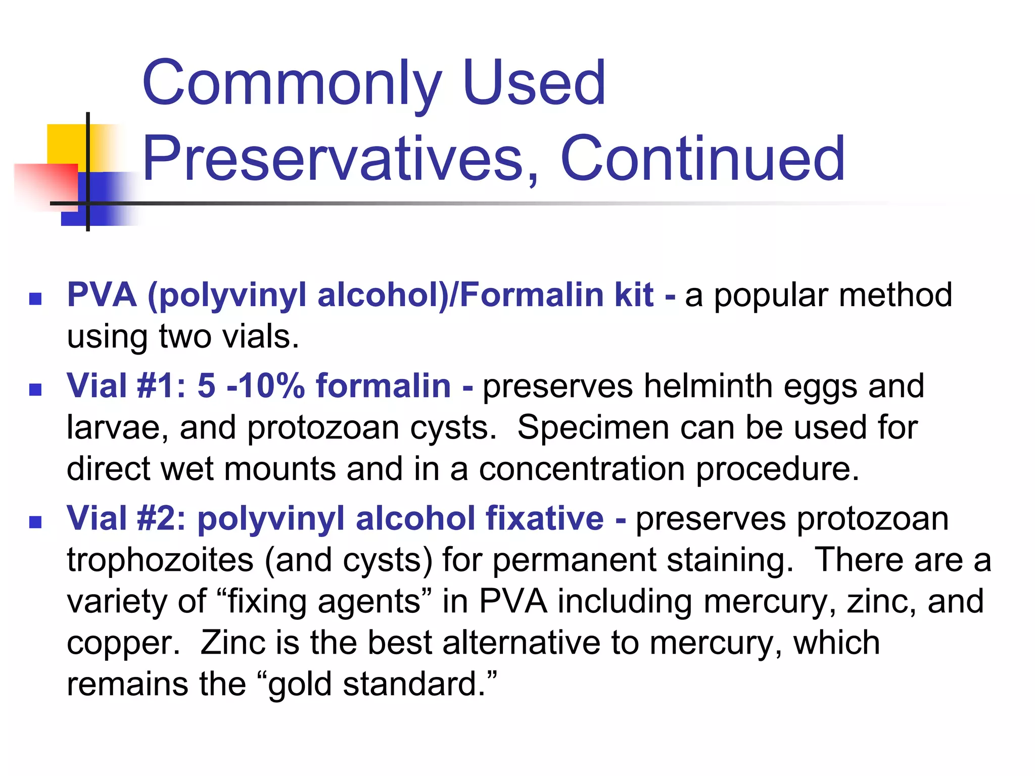
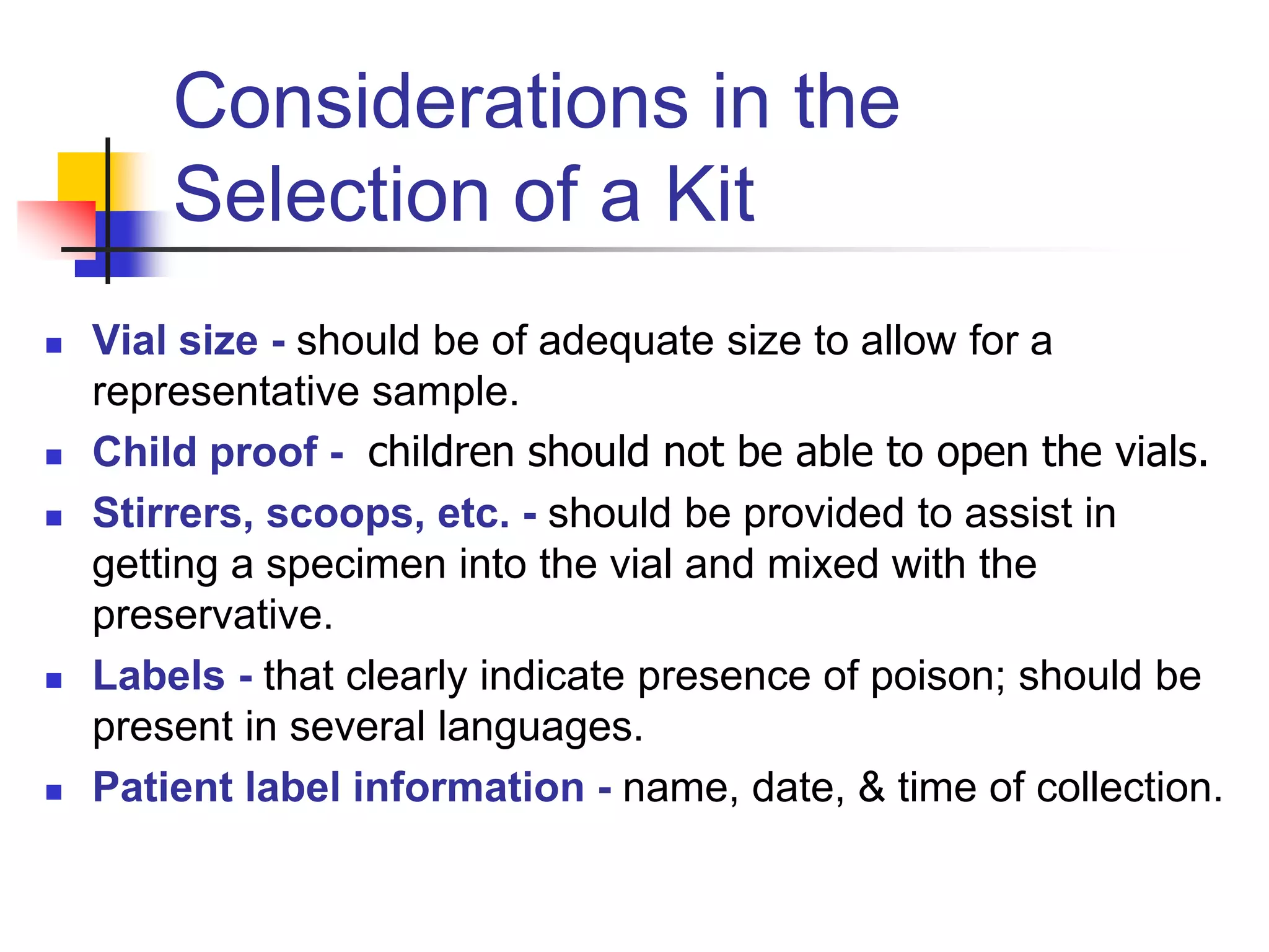
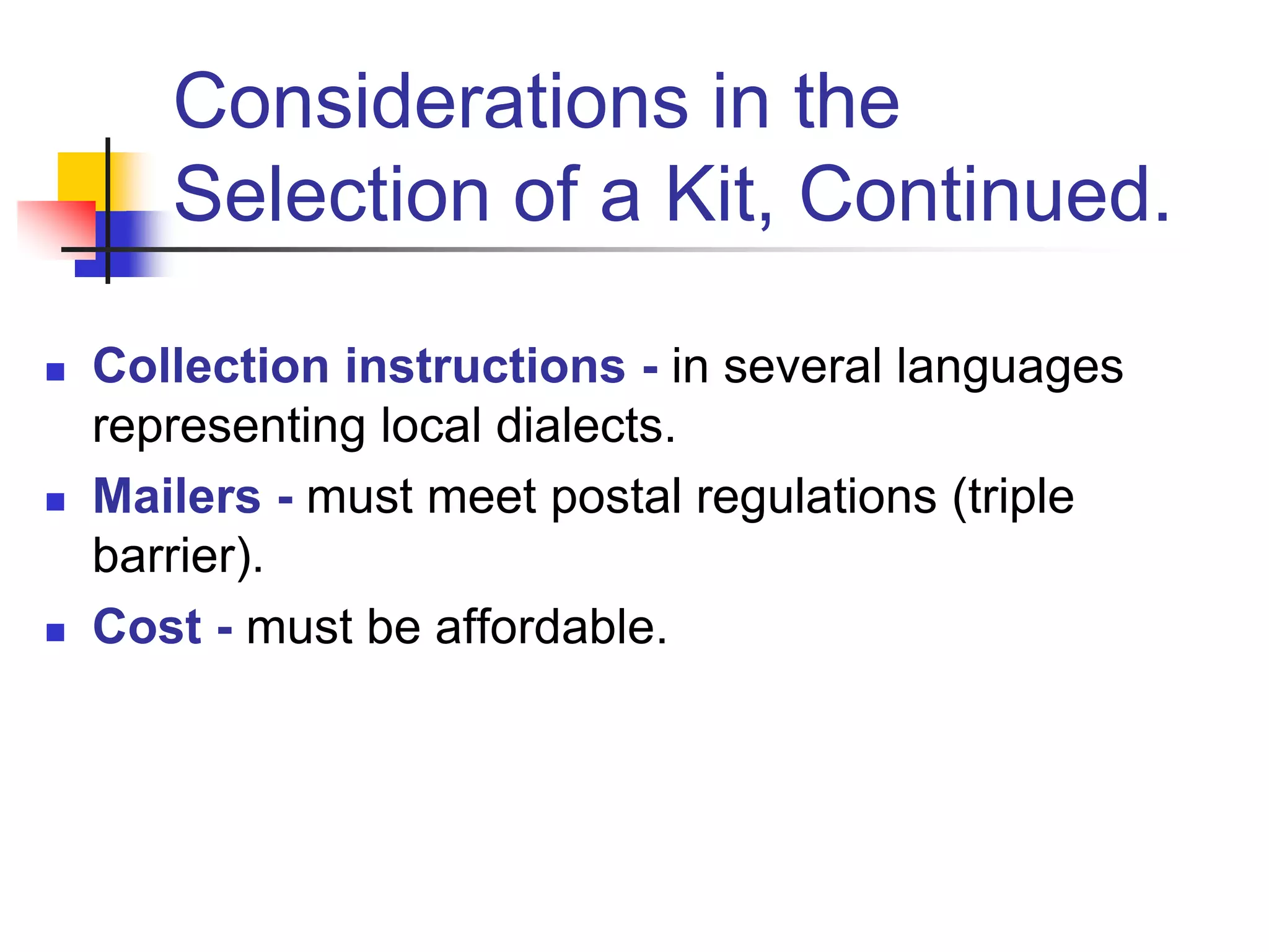
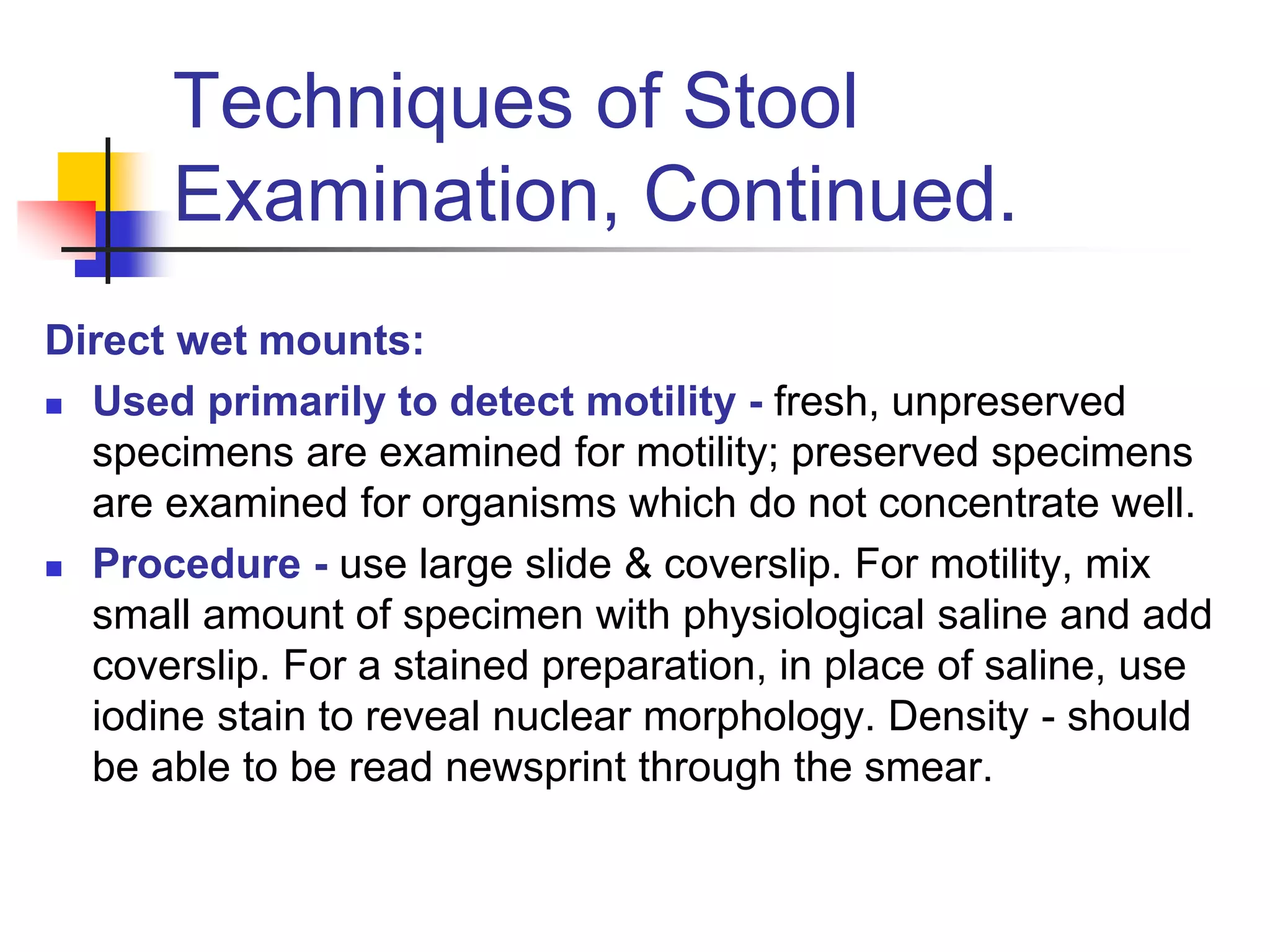
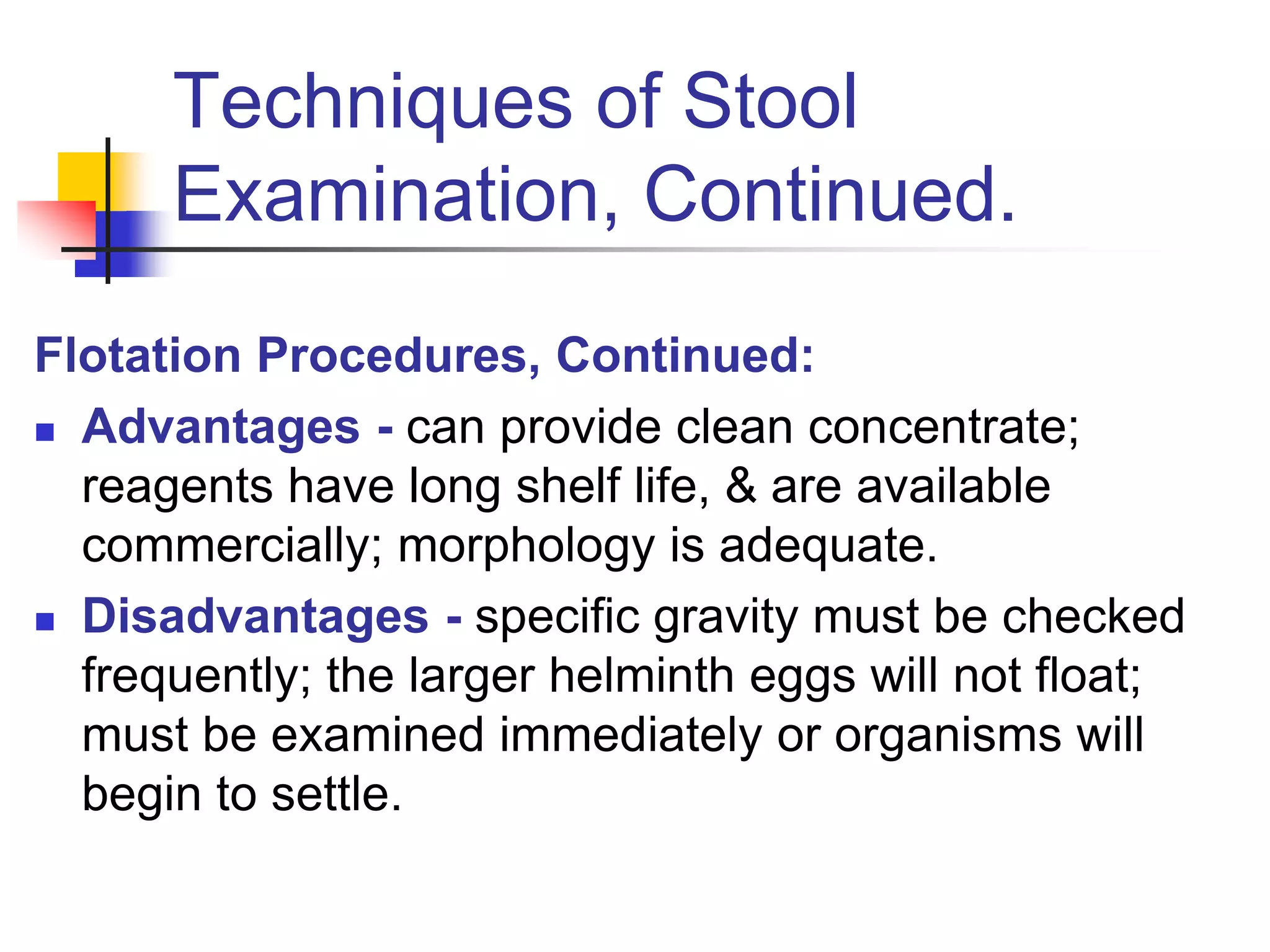
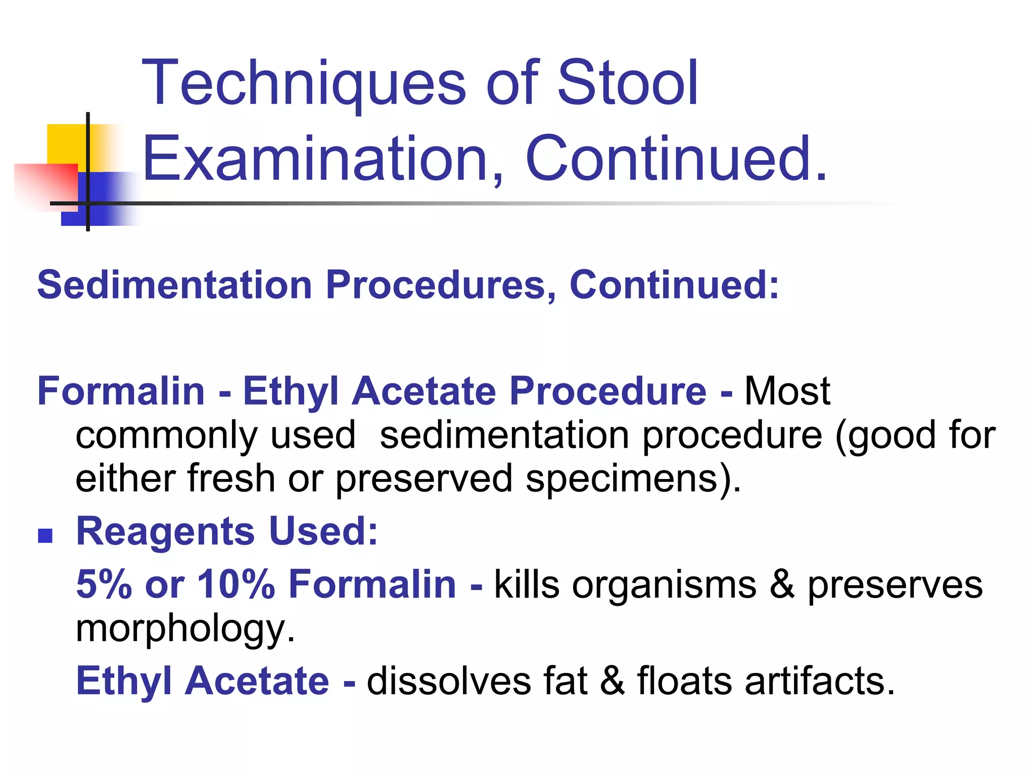
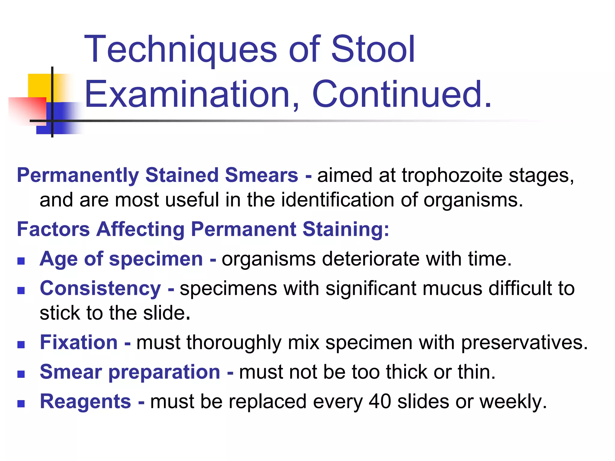
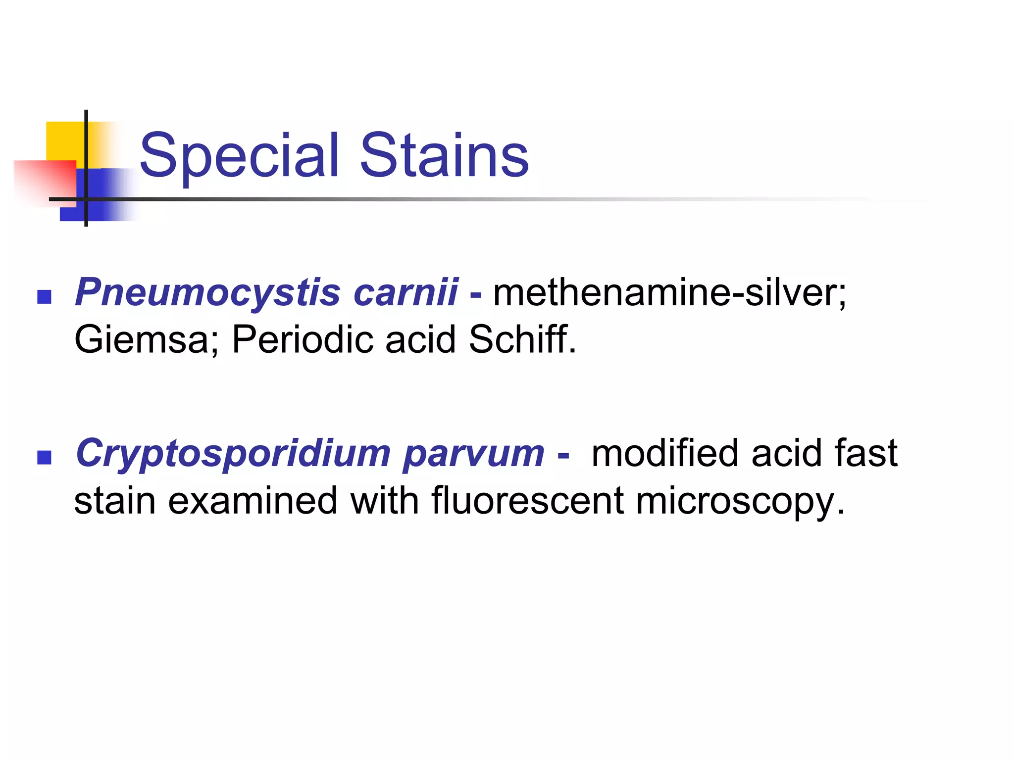
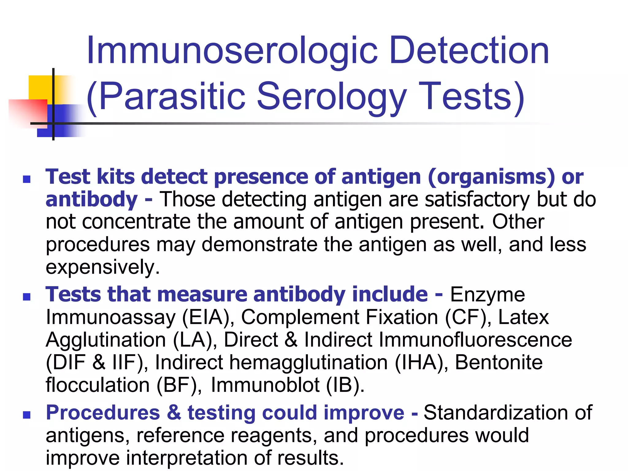
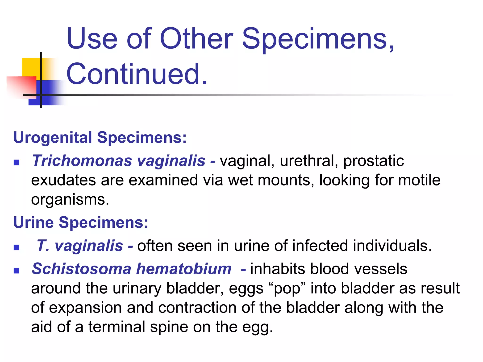
The document provides an overview of parasitology and techniques for diagnosing parasitic infections through stool examination. It discusses factors required for reliable diagnosis such as travel history and appropriate specimen collection. It also describes various stool examination techniques including direct wet mounts, concentration methods, and permanent staining. Flotation and sedimentation are outlined as concentration procedures. Considerations for stool collection kits and preservatives are also summarized.
This document provides an overview of medical parasitology and laboratory diagnosis of parasitic infections. It discusses key topics such as the definition of parasitology and medical parasitology. Various specimen types that can be used for diagnosis are described, including stool, blood, urine and others. Main diagnostic methods covered include microscopy, concentration techniques, staining of smears, and newer molecular methods. Specific procedures for examining common specimen types like stool, blood, and urine are outlined.
Strategies Novartis can use to GROW from a Billion Dollar Company to a Trillion Dollar Company like Alphabet Inc
Novartis is a leading healthcare company which is situated in Switzerland and uses digital technologies and innovative science to come up with transformative ways of treatment in areas of great medicinal needs. This article explains what Novartis strategies and what they should employ so that they can rise from a billion dollar company to a trillion dollar company like the Google Alphabet Inc.
Novartis was formed in March 1996 by the merging of pharmaceutical and agrochemical divisions of Ciba-Geigy and Sandoz companies. Thanks to the merging of the two companies, Novartis is one of the biggest pharmaceutical companies in the world. Novartis is one of the largest companies which achieved a great milestone within a few decades. Novartis as a whole is divided into three major divisions: Sandoz (generics), Innovative Medicines and Alcon (eyecare). Novartis is also involved in collaborative research projects that are publicly funded.
Below are some of Novartis best selling drugs and their revenue
1.Cosenty – This is the top selling drug with a revenue of 4.788 billion dollars
2.Enfresto – This has a revenue of 4.644 billions dollars
3.Promacta – This has a revenue 0f 2.088 billion dollars
Medicine manufactured by Novartis and their uses
Medicine Medicine use
Cosentyx Used to treat psoriatic arthritis
Entresto Used to treat heart failure
Lucentis Used to block abnormal vessel growth in the back of the eye
Tasigna Used to treat chronic myelogenous leukemia which has the Philadelphia chromosome
Jakavi Used to treat myelofibrosis, polycythemia vera and graft-versus-host disease
Promacta Used to treat patients with abnormal low platelet count
Sandostatin Used to treat patients with tumor experiencing symptoms like flushing and diarrhea
Xolair Used to treat moderate and severe asthma
Gilenya Used to treat multiple sclerosis
How Novartis became one of the biggest pharmaceutical companies in the world
1.Market control through partnership
Geigy, Sandoz and Ciba combined their power so that they can compete with strong foreign firms and formed a cartel called the Basal Syndicate or Basal IG. Basal IG secured most of the manufacturing facilities all over the US and across Europe. It later joined with IG Farben and other chemical companies to form a big cartel called the Quadrapartite Cartel which dominated all of the European market and enjoyed the profits made from the joint manufacturing.
2.Growth acceleration through mergers
Since competition was very rampant in the pharmaceutical industry, Ciba and Geigy decided to merge with Sandoz AG to form Novartis. With this merge, Novartis became one of the growing giants in the pharmaceutical industry. This made Novartis gain a lot of fame and build a strong reputation over other companies. Novartis majored on agrochemical and pharmaceutical industries which made it easy to focus on a specific mar
Medical parasitology : study of parasites that infect human, diseases caused by them, clinical picture, their diagnosis, treatment and prevention as well as controls.
It involves drug development, epidemiological studies and study of zoonoses.
To know various terms related to parasitology.
To know about general parasites and parasitic infections.
To get knowledge about laboratory diagnosis and its importance.
To gain idea about general epidemiological aspects of parasites that affect human.
Apply basic methods of specimen collection , preservation and processing in lab.
To prevent ourselves from these infections and apply control measures.
Quality control procedures must be followed for specimen collection, preparation of reagents, laboratory techniques, and examination of results to ensure accurate diagnosis of parasitic infections. Specimens like stool and blood must be properly collected and handled to prevent deterioration. Reagents have expiration dates and must be prepared and stored correctly. Laboratory techniques must be performed carefully according to standardized procedures. Examination of final samples, like blood films, requires a quality sample and careful evaluation to avoid errors. Proper specimen collection times are important for certain parasites like malaria and filaria detected in blood.
This document provides guidance on sampling methods for different animal diseases. It discusses the appropriate samples, packaging, and transport needed for accurate diagnosis of various conditions. Specific guidance is given for sampling blood, serum, swabs, feces, tissues, and other specimen types. Common diseases that can be diagnosed from different sample types are also outlined. The overall message is that collecting samples following these standards will provide veterinary laboratories the best opportunity for accurate diagnosis.
Parasitology is the study of parasites and parasitism. It explores aspects of parasite biochemistry, physiology, biology, and immunology. Key terms include: parasite, host, endoparasite, ectoparasite, reservoir host, vector, carrier, direct and indirect life cycles.
Helminthes are divided into three phyla: Nematodes (roundworms), Cestodes (tapeworms), and Trematodes (flukes). Important cestodes include Moniezia found in sheep and cattle. Key trematodes are Fasciola hepatica (liver fluke) and Paramphistomum (rumen fluke). Important nematodes are Haemonchus
LUMEN DWELLING FLAGELLATES - GIARDIA
REFS:
INTERNATIONALLY ACCEPTED BOOK OF MEDICAL PARASITOLOGY BY K. D. CHATTERJEE
TEXT BOOK OF MEDICAL PARASITOLOGY BY PANIKER
IMAGE SOURCES : FROM INTERNET
This document provides information about parasitology practice and laboratory diagnosis of parasites. It discusses different types of specimens that can be examined for parasites, including stool, blood, skin, sputum, vaginal discharge, and tissue biopsies. For stool samples, it describes proper collection and preservation methods. It also lists the common parasites that may be observed in different specimen types. Methods for microscopic examination of stool samples include direct smears and concentration techniques like floatation, sedimentation, and others.
The document provides an overview of parasitology, including definitions of key terms like parasite and host. It discusses the different types of parasite life cycles and relationships with hosts, how parasites can negatively impact hosts, examples of common internal and external parasites of small animals and livestock, and methods for diagnosing parasites via fecal examination. It also outlines the life cycle of fleas.
Routine stool examination provides important information about gastrointestinal health and disease. A stool sample is examined macroscopically for characteristics like color, consistency, and presence of blood or mucus. Microscopic examination looks for items like white blood cells, red blood cells, parasites, fat, and bacteria. Chemical tests can detect occult blood, excess fat, sugars, and other markers. Proper collection and preservation of stool samples is important for accurate examination and detection of parasites. Both wet mount microscopy and concentration techniques may be used depending on the suspected condition. Stool examination aids in diagnosis of gastrointestinal infections, inflammation, malignancy, and malabsorption disorders.
The document provides an introduction to medical parasitology. It defines parasites and the relationships between parasites and their hosts. It discusses the classification of parasites according to their habitat, life cycle dependence on the host, and pathogenicity. It also covers the transmission, diagnosis, and importance of human parasites. The key parasites discussed include protozoa that infect the intestines, blood, and tissues, as well as helminthic worms including nematodes, cestodes, and trematodes.
This document summarizes key information about parasitic infections, including their clinical presentation, laboratory diagnosis, and most common pathogens. It describes how parasitic infections are typically diagnosed via microscopic examination of stool, blood, and tissue specimens. The document then outlines the various protozoan and helminthic pathogens that can infect the intestinal tract and other body sites, providing details on morphology, life cycles, symptoms, and treatment for major parasites like Entamoeba histolytica, Giardia lamblia, and Cryptosporidium.
This document discusses 7 common myths in microbiology and cleanroom practices. It summarizes that colony forming units may not always accurately represent bacterial numbers due to variability in culture methods. Microbiology cabinets are not always laminar due to disruptions in airflow. Media growth promotion testing cannot always be skipped and relies on appropriate control strains. Microorganisms in cleanrooms are rarely free floating and are usually attached to particles. Environmental monitoring parameters cannot have universal pre-set conditions due to variability in microorganisms and methods. Bunsen burners are not always needed to create aseptic spaces and can increase risks of contamination. Identification results from phenotypic tests are not always reliable due to limitations in databases and possibility of mixed cultures or changes in phenotypes.
Fecal examination is commonly used to diagnose parasitic infections in animals. The process involves collecting a fresh fecal sample, preparing it using flotation or centrifugation with a flotation medium, and examining it under a microscope. Centrifugation speeds up the process by forcing heavier materials to the bottom and lighter parasite eggs to the top for easier identification. Examination of properly collected and prepared fecal samples can reveal evidence of parasitic infections and provide a diagnosis.
1. The document discusses intestinal helminthic infections, including intestinal nematodes like Ascaris lumbricoides, hookworms, and Trichuris trichiura.
2. It provides details on the morphology, life cycle, transmission, pathogenesis, clinical features, diagnosis, and treatment of Ascaris lumbricoides infection.
3. Laboratory diagnosis of ascariasis involves detection of Ascaris eggs in stool samples or detection of adult worms or larvae in stool, sputum, or tissue samples through microscopic examination, PCR, or imaging. Common treatments include albendazole, mebendazole, or ivermectin.
This document summarizes flagellates, including their classification, morphology, and life cycles. It focuses on Giardia intestinalis and Trichomonas vaginalis. G. intestinalis has trophozoite and cyst stages, with the cyst being infective. It causes giardiasis by damaging the intestinal epithelium. T. vaginalis only exists as a trophozoite and causes trichomoniasis through overgrowth in the vagina when pH increases. Both can be diagnosed via microscopy of stool or vaginal samples and treated with metronidazole or tinidazole.
: Parasitic water pollution in the Nile River (Schistosoma & Giardia lamblia)MenrvaSorial
Causative organism.
Geographical distribution.
Epidemiology & Risk factors.
Mode of Transmission.
Vector (if available).
Habitat.
Life cycle (including infective stage, Diagnostic stage, Final host, Intermediate host and Reservoir).
-According to your lab group assignment topic, you must mention at least two examples (Causative organisms) for the required type of parasitic infection and their prevalence in Egypt. -Then discuss briefly the mentioned examples covering all the following points:
As a pharmacist, how could you identify and confirm a patient with such disease?
(NB: Identification and confirmation include the signs and symptoms and the diagnostic tests in details)
What are the therapeutic options available (suggest a line of treatment).
How can we prevent & control such disease?
The document discusses stool examination, including indications such as detecting parasites, evaluating diarrhea, and differentiating between infectious bacteria. It describes the microscopic examination of stool to identify parasites, bacteria, and abnormalities, noting key features of various parasites like Entamoeba histolytica, Giardia intestinalis, and helminths. Precautions for stool collection and preparation of slides for microscopic examination are also outlined.
This document discusses parasitic infections that can cause diarrhea. It focuses on the laboratory diagnosis of parasitic infections through microscopic examination of stool and other specimens. The most common protozoan parasites found in stool are described in detail, including Entamoeba histolytica, Giardia lamblia, Cryptosporidium species, and others. Diagnostic methods for specific parasites are also outlined.
This document provides information on diagnosing diseases in aquaculture. It discusses the importance of understanding normal conditions to detect abnormalities. The diagnosis process aims to determine the accurate cause of disease to select proper treatment. Key steps include taking a case history, conducting clinical, post-mortem, laboratory, histopathological, and molecular examinations. Signs of disease may be behavioral, physical on external/internal examination. Mortality can occur in acute outbreaks or chronic patterns over time. Accurate diagnosis is needed to effectively treat diseases affecting aquaculture.
Entamoeba histolytica is a protozoan parasite that can cause intestinal amoebiasis and extra-intestinal disease in humans. It has a simple life cycle involving an infective cyst stage that is transmitted between hosts. In the intestine, it exists as a trophozoite that can invade and destroy intestinal tissue, potentially spreading to the liver to cause amoebic liver abscesses. Diagnosis involves microscopic identification of trophozoites or cysts in stool or abscess samples, as well as antigen detection or molecular tests. It remains an important cause of morbidity and mortality worldwide.
Prevalence of Aeromonas Species Among Patients Attending General Hospital Owerripijans
A total of two hundred (200) stool samples were collected from patients attending General Hospital Owerri
and screened for the presence of Aeromonas species. Out of the two hundred (200) stool samples, one
hundred and fifty (150) were collected from diarrheal patients while fifty (150) were collected from nondiarrheal patients. Aeromonas species were only isoloated from diarrheal patients stool samples.The
prevalence of Aeromonas species in diarrheal patients was 5.3%. Aeromonas species were found to be
highly (100%) susceptible to ceftazidime, followed by cefotaxime (85%), then Augmentin (75%) and
Gentamicin (65%), but highly (100%) resistant to Ampicillin and Tretracyclin following by contrimoxazole
(83%). This observation could probably indicates that Aeromonas as enteropathogen associated with
diarrheal and should be considered amongst the causative agents of diarrheal.
Typhoidal and non typhoidal salmonella.pptxVaisHali822687
This document discusses typhoidal and non-typhoidal salmonella. It begins by introducing salmonella as a gram-negative bacteria and describes its classification. It then discusses the different serotypes of salmonella including S. Typhi, S. Paratyphi A, B, and C which cause enteric fever. The document summarizes the pathogenesis, clinical features, diagnosis, and treatment of typhoidal salmonellosis. It provides details on the various laboratory techniques used to identify and diagnose salmonella such as culture-based methods, biochemical testing, and serological tests.
The document provides information on tapeworm parasites (order Cestoidea), including their classification, morphology, life cycles, transmission, clinical features, diagnosis and treatment. It discusses several important tapeworm species that can infect humans, including Taenia saginata, Taenia solium, Hymenolepis nana, Echinococcus granulosus, and Diphyllobothrium latum. Key details on the morphology, life cycles, epidemiology and clinical manifestations of each species are provided.
Zoonotic parasite; Toxoplasma is an opportunistic pathogen.
Infects animals, cattle, birds, rodents, pigs, and sheep.
and humans.
Causes the disease Toxoplasmosis
Collection and Handling of Specimens for Laboratory DiagnosisPerez Eric
This document provides guidance on collecting, handling, and transporting specimens for laboratory diagnosis of animal diseases. It discusses the purposes of collecting samples, such as for direct examination, isolation of microorganisms, serological and molecular testing. Key steps include using proper protective equipment, collecting sufficient samples before treatment, using sterile containers, maintaining cold chain transport, and providing epidemiological information. A checklist is provided for field sample collection kits and samples that should always be collected from various tissues and body fluids. Proper handling and rapid transport of samples to the laboratory is emphasized.
This document provides information on various protozoan parasites that infect the gastrointestinal tract, known as enteric coccidia. It defines protozoa and describes their characteristic features including morphology, locomotion, reproduction, and classification. It then focuses on the phylum Apicomplexa, outlining the main characteristics and providing examples such as Eimeria, Isospora, Cryptosporidium, and Cyclospora. The document discusses the life cycles, pathogenesis, diagnosis and control of these important enteric coccidia, emphasizing their clinical significance and the different disease syndromes they can cause in hosts.
This document discusses several intestinal parasites including Entamoeba histolytica, Giardia lamblia, Balantidium coli, Ascaris lumbricoides, Enterobius vermicularis, and Taenia solium. For each parasite, it describes the causative agent, reservoirs of infection, modes of transmission, clinical presentation, diagnosis, and prevention/control measures. The document provides information on the life cycles, pathogenic forms, symptoms caused, and public health approaches to reduce transmission of these common intestinal protozoan and helminth infections.
404414_INTESTINAL AND LUMINAL PROTOZOA.pptTofikMohammed3
The document discusses intestinal and luminal protozoa. It begins by defining different types of parasites and their hosts. It then provides a taxonomic classification of protozoa, dividing them into four main phyla: Mastigophora, which move using flagella; Sarcodina, which move using pseudopodia; Apicomplexa, which have no organelle for movement; and Ciliophora, which move using cilia. It focuses on Entamoeba histolytica, describing its lifecycle involving a trophozoite stage and a cyst stage, adaptations like its contractile vacuole, pathogenesis involving digestion of host cells, and diagnosis and treatment.
The document describes the structure and histology of the urinary system. It discusses the anatomy of the kidney, including the renal cortex containing the proximal convoluted tubule (PCT) and distal convoluted tubule (DCT), as well as the renal medulla containing loop of Henle. It also describes the ureter, urinary bladder, and urethra, noting the transitional epithelium lining these structures. Key cellular features of each component are highlighted, along with their functional significance in the urinary system.
The document provides information about the posterior abdominal wall and related structures. It discusses the bones that make up the posterior abdominal wall including the lumbar vertebrae, sacrum, and ilium. It also describes muscles of the posterior abdominal wall such as the psoas major, quadratus lumborum, and transversus abdominis. Additionally, it lists structures that are located in the posterior abdomen including the abdominal aorta and related blood vessels.
Calcium and phosphate homeostasis is regulated by parathyroid hormone (PTH) and vitamin D. PTH stimulates intestinal calcium absorption and bone resorption to increase calcium levels while vitamin D enhances intestinal calcium and phosphate absorption. Drugs used to regulate bone mineral homeostasis include PTH, vitamin D, calcitonin, bisphosphonates, and estrogens. These agents work by affecting bone formation/resorption as well as intestinal calcium absorption and renal calcium handling. Adverse effects include hypercalcemia, gastrointestinal issues, and bone pain.
Urticaria is a common skin disorder caused by localized mast cell degranulation, leading to itchy wheals that typically develop and fade within hours. Acute eczematous dermatitis presents as erythematous papules and plaques that can become crusted or scaled due to conditions like atopic dermatitis. Psoriasis is a chronic inflammatory disease characterized by well-demarcated salmon-colored plaques covered in silver scale. Lichen planus features purple, pruritic planar papules in a symmetric distribution. Common benign skin tumors include seborrheic keratoses, actinic keratoses, and melanocytic nevi, while squamous cell
Bacterial Diseases of the Respiratory System.pptxTofikMohammed3
This document discusses several bacterial diseases of the respiratory system, including streptococcal diseases like strep throat caused by Streptococcus pyogenes. It describes the properties, classification, and diseases associated with different streptococcal species. It also covers Corynebacterium diphtheriae which causes diphtheria, as well as pneumococcal pneumonia caused by Streptococcus pneumoniae. Other topics include mycoplasmal pneumonia from Mycoplasma pneumoniae, Klebsiella pneumoniae infections, and whooping cough from the bacterium Bordetella pertussis.
1. The document discusses obstructive lung diseases, which are characterized by increased resistance to airflow due to obstruction in the airways. The two main categories are chronic obstructive pulmonary disease (COPD) and asthma.
2. COPD commonly results from cigarette smoking and is characterized by irreversible airflow obstruction. The two main disorders that constitute COPD are emphysema and chronic bronchitis, which often overlap.
3. Emphysema involves destruction of alveolar walls leading to enlarged air spaces. Cigarette smoke and an imbalance of proteases cause lung damage and inflammation. On pathology, emphysema shows destruction of alveolar walls without fibrosis.
Shock is a life-threatening medical condition caused by inadequate blood flow throughout the body, which can lead to organ damage and death if not treated rapidly. Shock can result from various causes that impair blood flow such as blood loss, heart problems, infection, or neurological issues. The stages of shock progress from compensated to progressive to irreversible, with signs and symptoms ranging from anxiety and pale skin to organ failure and death.
This document discusses the embryology of the respiratory system. It begins with the development of the laryngotracheal diverticulum from the foregut during the 4th week. This divides into the trachea and esophagus. Lung buds form and divide to form the bronchi. The larynx, trachea, lungs and associated structures continue developing through pseudoglandular, canalicular, terminal sac and alveolar stages. Surfactant production is critical for lung maturation. Common congenital anomalies include tracheoesophageal fistula and lung hypoplasia.
This document provides an introduction to basic first aid. It discusses the history and need for first aid, defines first aid, and outlines the roles and responsibilities of first aid providers. It describes the aims of first aid as preserving life, preventing complications, and promoting healing. It emphasizes the importance of first aid training and lists some key benefits such as helping oneself and others during emergencies. The document provides guidance on general procedures for providing first aid, including assessing safety, securing the scene, and comforting victims. It also outlines characteristics important for first aiders such as being resourceful, tactful, and sympathetic.
The document provides an overview of clinical anatomy of the upper limb, covering common fractures, nerve injuries, and occlusions of blood vessels. It describes fractures of bones such as the clavicle, scapula, humerus, radius, ulna, and bones of the hand. Nerve injuries discussed include brachial plexus injuries, radial nerve injuries, and median/ulnar nerve injuries. Specific conditions like carpal tunnel syndrome and Dupuytren's contracture are also summarized.
This document provides an overview of grammar topics covered in an English language course, including modals and infinitives for giving advice, the present perfect tense, and conditionals. For modals and infinitives, it gives examples of using should, ought to, and had better to express advice. For the present perfect tense, it discusses uses with already, just, yet, for, since, so far, ever, and never. It then covers four types of conditionals - first (probable), second (improbable), third (impossible), and zero - providing examples for each.
This document summarizes several neuromuscular diseases. It first describes myasthenia gravis as an autoimmune disorder causing weakness that worsens with exertion, often involving extraocular muscles. It is diagnosed through clinical exams, autoantibody identification, and electrophysiology. Dermatomyositis is then covered as a systemic autoimmune disease causing muscle weakness and skin changes, often affecting children. Inflammatory myopathies like polymyositis and inclusion body myositis are discussed alongside toxic and muscular dystrophies like Duchenne muscular dystrophy. Duchenne is severe and early-onset while Becker is milder with later onset; both involve mutations disrupting the dystrophin
The document summarizes the development of the musculoskeletal system from mesenchymal tissues. It describes how:
1. Somites differentiate into sclerotome which forms bones and dermomyotome which forms muscles and dermis.
2. The skull develops from both endochondral and intramembranous ossification, with parts originating from neural crest and paraxial mesoderm.
3. The vertebral column develops through endochondral ossification, forming cartilage models that are later replaced with bone.
4. Ribs develop from costal processes of vertebrae, while the sternum develops from sternal bars.
5. Muscles develop from dermomyotome splitting
This document describes the major arteries, veins and lymphatic drainage of the lower limb. It discusses the gluteal, internal pudendal, obturator, femoral, profunda femoris, popliteal, anterior tibial, posterior tibial arteries and their branches. It also describes the great saphenous vein, small saphenous vein, femoral vein and lymphatic drainage of the lower limb.
The document summarizes the major nerves of the lower limb, including the femoral nerve, obturator nerve, sciatic nerve, tibial nerve, and common fibular nerve. It describes the formation, course, and branches of each nerve as well as the muscles and skin areas they innervate. The tibial and common fibular nerves are terminal branches of the sciatic nerve. In the foot, the tibial nerve bifurcates into the medial and lateral plantar nerves, which supply intrinsic foot muscles and skin.
The document provides the code of medical ethics for doctors in Ethiopia as published by the Ethiopian Medical Association in March 2010. It outlines ethical guidelines for doctors in their relationships with patients, other medical professionals, and the community. The code covers issues like maintaining confidentiality, obtaining informed consent, non-discrimination in treatment, avoiding harm, upholding professional standards, and other duties and obligations of doctors. It aims to give doctors appropriate guidance on acceptable professional behavior.
Medical ethics examines the moral issues that arise in medicine. It has a long history dating back to ancient times when diseases were viewed as supernatural. Over time, medicine became more scientific and data-driven. In Ethiopia, modern medicine was introduced in the 16th century and hospitals were established starting in the early 20th century. There are several frameworks for analyzing medical ethics issues, including utilitarianism which focuses on producing the greatest benefit for the greatest number, deontology which emphasizes moral duties and rules, and virtue ethics which focuses on good character.
Cholinergic drugs stimulate the parasympathetic nervous system by mimicking the effects of acetylcholine. They are used to treat conditions like glaucoma, urinary retention, myasthenia gravis, and dry mouth. Common side effects include increased salivation, sweating, nausea, and diarrhea due to overstimulation of muscarinic receptors. Nurses monitor patients for therapeutic effects like improved bowel and bladder function while also watching for potential adverse effects. Dosing is important to maximize benefits and avoid complications.
The autonomic nervous system regulates involuntary body functions and is divided into the sympathetic and parasympathetic divisions. The sympathetic division prepares the body for fight or flight while the parasympathetic division controls functions during rest. Autonomic disorders can cause a variety of symptoms like dizziness, sweating issues, digestive problems, and urinary/defecation issues. Diagnosis involves tests like tilt table testing and sweat testing. Fainting occurs when blood flow to the brain is reduced, often due to standing up, and more than 1/3 of people who faint may faint again within 3 years. Upright posture shifts blood to the lower body, so the body has mechanisms to maintain blood pressure and flow to the brain
karnataka housing board schemes . all schemesnarinav14
The Karnataka government, along with the central government’s Pradhan Mantri Awas Yojana (PMAY), offers various housing schemes to cater to the diverse needs of citizens across the state. This article provides a comprehensive overview of the major housing schemes available in the Karnataka housing board for both urban and rural areas in 2024.
Jennifer Schaus and Associates hosts a complimentary webinar series on The FAR in 2024. Join the webinars on Wednesdays and Fridays at noon, eastern.
Recordings are on YouTube and the company website.
https://www.youtube.com/@jenniferschaus/videos
Contributi dei parlamentari del PD - Contributi L. 3/2019Partito democratico
DI SEGUITO SONO PUBBLICATI, AI SENSI DELL'ART. 11 DELLA LEGGE N. 3/2019, GLI IMPORTI RICEVUTI DALL'ENTRATA IN VIGORE DELLA SUDDETTA NORMA (31/01/2019) E FINO AL MESE SOLARE ANTECEDENTE QUELLO DELLA PUBBLICAZIONE SUL PRESENTE SITO
1. Introduction to Parasitology
Required for reliable diagnosis of infection:
Knowledge of patient - travel history, day care
attendance, refugee?
Appropriate specimen - number, frequency, time
of collection.
Use of appropriate procedures - flotation,
sedimentation, staining, etc.
Adequate training of technologist - college
courses, workshops, continuing education.
2. North Americans Do Not Suffer
From a Multitude of Parasites
High standards of education - better housing, higher
standard of living.
General good health - poor health = more susceptible to
disease.
Nutrition - adequate diet.
Sanitation - sewers and septic systems keeps raw sewage
out of streams.
Temperate climate - parasites do better in the warmth of
the tropics.
Absence of certain vectors - intermediate hosts such as
the tsetse fly, certain snails, etc.
3. Reasons We Have Parasitic
Infections in This Country
Increased travel - in some areas of the world
parasitic diseases are very common.
Low understanding about parasitic infections -
results in an increased likelihood of transmission.
4. Definitions
Parasite - one animal deriving its sustenance from another
without making compensation. The uncompensated animal
is the host.
Parasitology - the science or study of host-parasite
relationships.
Medical parasitology - study of parasites which infect
humans.
Host - the partner providing food and/or protection. Some
parasites require more than one host to complete their life
cycle; Or may not require a host during some stage(s).
5. Definitions, Continued.
Types of Hosts
Definitive host - the host in which sexual maturity
and reproduction takes place.
Intermediate host - the host in which the parasite
undergoes essential development.
Reservoir (carrier) host - the host harboring a
parasite in nature, serving as a source of infection
for other susceptible hosts. Reservoir hosts show
no sign or symptom of disease.
Paratenic host - an accidental host serving as a
holding place for a parasite.
6. Definitions, Continued.
Vector - “carrier” of a parasite from one host to another.
Often an insect.
Symbiosis - “living together,” a close association between
two organisms.
Mutualism - both organisms are benefited (bacteria in
bowel).
Commensalism - “eating at the same table;” One organism
is benefited, the other is unaffected.
Parasitism - one organism is benefited at the expense of
another (the host).
7. A Successful Parasite
A parasite is successful - when it is in delicate
balance with the host. If the balance is upset, the
host may destroy or expel the parasite; If the host is
overly damaged, it may die - as will the parasite.
Parasitology is important - because this balance
is not always maintained.
8. Parasitic Damage to Host:
Trauma - damage to tissues, intestine, liver, eye.
Lytic action - activity of enzymes elaborated by
organism.
Tissue response - localized inflammation,
eosinophilia.
Blood loss - heavy infection with hookworm may
cause anemia.
Secondary infections - weakened host
susceptible to bacterial infection, etc.
9. Modes of Infection
Filth-borne or contaminative - where personal hygiene and
community sanitation lacking. Infectious stages remain
viable for long periods in contaminated soil.
Soil or water-borne - water or dirt which can contain eggs,
etc.; Larvae can penetrate skin of bare feet or enter skin in
infested water.
Food-borne - inadequately cooked beef, pork, fish, shell
fish.
Arthropod-borne - the most difficult of all to control.
Mosquitoes transmitting malaria, etc.
10. Collection, Processing, &
Examination of Specimens
Microscope - objectives must be calibrated in order
to insure accurate measurement of organisms.
Centrifuges - swinging bucket type is required.
11. Types of specimens which can be examined for
diagnosis of parasites:
Natural secretions - feces, sputum, and urine are
used to detect lumen dwelling parasites of GI,
pulmonary and genitourinary tracts.
Blood - usual specimen for detection of blood and
tissue parasites, along with tissue biopsies,
aspirates, etc.
Collection, Processing, &
Examination of Specimens,
continued.
12. Collection, Processing, &
Examination of Specimens,
continued.
Intestinal dwelling parasites - fecal specimens.
Patient preparation - must avoid substances
which can interfere with stool examination. No anti-
microbial medications should be taken during the
10 days prior to collection of specimens.
Medications containing antimicrobial agents –
Bismuth, Barium, Mineral oil, Kaolin, Anti-diarrheal
preparations, Laxatives.
Contaminant free specimens - no urine, water or
dirt - these may destroy organisms, or could
contain confusing free-living organisms.
13. Types of Stool Specimens.
Liquid specimens - trophozoite stages are more likely to be
present.
Procedures - direct wet mounts for detecting motility;
permanent stains exhibit the best morphology.
Must be examined within 30 minutes of passage or placed
into an appropriate preservative.
Freshly passed specimens are necessary in order to recover
motile trophozoites.
Cyst formation will not occur once the organism is outside
the body; trophozoites disintegrate rapidly after passage.
14. Semi-formed specimens (soft specimens) - all
stages may be present.
Procedures - one should perform all procedures
(direct examinations, concentration, acid fast, &
permanent stain).
Types of Stool Specimens,
Continued.
15. Types of Stool Specimens,
Continued.
Formed specimens - cysts of protozoa and
eggs/larvae of helminths may be present.
Procedures - direct wet mounts will serve to detect
those organisms which do not concentrate well.
Concentration is necessary to detect light
infections, but permanent stains are equivocal. If
blood or mucous is present on the specimen, it
should be removed and stained with a permanent
staining procedure.
16. Collection Methods
Submit fresh specimens directly to lab - these
must be examined within 30 minutes to one hour.
Store these at room temperature if examined within
30 minutes of collection. Refrigerate them
otherwise, but never incubate these specimens.
Commercially prepared preservation kits - use a
kit if not able to examine within 30 minute frame.
Preservation will insure the integrity of the
specimen.
17. Fixatives and Preservatives
Introduction.
An ideal preservative would preserve all diagnostic
stages, and not interfere with concentration &
staining techniques.
If a specimen cannot be processed immediately, at
least prepare a slide for permanent staining on the
day it is received. Then it can be stained during the
next work day, avoiding delaying results.
18. Commonly Used
Preservatives
MIF (merthiolate - iodine - formalin) - for wet
smear & concentration only. Cannot permanent
stain.
SAF (sodium acetate - acetic acid) - OK for
concentration, can permanent stain with iron
hemotoxylin only, trichrome stain will not produce
satisfactory results.
19. Commonly Used
Preservatives, Continued
PVA (polyvinyl alcohol)/Formalin kit - a popular method
using two vials.
Vial #1: 5 -10% formalin - preserves helminth eggs and
larvae, and protozoan cysts. Specimen can be used for
direct wet mounts and in a concentration procedure.
Vial #2: polyvinyl alcohol fixative - preserves protozoan
trophozoites (and cysts) for permanent staining. There are a
variety of “fixing agents” in PVA including mercury, zinc, and
copper. Zinc is the best alternative to mercury, which
remains the “gold standard.”
20. Commonly Used
Preservatives, Continued
Schaudinn's Fixative - used for staining fresh
specimens, contains mercury.
10% buffered formalin - for concentration only.
21. Stool Collection Kits
Ideally, a kit should have three (3) containers:
Formalin (5% or 10%) - for concentration.
PVA-Fixative - for permanent staining.
Clean vial - for unpreserved portion culture and
assessment.
22. Considerations in the
Selection of a Kit
Vial size - should be of adequate size to allow for a
representative sample.
Child proof - children should not be able to open the vials.
Stirrers, scoops, etc. - should be provided to assist in
getting a specimen into the vial and mixed with the
preservative.
Labels - that clearly indicate presence of poison; should be
present in several languages.
Patient label information - name, date, & time of collection.
23. Collection instructions - in several languages
representing local dialects.
Mailers - must meet postal regulations (triple
barrier).
Cost - must be affordable.
Considerations in the
Selection of a Kit, Continued.
24. Specimen Collection
Collect in a clean container - without urine or water (these
may be damaging to trophozoites).
Minimum number of specimens - due to irregular shedding
patterns of parasites, a series of three normally passed
specimens is preferred.
Frequency of collection - collect on alternate days. Never
on same day.
No laxatives permitted - these can mask infections or
damage organisms.
Date and time of collection - important, should be required
information.
25. Techniques of Stool
Examination
Gross examination - grade consistency of the
specimen; decide on best methods of examination
to allow for detecting most likely stages/parasites.
Look for worms, segments of worms. Remove these
for separate identification. If present, blood and/or
mucous should be examined with wet mounts &
permanent stains.
26. Techniques of Stool
Examination, Continued.
Direct wet mounts:
Used primarily to detect motility - fresh, unpreserved
specimens are examined for motility; preserved specimens
are examined for organisms which do not concentrate well.
Procedure - use large slide & coverslip. For motility, mix
small amount of specimen with physiological saline and add
coverslip. For a stained preparation, in place of saline, use
iodine stain to reveal nuclear morphology. Density - should
be able to be read newsprint through the smear.
27. Techniques of Stool
Examination, Continued.
Direct wet mounts, Continued:
Advantages - will reveal helminth eggs and larvae; may
reveal motile trophozoite and nonmotile cysts; will reveal
other cells indicative of an inflammatory process,
(macrophages, leucocytes).
Disadvantages - does not lend itself to oil immersion
examination; may not reveal adequate morphology causing
misinterpretation; if preparation is too thick, organisms will
be missed; cannot be used on PVA-preserved specimens
(iodine stain coagulates the PVA).
28. Techniques of Stool
Examination, Continued.
Stained Wet Mounts:
Liquid specimens - more likely to contain
trophozoites, buffered methylene blue may be
used. Iodine is too harsh for trophozoites - often
damages morphology of nucleus.
Formed specimens - more likely to contain only
cysts, popular stains include Dobell, Lugol, or
D'Antoni iodine stains. Do not make smears too
thick; examine them systematically.
29. Techniques of Stool
Examination, Continued.
Concentration Techniques - Purpose:
Reduce background fecal debris
Increase relative number of parasites
Preserve morphology of parasites
30. Techniques of Stool
Examination, Continued.
Types of concentration procedures:
Flotation Procedures - Floats parasites free of
fecal debris by using a solution having a specific
gravity greater than the parasites, and less than
background fecal matter. Can lose operculated
eggs, and other larger eggs since they are too
heavy to float.
31. Techniques of Stool
Examination, Continued.
Flotation Procedures, Continued:
Advantages - can provide clean concentrate;
reagents have long shelf life, & are available
commercially; morphology is adequate.
Disadvantages - specific gravity must be checked
frequently; the larger helminth eggs will not float;
must be examined immediately or organisms will
begin to settle.
32. Techniques of Stool
Examination, Continued.
Types of concentration procedures, Continued:
Sedimentation Procedures - Concentrate
diagnostic stages in sediment. Use of ethyl acetate
cleans the specimen by dissolving and floating fat.
33. Techniques of Stool
Examination, Continued.
Sedimentation Procedures, Continued:
Formalin - Ethyl Acetate Procedure - Most
commonly used sedimentation procedure (good for
either fresh or preserved specimens).
Reagents Used:
5% or 10% Formalin - kills organisms & preserves
morphology.
Ethyl Acetate - dissolves fat & floats artifacts.
34. Techniques of Stool
Examination, Continued.
Formalin - Ethyl Acetate Procedure, Continued.
Advantages - allows the recovery of all helminth eggs,
larvae, and protozoan cysts; easy to perform; can be read
anytime following concentration; several stopping places
exist in the procedure.
Disadvantages - must exercise caution since ethyl acetate
is flammable; preparations not as clean as with floatation;
amount of specimen used in relation to reagents must be
carefully monitored (too much specimen can interfere with
efficiency of cleansing & concentration).
35. Techniques of Stool
Examination, Continued.
Permanently Stained Smears - aimed at trophozoite stages,
and are most useful in the identification of organisms.
Factors Affecting Permanent Staining:
Age of specimen - organisms deteriorate with time.
Consistency - specimens with significant mucus difficult to
stick to the slide.
Fixation - must thoroughly mix specimen with preservatives.
Smear preparation - must not be too thick or thin.
Reagents - must be replaced every 40 slides or weekly.
36. Techniques of Stool
Examination, Continued.
Permanent Staining Procedures:
Iron Hematoxylin Procedure - provides excellent
morphology; time consuming and difficult to
perform.
Wheatley’s Trichrome Stain Procedure - rapid
procedure and technically easier to perform; stable
reagents; more reproducible results.
37. Special Stains
Pneumocystis carnii - methenamine-silver;
Giemsa; Periodic acid Schiff.
Cryptosporidium parvum - modified acid fast
stain examined with fluorescent microscopy.
38. Immunoserologic Detection
(Parasitic Serology Tests)
Test kits detect presence of antigen (organisms) or
antibody - Those detecting antigen are satisfactory but do
not concentrate the amount of antigen present. Other
procedures may demonstrate the antigen as well, and less
expensively.
Tests that measure antibody include - Enzyme
Immunoassay (EIA), Complement Fixation (CF), Latex
Agglutination (LA), Direct & Indirect Immunofluorescence
(DIF & IIF), Indirect hemagglutination (IHA), Bentonite
flocculation (BF), Immunoblot (IB).
Procedures & testing could improve - Standardization of
antigens, reference reagents, and procedures would
improve interpretation of results.
39. Identification by Molecular
Methods
Nucleic acid probes and molecular techniques
to aid in detection of malaria, toxoplasmosis,
amebiasis and leishmaniasis are available for
diagnostic and epidemiologic purposes.
40. Quantitative Worm Egg Count
This test is best used to estimate worm burden.
This must be performed on unpreserved
specimens. Preservatives dilute the specimen,
eliminating the ability to calculate “eggs per
gram” of feces.
41. In Vitro Cultivation of
Parasites
Primarily used for blood & tissue protozoa.
Can culture for intestinal protozoa, but not
generally done due to time and sensitivity of
test issues.
42. Animal Inoculation
Not routinely done - expensive, time consuming, lacks
sensitivity.
Use - primarily used for isolation of blood and tissue
parasites (trypanosomes, etc.).
Xenodiagnosis - may be considered a “special case” of
animal inoculation. The term was originally applied to the
diagnosis of Chagas' disease. After placing uninfected
reduviid bugs on a patient suspected of having the disease,
and allowing them to feed, the bugs are examined for
developmental stages of the parasite Trypanosoma cruzi.
43. Use of Other Specimens
Anal Swabs / Scotch Tape Preparation for
Enterobius vermicularis - must be collected in
the morning prior to bathing or bowel movement;
used for diagnosis of pinworm infections but other
helminth eggs can be seen also, especially Taenia
spp. eggs.
Procedure - make impressions with sticky paddle
or clear cellophane tape around the anus of the
patient; examine for eggs under the microscope.
44. Use of Other Specimens,
Continued.
Urogenital Specimens:
Trichomonas vaginalis - vaginal, urethral, prostatic
exudates are examined via wet mounts, looking for motile
organisms.
Urine Specimens:
T. vaginalis - often seen in urine of infected individuals.
Schistosoma hematobium - inhabits blood vessels
around the urinary bladder, eggs “pop” into bladder as result
of expansion and contraction of the bladder along with the
aid of a terminal spine on the egg.
45. Use of Other Specimens,
Continued.
Sputum Specimens:
Paragonimus westermani (the lung fluke) - the worm lives
in lung tissue; eggs are shed into alveoli, and are present in
the sputum.
Ascaris lumbricoides, Strongyloides stercoralis, and
Hookworm – larvae can be present in sputum as a result of
lung migration.
Entamoeba histolytica – trophozoites may be present as a
result of pulmonary amebic abscesses.
Pneumocystis carinii - organisms may be seen in the
sputum of AIDS patients.
46. Use of Other Specimens,
Continued.
Aspirates and Biopsies:
Aspiration of duodenal contents - Can be examined for
Giardia lamblia and Strongyloides stercoralis.
Entero-Test - a capsule containing a free-wheeling piece of
yarn used to sample duodenal contents.
Sigmoidoscopy/tissue biopsies - material from mucosa
may be examined for parasites.
Abscess aspirates - usually for extra-intestinal amoebiasis
(wall of abscess is best area to examine).
47. Use of Other Specimens,
Continued.
Biopsies:
Muscle - Direct microscopic examination for
presence of Trichinella spiralis larvae.
Intestinal or bladder mucosa - Direct
microscopic examination for Schistosoma spp.
eggs.
48. Procedures for Detecting
Blood Parasites
Collection of Blood Samples:
Finger, heel or earlobe sticks - preferred for thick and thin
blood smears.
EDTA samples - best if smear made within one hour.
Sodium citrate specimens - used for larger amounts of
blood to be used in concentration or cultivation.
Clot tubes - for serological procedures; lets clot retract.
49. Procedures for Detecting
Blood Parasites, Continued.
Examination of Blood Samples:
Wet Mounts - screening for motile organisms
(trypanosomes & filariae).
Permanent Stained Smears:
Stains - are methylene blue-eosin based.
Wright's stain - alcohol based, can not adjust pH.
Giemsa - water based, can adjust pH; preferred
stain for malaria examination.
50. Procedures for Detecting
Blood Parasites, Continued.
Thick Blood Films:
Will detect - malaria parasites, trypanosomes, and
microfilariae.
Preparation - 3 or 4 drops of blood stirred together to size
of a dime; must dehemoglobinize in buffered water prior to
staining with Wright’s stain (not necessary if using Giemsa
stain); newsprint just legible through smear.
Advantage - concentrates blood, picks up light infections.
Disadvantage - infected red blood cells are lost; more
experience is needed to recognize organisms. Must dry
overnight before staining.
51. Procedures for Detecting
Blood Parasites, Continued.
Thin Blood Films:
Will detect - malaria parasites, trypanosomes, and
microfilariae.
Preparation - same as for CBC differential.
Advantages - allows for observation of infected red
blood cell. Morphology of organisms is better.
Disadvantages - must examine for 30 minutes or
100 fields. Light infections may be missed.