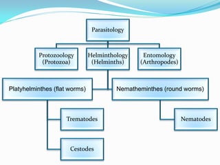
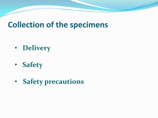
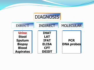
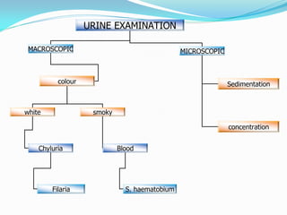
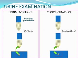
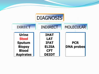
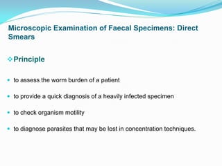
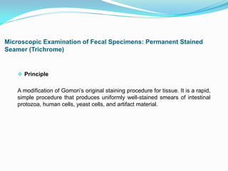
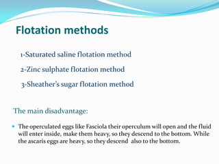
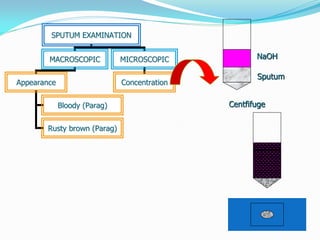
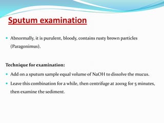
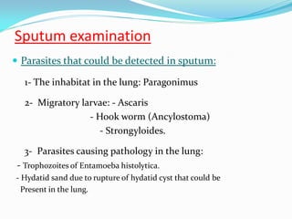
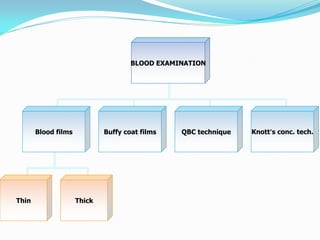
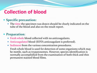
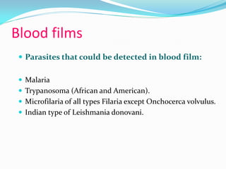
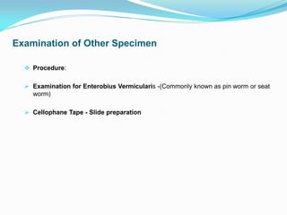
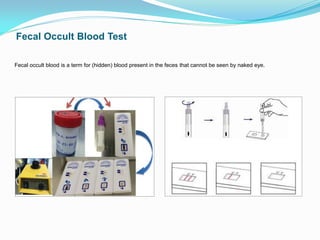
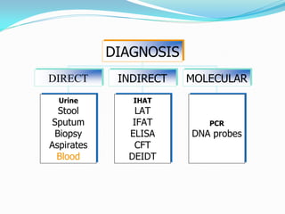
Parasitology
Protozoology
(Protozoa)
Helminthology
(Helminths)
Platyhelminthes (flat worms)
Trematodes
Cestodes
Nematheminthes (round worms)
Nematodes
Entomology
(Arthropodes)
Collection of the specimens
• Delivery
• Safety
• Safety precautions
DIAGNOSIS
DIRECT INDIRECT MOLECULAR
Urine
Stool
Sputum
Biopsy
Blood
Aspirates
PCR
DNA probes
IHAT
LAT
IFAT
ELISA
CFT
DEIDT
Urine examination
Terminal drops of urine.
Parasites detected in urine:
Helminths:
Schistosoma haematobium eggs.
Enterobius vermicularis eggs in female patients.
Microfilaria of Wuchereria bancrofti.
Protozoa:
Trichomonas vaginalis trophozoite in female patients.
URINE EXAMINATION
MACROSCOPIC MICROSCOPIC
colour
white smoky
Sedimentation
concentration
Chyluria Blood
Filaria S. haematobium
URINE EXAMINATION
SEDIMENTATION CONCENTRATION
15-20 min Centrifuge (2 min)
Clean conical
glass receptacle
Urine examination
Other techniques:
Membrane Filter technique:
For concentration of Schistosoma eggs
Fill a syringe with urine, pass the urine through a filter.
Remove the filter and place it on the slide, and examine
it microscopically.
Iodine mounts of Faecal specimens
• You will see a lot more than just
parasites!
• 1 drop of mixed specimen + 1 drop of
iodine for contrast
• Kohler illumination will also give you
the right contrast to see the parasites
among the other constituents
• http://dpd.cdc.gov/dpdx/
DIAGNOSIS
DIRECT INDIRECT MOLECULAR
Urine
Stool
Sputum
Biopsy
Blood
Aspirates
PCR
DNA probes
IHAT
LAT
IFAT
ELISA
CFT
DEIDT
 Collection method:
 Number of specimens:
 Collection time:
 Specimen type and stability:
 Liquid specimens
 Soft (semi-formed) specimens
 Formed specimens
 Preservation of stool specimens:
 Aim:
1- To preserve protozoan morphology.
2- To prevent the continued development of some helminthic
eggs and larvae.
 The most common preservative used is 10% formalin.
Microscopic Examination of Faecal Specimens: Direct
Smears
Principle
 to assess the worm burden of a patient
 to provide a quick diagnosis of a heavily infected specimen
 to check organism motility
 to diagnose parasites that may be lost in concentration techniques.
ParaTest (Kit) for Fecal Examination for Intestinal Parasites
 Principle
A new way of doing Natural Sedimentation Technique using 4ml of 5% buffered
formalin in special container covered with pointed end with small cover, the cover line
with a fine filter and the pointed tip is open working as a dropper.
Microscopic Examination of Fecal Specimens: Permanent Stained
Seamer (Trichrome)
 Principle
A modification of Gomori’s original staining procedure for tissue. It is a rapid,
simple procedure that produces uniformly well-stained smears of intestinal
protozoa, human cells, yeast cells, and artifact material.
Flotation methods
1-Saturated saline flotation method
2-Zinc sulphate flotation method
3-Sheather’s sugar flotation method
The main disadvantage:
 The operculated eggs like Fasciola their operculum will open and the fluid
will enter inside, make them heavy, so they descend to the bottom. While
the ascaris eggs are heavy, so they descend also to the bottom.
SPUTUM EXAMINATION
MACROSCOPIC MICROSCOPIC
Appearance Concentration
Bloody (Parag)
Rusty brown (Parag)
NaOH
Sputum
Centfifuge
Sputum examination
 Abnormally, it is purulent, bloody, contains rusty brown particles
(Paragonimus).
Technique for examination:
 Add on a sputum sample equal volume of NaOH to dissolve the mucus.
 Leave this combination for a while, then centrifuge at 200xg for 5 minutes,
then examine the sediment.
Sputum examination
 Parasites that could be detected in sputum:
1- The inhabitat in the lung: Paragonimus
2- Migratory larvae: - Ascaris
- Hook worm (Ancylostoma)
- Strongyloides.
3- Parasites causing pathology in the lung:
- Trophozoites of Entamoeba histolytica.
- Hydatid sand due to rupture of hydatid cyst that could be
Present in the lung.
BLOOD EXAMINATION
Blood films Buffy coat films QBC technique Knott’s conc. tech.
Thin Thick
Collection of blood
 Specific precaution:
 The time the specimen was drawn should be clearly indicated on the
tube of the blood and also on the result report.
 Preparation:
 Fresh whole blood collected with no anticoagulants.
 Anticoagulated blood (EDTA anticoagulant is preferred).
 Sediment from the various concentration procedures.
Fresh whole blood is used for detection of some organisms which may
be motile, such as trypanosomes. However, species identification is
normally accomplished from the examination of both thick and thin
permanent stained blood films.
Blood films
 Two types of blood films can be made, thin and thick blood
films:
 Thick films
 Thin films
 Thin  Thick
Bld drop
spread
Air dry
methyl alcohol
Geimsa
Air dry
Geimsa
Circular motion
Blood films
 Parasites that could be detected in blood film:
 Malaria
 Trypanosoma (African and American).
 Microfilaria of all types Filaria except Onchocerca volvulus.
 Indian type of Leishmania donovani.
Examination of Other Specimen
 Procedure:
 Examination for Enterobius Vermicularis -(Commonly known as pin worm or seat
worm)
 Cellophane Tape - Slide preparation
 Sigmoidscopy Material
 Duodenal Materials
 Wet Preparation
 Aspirate
 Examination of aspirated material has become a valuable diagnostic aid and may reveal parasites
when other methods have failed.
Examination of Other Specimen
 Lung and Liver
 Aspiration from lung and liver could be examined for:
1- Pneumocytosis
2- Amoebiasis:
 Technique: The use of proteolytic enzymes is recommended to free the
organisms from the aspirate material
3- Hydatid Disease
 Lymph nodes, Spleen, Liver, Bone Marrow and Spinal Fliud: Aspirated material
may be examined for presence of trypanosomes, leishmanial forms and amoebae.
 Cutaneous Ulcers
 Leishmaniasis
 NNN Medium: (Novy-Mac Neal-Nicolle)
Examination of Other Specimen
Fecal Occult Blood Test
Fecal occult blood is a term for (hidden) blood present in the feces that cannot be seen by naked eye.
DIAGNOSIS
DIRECT INDIRECT MOLECULAR
Urine
Stool
Sputum
Biopsy
Aspirates
Blood
PCR
DNA probes
IHAT
LAT
IFAT
ELISA
CFT
DEIDT

Parasitology lab diagnostic

  • 2.
  • 3.
    Collection of thespecimens • Delivery • Safety • Safety precautions
  • 4.
  • 5.
    Urine examination Terminal dropsof urine. Parasites detected in urine: Helminths: Schistosoma haematobium eggs. Enterobius vermicularis eggs in female patients. Microfilaria of Wuchereria bancrofti. Protozoa: Trichomonas vaginalis trophozoite in female patients.
  • 6.
    URINE EXAMINATION MACROSCOPIC MICROSCOPIC colour whitesmoky Sedimentation concentration Chyluria Blood Filaria S. haematobium
  • 7.
    URINE EXAMINATION SEDIMENTATION CONCENTRATION 15-20min Centrifuge (2 min) Clean conical glass receptacle
  • 8.
    Urine examination Other techniques: MembraneFilter technique: For concentration of Schistosoma eggs Fill a syringe with urine, pass the urine through a filter. Remove the filter and place it on the slide, and examine it microscopically.
  • 9.
    Iodine mounts ofFaecal specimens • You will see a lot more than just parasites! • 1 drop of mixed specimen + 1 drop of iodine for contrast • Kohler illumination will also give you the right contrast to see the parasites among the other constituents • http://dpd.cdc.gov/dpdx/
  • 10.
  • 11.
     Collection method: Number of specimens:  Collection time:
  • 12.
     Specimen typeand stability:  Liquid specimens  Soft (semi-formed) specimens  Formed specimens
  • 13.
     Preservation ofstool specimens:  Aim: 1- To preserve protozoan morphology. 2- To prevent the continued development of some helminthic eggs and larvae.  The most common preservative used is 10% formalin.
  • 14.
    Microscopic Examination ofFaecal Specimens: Direct Smears Principle  to assess the worm burden of a patient  to provide a quick diagnosis of a heavily infected specimen  to check organism motility  to diagnose parasites that may be lost in concentration techniques.
  • 15.
    ParaTest (Kit) forFecal Examination for Intestinal Parasites  Principle A new way of doing Natural Sedimentation Technique using 4ml of 5% buffered formalin in special container covered with pointed end with small cover, the cover line with a fine filter and the pointed tip is open working as a dropper.
  • 16.
    Microscopic Examination ofFecal Specimens: Permanent Stained Seamer (Trichrome)  Principle A modification of Gomori’s original staining procedure for tissue. It is a rapid, simple procedure that produces uniformly well-stained smears of intestinal protozoa, human cells, yeast cells, and artifact material.
  • 17.
    Flotation methods 1-Saturated salineflotation method 2-Zinc sulphate flotation method 3-Sheather’s sugar flotation method The main disadvantage:  The operculated eggs like Fasciola their operculum will open and the fluid will enter inside, make them heavy, so they descend to the bottom. While the ascaris eggs are heavy, so they descend also to the bottom.
  • 18.
    SPUTUM EXAMINATION MACROSCOPIC MICROSCOPIC AppearanceConcentration Bloody (Parag) Rusty brown (Parag) NaOH Sputum Centfifuge
  • 19.
    Sputum examination  Abnormally,it is purulent, bloody, contains rusty brown particles (Paragonimus). Technique for examination:  Add on a sputum sample equal volume of NaOH to dissolve the mucus.  Leave this combination for a while, then centrifuge at 200xg for 5 minutes, then examine the sediment.
  • 20.
    Sputum examination  Parasitesthat could be detected in sputum: 1- The inhabitat in the lung: Paragonimus 2- Migratory larvae: - Ascaris - Hook worm (Ancylostoma) - Strongyloides. 3- Parasites causing pathology in the lung: - Trophozoites of Entamoeba histolytica. - Hydatid sand due to rupture of hydatid cyst that could be Present in the lung.
  • 21.
    BLOOD EXAMINATION Blood filmsBuffy coat films QBC technique Knott’s conc. tech. Thin Thick
  • 22.
    Collection of blood Specific precaution:  The time the specimen was drawn should be clearly indicated on the tube of the blood and also on the result report.  Preparation:  Fresh whole blood collected with no anticoagulants.  Anticoagulated blood (EDTA anticoagulant is preferred).  Sediment from the various concentration procedures. Fresh whole blood is used for detection of some organisms which may be motile, such as trypanosomes. However, species identification is normally accomplished from the examination of both thick and thin permanent stained blood films.
  • 23.
    Blood films  Twotypes of blood films can be made, thin and thick blood films:  Thick films  Thin films
  • 24.
     Thin Thick Bld drop spread Air dry methyl alcohol Geimsa Air dry Geimsa Circular motion
  • 25.
    Blood films  Parasitesthat could be detected in blood film:  Malaria  Trypanosoma (African and American).  Microfilaria of all types Filaria except Onchocerca volvulus.  Indian type of Leishmania donovani.
  • 26.
    Examination of OtherSpecimen  Procedure:  Examination for Enterobius Vermicularis -(Commonly known as pin worm or seat worm)  Cellophane Tape - Slide preparation
  • 27.
     Sigmoidscopy Material Duodenal Materials  Wet Preparation  Aspirate  Examination of aspirated material has become a valuable diagnostic aid and may reveal parasites when other methods have failed. Examination of Other Specimen
  • 28.
     Lung andLiver  Aspiration from lung and liver could be examined for: 1- Pneumocytosis 2- Amoebiasis:  Technique: The use of proteolytic enzymes is recommended to free the organisms from the aspirate material 3- Hydatid Disease
  • 29.
     Lymph nodes,Spleen, Liver, Bone Marrow and Spinal Fliud: Aspirated material may be examined for presence of trypanosomes, leishmanial forms and amoebae.  Cutaneous Ulcers  Leishmaniasis  NNN Medium: (Novy-Mac Neal-Nicolle) Examination of Other Specimen
  • 30.
    Fecal Occult BloodTest Fecal occult blood is a term for (hidden) blood present in the feces that cannot be seen by naked eye.
  • 31.