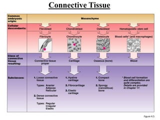
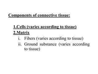
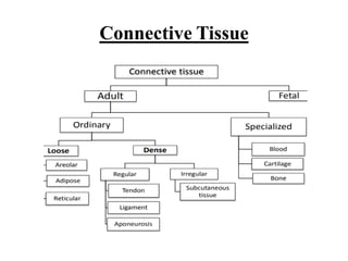
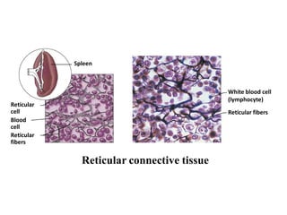
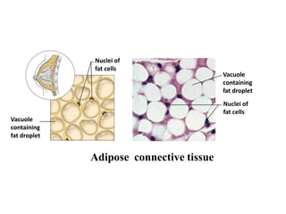
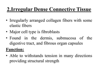
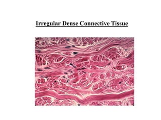
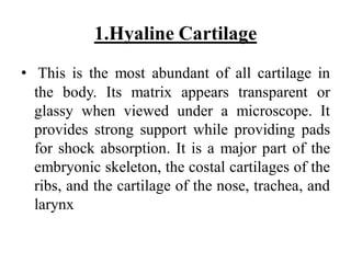
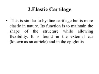
University of Duhok
College of Health Sciences
Dep. of Medical Laboratories & Anesthesia
Biology/1st Stage
Lecture 8
Dr.: Shameeran S. Ismael
BVM & S, M.Sc Medical Microbiology(Parasitology),
PhD Molecular Parasitology
Types of Tissue Continued:
II. Connective Tissue (support)
Connective Tissue
• Found throughout the body; most abundant
tissue and widely distributed in primary
tissues
• Common embryonic origin – mesenchyme
Main classes
 Connective tissue proper
 Cartilage
 Bone tissue
 Blood
Connective Tissue
Function:
• Binds structures together
• Provides support & protection
• Fills spaces
• Produces blood cells
• Stores fat (For energy, insulation, organ
protection)
• Transportation
Connective Tissue
Figure 4.5
Components of connective tissue:
1.Cells (varies according to tissue)
2.Matrix
i. Fibers (varies according to tissue)
ii. Ground substance (varies according
to tissue)
Components of Connective Tissue:
Cells
 Each Type of connective tissue has to be made
by a certain cell type. In an immature stage each
cell below secrets the fibers needed for its
connective tissue.
 Fibroblasts – connective tissue proper
 Chondroblasts – cartilage
 Osteoblasts – bone
 Hematopoietic stem cells – blood
 White blood cells, plasma cells, macrophages,
and mast cells
Fibers of the Matrix
1. White fibers or Collagenfibers: contain
collagen which gives the fibers flexibility and
strength
2. Yellow fibers or Elastic Fibers: contain
elastin more elastic than collagen but not as
strong
3. Reticular fibers: thin, highly branched
collagenous fibers that provide support
Embryonic connective tissue
Embryonic connective tissue(Mesenchyme):
 Gel-like ground substance with fibers and star-
shaped mesenchymal cells
 Gives rise to all other connective tissues
 Found in the embryo
A/Connective tissue proper
I. Loose Connective Tissue
 Areolar
 Reticular
 Adipose
II.Dense Connective Tissue
 Regular
 Irregular
 Elastic
Connective Tissue
I. Loose Connective Tissue
1.Areolar Connective Tissue
 Gel-like matrix with all three connective tissue
fibers types (collagen, reticular, elastic) for support
 Ground substance is made up by glycoproteins
also made and secreted by the fibroblasts.
 Widely distributed throughout the body
Function
 Wraps and cushions organs
 Holds and conveys tissue fluid
 Important role in inflammation Main battlefield in
fight against infection
Mucosa
epithelium
Lamina
propria
Fibers of
matrix
Nuclei of
fibroblasts
Elastic
fibers
Collagen
fibers
Fibroblast
nuclei
Areolar connective tissue
2.Reticular connective tissue
 Loose ground substance with reticular fibers
 Reticular cells lie in a fiber network
 Found in lymph nodes, bone marrow, and
the spleen
Function:
 Forms a soft internal skeleton, or stroma,
that supports other cell types
Spleen
Reticular connective tissue
Reticular
cell
Blood
cell
Reticular
fibers
White blood cell
(lymphocyte)
Reticular fibers
 Matrix similar to areolar connective tissue with
closely packed adipocytes
 Found under skin, around kidneys, within
abdomen, and in breasts
Function:
 Reserves food stores, insulates against heat loss,
and supports and protects
 Local fat deposits serve nutrient needs of highly
active organs
3. Adipose connective tissue
Nuclei of
fat cells
Vacuole
containing
fat droplet
Vacuole
containing
fat droplet
Nuclei of
fat cells
Adipose connective tissue
II.Dense Connective Tissue
1.Regular Dense Connective Tissue
• Parallel collagen fibers with a few elastic
fibers
• Major cell type is fibroblasts
• Found in tendons, ligaments, and aponeuroses
Function:
• Attaches muscles to bone or to other muscles,
and bone to bone
Regular Dense Connective Tissue
2.Irregular Dense Connective Tissue
• Irregularly arranged collagen fibers with some
elastic fibers
• Major cell type is fibroblasts
• Found in the dermis, submucosa of the
digestive tract, and fibrous organ capsules
Function:
• Able to withstands tension in many directions
providing structural strength
Irregular Dense Connective Tissue
3. Elastic Dense Connective Tissue
 Elastic fibers consist of an amorphous protein
called elastin and numerous protein
microfibrils embedded in it.
 Elastic fibers are collected in thick, wavy,
parallel bundles & separated by loose
collagenous tissue with fibroblasts.
 Ground substance is sparse.
.
 Predominates in the ligamentum flava of the
vertebral column & the suspensory ligament of
the penis
Function:
Elastic connective tissue provides flexible
support.
Elastic Dense Connective Tissue (blood vessel wall)
Internal
elastic
lamina
B/ Cartilage
• This is a flexible connective tissue found in many
areas in the bodies of humans and other animals,
including the joints between bones, the rib cage,
the ear, the nose, the elbow, the knee, the ankle,
the bronchial tubes, and the intervertebral discs
Components of cartilage
1. Cells- chondrocytes : Cartilage is composed
of specialized cells called chondroblasts
2. Ground substance/ matrix
3. Fibres- collagen fibres
• Unlike other connective tissues, cartilage
does not contain blood vessels
• Cartilage is composed of specialized cells
called chondroblasts and,
Function of Cartalage:
• Due to extracellular matrix (ECM) , it allows
the tissue to bear mechanical stress
• In respiratory tracts, ears and nose, cartilage
forms framework supporting soft tissues
• Cartilage provides shock absorbing and sliding
regions with in joints and facilitate bone
movements due to smooth lubricated surface
and resiliency
Types of Cartilage:
• There are three types:
1. Hyaline cartilage
2. Elastic cartilage
3. Fibrocartilage, which differ in the relative
amounts of these three main components.
• Cartilage is strong due to the collagen fibers
within its matrix, and it is resilient due to a gel
matrix. Cartilage is also found in the body as a
cushion within the skeletal system.
1.Hyaline Cartilage
• This is the most abundant of all cartilage in
the body. Its matrix appears transparent or
glassy when viewed under a microscope. It
provides strong support while providing pads
for shock absorption. It is a major part of the
embryonic skeleton, the costal cartilages of the
ribs, and the cartilage of the nose, trachea, and
larynx
2.Elastic Cartilage
• This is similar to hyaline cartilage but is more
elastic in nature. Its function is to maintain the
shape of the structure while allowing
flexibility. It is found in the external ear
(known as an auricle) and in the epiglottis
3.Fibrocartilage
• This is a blend of hyaline cartilage and dense
regular connective tissue. Because it is
compressible and resists tension well,
Fibrocartilage is found where strong support
and the ability to withstand heavy pressure are
required. It is found in the intervertebral discs
of the bony vertebrae and knee meniscus.
Thanks for attention,,,

Connective tissue

  • 1.
    University of Duhok Collegeof Health Sciences Dep. of Medical Laboratories & Anesthesia Biology/1st Stage Lecture 8 Dr.: Shameeran S. Ismael BVM & S, M.Sc Medical Microbiology(Parasitology), PhD Molecular Parasitology
  • 2.
    Types of TissueContinued: II. Connective Tissue (support)
  • 3.
    Connective Tissue • Foundthroughout the body; most abundant tissue and widely distributed in primary tissues • Common embryonic origin – mesenchyme Main classes  Connective tissue proper  Cartilage  Bone tissue  Blood
  • 4.
    Connective Tissue Function: • Bindsstructures together • Provides support & protection • Fills spaces • Produces blood cells • Stores fat (For energy, insulation, organ protection) • Transportation
  • 5.
  • 6.
    Components of connectivetissue: 1.Cells (varies according to tissue) 2.Matrix i. Fibers (varies according to tissue) ii. Ground substance (varies according to tissue)
  • 7.
  • 8.
    Cells  Each Typeof connective tissue has to be made by a certain cell type. In an immature stage each cell below secrets the fibers needed for its connective tissue.  Fibroblasts – connective tissue proper  Chondroblasts – cartilage  Osteoblasts – bone  Hematopoietic stem cells – blood  White blood cells, plasma cells, macrophages, and mast cells
  • 9.
    Fibers of theMatrix 1. White fibers or Collagenfibers: contain collagen which gives the fibers flexibility and strength 2. Yellow fibers or Elastic Fibers: contain elastin more elastic than collagen but not as strong 3. Reticular fibers: thin, highly branched collagenous fibers that provide support
  • 10.
    Embryonic connective tissue Embryonicconnective tissue(Mesenchyme):  Gel-like ground substance with fibers and star- shaped mesenchymal cells  Gives rise to all other connective tissues  Found in the embryo
  • 11.
    A/Connective tissue proper I.Loose Connective Tissue  Areolar  Reticular  Adipose II.Dense Connective Tissue  Regular  Irregular  Elastic
  • 12.
  • 13.
  • 14.
    1.Areolar Connective Tissue Gel-like matrix with all three connective tissue fibers types (collagen, reticular, elastic) for support  Ground substance is made up by glycoproteins also made and secreted by the fibroblasts.  Widely distributed throughout the body Function  Wraps and cushions organs  Holds and conveys tissue fluid  Important role in inflammation Main battlefield in fight against infection
  • 15.
  • 16.
    2.Reticular connective tissue Loose ground substance with reticular fibers  Reticular cells lie in a fiber network  Found in lymph nodes, bone marrow, and the spleen Function:  Forms a soft internal skeleton, or stroma, that supports other cell types
  • 17.
  • 18.
     Matrix similarto areolar connective tissue with closely packed adipocytes  Found under skin, around kidneys, within abdomen, and in breasts Function:  Reserves food stores, insulates against heat loss, and supports and protects  Local fat deposits serve nutrient needs of highly active organs 3. Adipose connective tissue
  • 19.
    Nuclei of fat cells Vacuole containing fatdroplet Vacuole containing fat droplet Nuclei of fat cells Adipose connective tissue
  • 20.
  • 21.
    1.Regular Dense ConnectiveTissue • Parallel collagen fibers with a few elastic fibers • Major cell type is fibroblasts • Found in tendons, ligaments, and aponeuroses Function: • Attaches muscles to bone or to other muscles, and bone to bone
  • 22.
  • 23.
    2.Irregular Dense ConnectiveTissue • Irregularly arranged collagen fibers with some elastic fibers • Major cell type is fibroblasts • Found in the dermis, submucosa of the digestive tract, and fibrous organ capsules Function: • Able to withstands tension in many directions providing structural strength
  • 24.
  • 25.
    3. Elastic DenseConnective Tissue  Elastic fibers consist of an amorphous protein called elastin and numerous protein microfibrils embedded in it.  Elastic fibers are collected in thick, wavy, parallel bundles & separated by loose collagenous tissue with fibroblasts.  Ground substance is sparse. .
  • 26.
     Predominates inthe ligamentum flava of the vertebral column & the suspensory ligament of the penis Function: Elastic connective tissue provides flexible support.
  • 27.
    Elastic Dense ConnectiveTissue (blood vessel wall) Internal elastic lamina
  • 28.
    B/ Cartilage • Thisis a flexible connective tissue found in many areas in the bodies of humans and other animals, including the joints between bones, the rib cage, the ear, the nose, the elbow, the knee, the ankle, the bronchial tubes, and the intervertebral discs
  • 29.
    Components of cartilage 1.Cells- chondrocytes : Cartilage is composed of specialized cells called chondroblasts 2. Ground substance/ matrix 3. Fibres- collagen fibres • Unlike other connective tissues, cartilage does not contain blood vessels
  • 30.
    • Cartilage iscomposed of specialized cells called chondroblasts and,
  • 31.
    Function of Cartalage: •Due to extracellular matrix (ECM) , it allows the tissue to bear mechanical stress • In respiratory tracts, ears and nose, cartilage forms framework supporting soft tissues • Cartilage provides shock absorbing and sliding regions with in joints and facilitate bone movements due to smooth lubricated surface and resiliency
  • 32.
    Types of Cartilage: •There are three types: 1. Hyaline cartilage 2. Elastic cartilage 3. Fibrocartilage, which differ in the relative amounts of these three main components.
  • 33.
    • Cartilage isstrong due to the collagen fibers within its matrix, and it is resilient due to a gel matrix. Cartilage is also found in the body as a cushion within the skeletal system.
  • 34.
    1.Hyaline Cartilage • Thisis the most abundant of all cartilage in the body. Its matrix appears transparent or glassy when viewed under a microscope. It provides strong support while providing pads for shock absorption. It is a major part of the embryonic skeleton, the costal cartilages of the ribs, and the cartilage of the nose, trachea, and larynx
  • 35.
    2.Elastic Cartilage • Thisis similar to hyaline cartilage but is more elastic in nature. Its function is to maintain the shape of the structure while allowing flexibility. It is found in the external ear (known as an auricle) and in the epiglottis
  • 36.
    3.Fibrocartilage • This isa blend of hyaline cartilage and dense regular connective tissue. Because it is compressible and resists tension well, Fibrocartilage is found where strong support and the ability to withstand heavy pressure are required. It is found in the intervertebral discs of the bony vertebrae and knee meniscus.
  • 37.