Laboratory diagnosis
of Parasitology
Dr.BALAJI.S
2nd
year PG
Department of Microbiology
SVMC, Tirupati.
Direct methods Immunological methods Molecular methods
Stool ICT PCR
Urine IHAT DNA probes
Blood LAT
Sputum IFAT
CSF ELISA
Tissue biopsy and CFT
Aspirates
Diagnostic methods
Parasites and their related disease
Entamoeba histolytica: Amoebic
dysentery
Giardia lamblia: Diarrhoea
Leishmania donovani: Kala-azar
Plasmodium: Malaria
Trypanosoma cruzi: Chagas disease
Toxoplasma gondii: Encephalomyelitis
Taenia solium: Cysticercosis
Echinococcus granulosus: Hydatid
disease
Ascaris lumbricoides: Diarrhoea
Ancyclostoma: Diarrhoea
Strongyloides: Diarrhoea
Trichuris trichiura: Diarrhoea
Enterobius vermicularis: Diarrhoea
Wuchereria bancrofti: Filaria
Trichomonas vaginalis: Vaginal infection
Examination of Stool
Collection of Fresh Stool Specimen
All stool specimens should be
collected in a suitable, clean, wide
mouthed container like a plastic
container
At least three stool specimens
collected on alternate days
The specimen should not be
contaminated with water, urine, or
disinfectants.
Liquid stools should be examined
or preserved within 30 minutes of
passage(trophozoites)
Soft stools should be examined or
preserved within 1 hour of passage
(trophozoites and cysts)
Formed stool should be examined
or preserved within 24 hours of
passage(cysts)
Preservations of specimens
5 or 10% formalin – preserves protozoal cysts, helminthic eggs and larvae
Merthiolate Iodine Formalin-used both as stain and preservative
Sodium acetate- Acetic acid formalin – fixative which gives better result
when stained with iron haematoxylin
Polyvinyl alcohol – preservative for protozoal cysts and trophozoites in
stool.
Macroscopic examination of stool
consistency Liquid stool – trophozoite (motile)
Semisolid stool – cysts and trophozoites
Formed stool – cyst forms
Helminthic eggs and larvae are found in any
type of stool.
Presence of
parasite
Adult worm – ascaris lumbricoides,
Ancylostoma duodenale, Trichuris trichiura,
Enterobius vermicularis, intestinal flukes.
Tapeworm segments.
Blood and mucus Suggestive of amoebic dysentery
Microscopic examination
The saline wet mount is the standard preparation
and is made by emulsifying a small quantity of
stool in a drop of (0.85%) saline placed on a slide
and applying a coverslip (22 mm × 22 mm) on top,
avoiding air bubbles.
The entire field under coverslip should be
systematically examined with low power objective
(10X) under low light intensity.
Any suspicious object may then be examined with
the high power objective.
Wet
mount
Use Advantage Disadvantage
Saline wet
mount
Preliminary
identification
of cysts
Demonstrate
helminthic
eggs, motile
trophozoites,
larvae
Can demonstrate
viable, motile
trophozoite
Chromatoid body
seen in cysts
Saline stained
cysts does not
show nuclei,
cytoplasm and
glycogen
material
Lugol’s
Iodine
Further
identification
of protozoal
cysts
Iodine stained
cysts show
brown dot nuclei,
yellowish
cytoplasm and
brown glycogen
material
Trophozoite
killed with
iodine so
motile
trophozoite not
seen
Parasites and their developmental stages found in stool
Cysts/Trophozoites Eggs Larvae Adult worm
Entamoeba histolytica
Giardia lamblia
Balantidium coli
Sarcocystis spp.
Cyclospora
cayetanensis
Cryptosporidium
parvum
Cestodes
Taenia spp.
Hymenolepsis nana
Hymenolepsis diminuta
Diphyllobothrium latum
Nematodes
Trichuris trichiura
Enterobius
vermicularis
Ascaris lumbricoides
Ancylostoma
duodenale
Necator americanus
Strongyloides
stercoralis
Taenia solium
Taenia saginata
Diphyllobothrium
latum
Ascaris
lumbricoides
Enterobius
vermicularis
Trichinella spiralis
Trematodes
Schistosoma spp.
Fasciolopsis buski
Fasciola hepatica
Clonorchis sinensis
Opisthorchis spp.
Bile stained eggs
Ascaris lumbricoides(fertilized egg)
Trichuris trichiura
Taenia species
Non bile stained eggs
Enterobius vermicularis
Hymenolepsis nana
Hymenolepsis diminuta
Diphyllobothrium latum
Permanent Stained Smears
The two methods - Wheatley’s trichrome stain and the iron-hematoxylin
stain.
Permanent stained smears are examined normally under oil immersion
(100X) objective.
Confirmation of the intestinal protozoan, both trophozoites and cysts, is the
primary purpose of this technique.
 Helminthic eggs and larvae take up too much stain and usually cannot be
identified.
Permanent smear can be prepared with both fresh and polyvinyl alcohol
preserved stool specimen.
Trichrome stain (Wheatley’s method)
The trichrome technique of Wheatley for stool
specimens is a modification of Gomori’s original
staining procedure for tissue.
It is a quicker and simpler method, which produces
uniformly well-stained smears of the intestinal
protozoa, human cells, yeast cells, and artefact
material in about 45 minutes or less.
Helminth eggs and larvae can not be stained.
Microsporidia spores can not be seen using the
Wheatley Trichrome Stained smear.
Trichrome stain Iron Hematoxylin stain
Principle Uses chromotrope dyes to give contrast
between cytoplasm, nuclei and chromatoid
bodies
Uses as a mordent with
hematoxylin to give sharp
nuclear detail
Staining time Shorter, easier,quicker Longer, more labor - intensive
Nucleus Moderate contrast Excellent contrast
Cytoplasm Good differentiation of cytoplasm and
chromatoid bodies
Cytoplasm appears darker less
contrast than nuclei
Appearance Cytoplasm : blue/green
Nuclei : red/purple
chromatoid bodies: red
Cytoplasm : Grey to black
Nuclei : Dark black /blue
Use Common for routine diagnosis of intestinal
protozoa
Used when precise nuclear
morphology is
required(research)
Hot method (Modified ZN
stain)
Cold method
(Kinyon’s stain)
Organisms
detected
Cryptosporidium, Isospora,
Cyclospora
Cryptosporidium,
Isospora, Cyclospora
Heating Recquired (carbol fuchsin
applied with heat)
Not recquired
Decolorizer 5% sulphuric acid(mild) 1% sulphuric acid
Appearance of
oocysts
Pink – red against blue
background
Pink – red against
blue background
Advantage Better penetration and
contrast
Simple, safe and
avoids heating
Concentration Methods
When the parasites are scanty in stools, routine microscopic examination
may fail to detect them.
It is then necessary to selectively concentrate the protozoan cysts and
helminth eggs and larvae.
Concentration may be done using fresh or preserved feces.
Technique Principle Types Advantage Disadvantage
Floatation method The suspending
fluids used are
denser(has high
specific gravity)
than the parasitic
forms, this helps in
separation of
helminthic eggs
and protozoal
cysts from faecal
debris, parasite
rise to surface.
Saturated salt
solution method
Zinc sulphate
floatation method
Saturated
magnesium
sulphate solution
method
Sugar floatation
method
Both surface film
and sediments can
be examined for
parasite
Some helminthic
eggs do not float
in salt solution
High specific
gravity of the fluid
may cause
distortion in the
morphology of
eggs and cysts.
Helminthic eggs which does not float in salt solution: operculated eggs of trematodes, eggs of Taenia
species and unfertilised eggs of Ascaris lumbricoides, larvae of Strongyloides stercoralis
Technique Principle Types Advantage Disadvantage
Sedimentation
method
Suspending fluids
used are lighter
than the parasitic
forms. So the
parasite sink to the
bottom so it can
be recovered from
the sediment.
Sedimentation by
gravity or by
centrifugation.
Formalin ether
sedimentation
method
Simple gravity
sedimentation
method
Formalin –Acetone
sedimentation
method
Easy to perform
More sensitive
Morphology of
eggs and cysts are
preserved
Formalin fixes the
eggs, larvae and
cysts so that they
become non-
infectious
Sediment
preparation
contains more
faecal debris
Ether is extremely
flammable and
highly volatile
Quantification of worm burden
There are two methods
Direct smear egg count
Stoll’s method
Direct smear method
Two mg of faeces is mixed in a small drop of saline on a slide and cover
the cover slip.
Examined under low power of the microscope and count the eggs and
calculate the number of eggs/gm of faeces.
Stoll’s technique for counting helminth egg
4 gm of stool and 56 mL of N/10 sodium hydroxide
0.075 mL on slid
Multiply result in 200
Number in 1 gm
NIH swab
Eggs are deposited in peri anal
and perineal skin.
The cellophane part is used for
swabbing by rolling over the
perianal area.
Spread over glass slide and
examined microscopically.
This procedure should be repeated
on three successive days.
Scotch cellulose adhesive tape method
The adhesive surface of the tape is then
pressed against the perineal skin at several
places.
Then placed adhesive – side – down on the
slide for examination.
A drop of toluene or xylol may be placed
between the tape and the slide to clear the
preparation.
Urine Examination
Large volume of urine samples should be
allowed to settle for 1–2 hours.
 About 50 mL of the bottom sediment of the
sample is taken for centrifugation.
The highly concentrated sediment after
centrifugation is examined for direct wet
mount microscopy.
May show eggs of Schistosoma and
Trichomonas vaginalis. Microfilaria may be
detected from chylous urine in lymphatic
filariasis.
Examination of Blood
Blood examination is the routine
diagnostic method in malaria,
filariasis, African trypanosomiasis,
and babesiosis.
Two methods
Wet preparation
Stained blood smear
Parasites found in peripheral blood film
Protozoa Nematodes
Plasmodium spp Wuchereria bancrofti
Babesia spp Brugia malayi
Trypanosoma spp Loa loa
Leishmania spp Mansonella spp
Wet preparation
A drop of anticoagulated blood is placed on
a clean glass and cover slip is put over it.
This preparation is examined
microscopically for parasite such as
Trypanosomes and Microfilariae.
Stained blood smears
Three types of blood smear
Thin blood smear
Thick blood smear
Thin and thick blood smear on
same slide
Features Thin smear Thick smear
Preparation Small drop of blood spread into
a thin film, fixed with methanol
Large drop of blood spread
thick, not fixed (RBC lysed
during staining)
Purpose Species identification Screening of presence of
malaria parasite (more sensitive)
Parasite density detection Detects parasite when
parasitemia is moderate to high
Detects parasites even at low
density
RBC morphology Preserved – useful for
differentiating plasmodium
species
Destroyed (RBCs hemolysed)
so species identification difficult.
Advantage Provides detailed morphology of
parasite and RBC
Increased sensitivity by
examining larger blood volume
Disadvantage Less sensitive at low
parasitemia
Cannot accurately identify
species alone
Staining of blood smears
Thick or thin smears are stained with
Romanowsky’s stain.
These stain include
Giemsa stain
Leishman’s stain
Field’s stain
Jaswant singh and Bhattacharjee stain
Theses stains are combination of
methylene blue and eosin.
Quantitative buffy coat (QBC):
This involves collection of the
blood in a capillary tube coated
internally with acridine orange
stain, centrifugation and then
examination of the buffy coat
region under fluorescence
microscopy.
This is extremely useful for the
detection of the malaria parasites
and microfilariae
Quantitative buffy coat (QBC):
Concentration of blood:
This is useful for the detection of microfilariae from the blood specimen.
Various concentration methods are:
Sedimentation technique
Gradient centrifugation
Membrane filtration
Knott concentration
Types of buffy coat concentration methods
Method procedure
Gradient centrifugation Venous blood is collected in heparinised tube
4ml of Ficoll – hypaque solution is taken in a 15ml centrifuge tube and
mixed with equal volume of blood
Centrifuged
Middle Ficoll – hypaque layer is examined for the Microfilariae.
Membrane centrifugation 5ml heparinised blood is collected in syringe.
Blood is filtered through the membranes.
Microfilaria sticking to the surface is examined.
Knott centrifugation 10ml of 2% formalin is collected in a 15ml centrifuge tube.
1ml of venous blood is collected aseptically and is add to the tube and
mixed thoroughly
Blood and formalin suspension is centrifuged at 200g for 2 mins
Sediment is collected and then examined for microfilariae
Diethylcarbamazine Provocation Test
Oral administration of diethylcarbamazine (DEC; 100 mg or 2 mg/kg of
body weight) brings about mobilization of microfilariae into peripheral
blood.
After 30 mins, the capillary blood is collected by finger prick for
demonstration of microfilariae by direct wet mount or staining.
This is a great advantage for surveys.
But the drug may cause febrile reactions, particularly in brugiasis.
Sputum Examination
Sputum is examined for the
detection of egg of paragonimus
westermani.
Parasites found in sputum
P.Westermani eggs Rarely migrating larvae of ascaris
E.Histolytica (trophozoites in case of pulmonary
abscess)
Rarely migrating larvae of strongyloides
Pneumocystis jirovecii Rarely migrating larvae of Ancylostoma duodenale
Rarely migrating larvae of necator americanus
CSF examination
Direct microscopic examination of
unstained and stained smears of
CSF is useful for detection of
Naegleria fowleri
Acanthamoeba species
Balamuthia mandrillaris
Trypanosoma brucei
Examination of aspirates
The aspirate from liver is useful in
diagnosis of amoebic liver abscess
and hydatid cyst.
Duodenal aspirates may reveal
trophozoites of Giardia lamblia.
Duodenal Capsule Technique (Entero test)
Entero test is a simple method of sampling duodenal contents.
 The device is composed of a length of nylon yarn coiled inside a gelatine
capsule.
 The end of the yarn is affixed to the patient’s face.
The capsule is then swallowed and the gelatin dissolves in the stomach.
The weighted string is carried into the duodenum by peristalsis.
 Bile-stained mucus is then retrieved after 3–4 hours and duodenal
contents adherent to the yarn is scrapped off and examined under
microscope. Usually 4–5 drops of material is obtained.
 Entero test is used for detecting trophozoites of Giardia, larvae of
Strongyloides, eggs of liver flukes, and oocysts of Isospora
Examination of biopsy
Muscle biopsy
Cysticerci of Taenia solium
Larvae of Trichinella spiralis
Sarcocysts of sarcocystis lindemani
Liver biopsy
Entamoeba histolytica
Leishmania donovani
Echinococcus granulosus
Brain biopsy
Entamoeba histolytica
Naegleria fowleri
Culture Methods
Many parasites can now be grown in
culture,
but this has not become a routine
diagnostic method in parasitic
infections.
It is sometimes employed for accurate
identification of the parasite species.
parasites Culture medium
Entamoeba
histolytica
Boeck and Drbhohlav
diphasic medium
Balantidium coli balamuth’s medium
Giardia lamblia Axenic culture
Trichomonas
vaginalis
Trypticase serum media
Acanthamoeba
spp.
Naegleria
fowleri
Agar plates
Leishmania
spp.
Trypanosoma
spp.
NNN(Novy – MacNeal –
Nicolle) medium
Plasmodium
spp.
RPMI medium(Roswell
Park Memorial Institute
Medium)
Animal inoculation
Animal inoculation is not a routine diagnostic
procedure in parasitic infection.
It is useful in some parasites such as
Toxoplasma gondii(intraperitoneal inoculation
of mice)
Leishmania donovani(intraperitoneal
inoculation of hamsters)
Trypanosoma species(intraperitoneal
inoculation or tail vein inoculation in mice)
Xenodiagnosis
This method involves the diagnostic infection of a vector, in which the parasite
multiplies and can be demonstrated.
In Trypanosoma cruzi diagnosis may be established by letting the vector reduviid
bug feed on suspected patients.
 In 4–5 weeks, live flagellate forms can be seen in the feces of the bugs.
Immunological Diagnosis
Amoebiasis
Invasive amoebiasis, particularly in liver
abscess, serology is very useful.
IHA is most widely employed.
Titers of 1:256 or more are significant in
cases of amoebic liver abscess and have
prognostic value.
 E. histolytica test was able to detect
galactose lectin (galnac) antigen in almost
all patients of amoebic liver abscess
Antigen detection in parasitic disease
Galactose lectin
antigen
Entamoeba histolytica
Giardia- specific
antigen 65
Giardia lamblia
WKK and rk39 antigen Leishmania donovani
HRP- 2 antigen Plasmodium falciparum
Vivax specific LDH Plasmodium vivax
200 kDa Ag and OG43A
antigen
Wuchereria bancrofti
Giardiasis
ELISA and indirect immunofluorescence test (IIF) have been developed for
detection of Giardia.
Commercially available ELISA (ProSpec T/Giardia) Kit detects Giardia-
specific antigen 65 (GAS 65).
The sensitivity of the test is 95% and specificity is 100%, when compared
to conventional microscopy.
Trypanosomiasis
Serological tests used to detect trypanosomiasis are IHA, indirect
fluorescent antibody (IFA), and ELISA.
Specific antibodies can be demonstrated by IFA and ELISA in CSF sample.
Specific antibodies can be identified in the serum by these tests about 2–3
weeks after infection.
Leishmaniasis
 IHA, Counter Immuno Electrophoresis (CIEP), and DOT-ELISA are
usually positive in Kala-azar.
 Complement test using Witebsky Klingenstien, and Kuhn (WKK) antigen
from the acid-fast Kedrowsky bacillus are relatively less sensitive.
IFA test is positive very early in the disease, even before the appearance
of symptoms and becomes negative within 6 months of cure.
rK 39 micro ELISA test is a qualitative immuno chromatographic assay for
detection of antibodies to Leishmania.
Malaria
IIF, ELISA, and IHA are sensitive and specific,
but are not useful for diagnosis of acute malaria
because antibodies persist for some years after
cure.
A negative test may, however help to exclude
malaria.
 Molecular assays such as antigen capture for
detection of HRP-2 and pLDH have been
applied for developing rapid dip-stick tests.
Toxoplasmosis
Serological tests offer the most useful diagnostic method in toxoplasmosis.
 The original Sabin-Feldman dye test, though very specific and sensitive,
is no longer in use.
 IIF, IHA, and CFT were other useful tests.
The dye test remains positive for life, while CFT becomes negative soon
after active infection.
At present, ELISA is routinely used in Toxoplasma serology. It is very
informative, as it provides titers of IgM and IgG antibodies separately for
better interpretation of the results.
Cryptosporidiosis
• IFA and ELISA using purified oocysts as antigens have been used to
detect circulating antibodies specific to Cryptosporidium parvum .
• but extensive cross-reactions limit their use in diagnosis.
Trichinosis
Bentonite flocculation slide tests and CFT become positive 3–4 weeks
after infection.
 IIF becomes positive even earlier.
 ELISA is also available.
Demonstration of seroconversion is diagnostic.
Filariasis
IHA and bentonite flocculation tests with antigen from Dirofilaria immitis
gives positive reaction in patients, and high titers in tropical pulmonary
eosinophilia.
But cross reactions are frequent.
Immunochromatographic filariasis card test (ICT) is a new and rapid filarial
antigen test that detects soluble Wuchereria bancrofti antigens in the
serum of infected humans.
Echinococcosis
 Several serological tests have been developed using hydatid fluid or
scolex antigens from hydatid cysts in sheep.
IHA, IIF, CIEP, and ELISA are very sensitive.
Cross-reactions occur with cysticercosis.
Skin test
Skin test Disease
Casoni’s test Hydatid disease
Montenegro test or leishmanin test Kala azar
Frenkel’s test Toxoplasmosis
Fairley’s test Schistosomiasis
Bachman intradermal test Trichinellosis
Molecular Methods
DNA probe is a highly sensitive method for the diagnosis of malaria.
It can detect even less than 10 parasite/µL of blood.
Drug resistances in malaria are detected now by PCR techniques.
 B1 gene of Toxoplasma gondii can be detected by PCR of the amniotic
fluid in case of congenital toxoplasmosis.
PCR have been developed for detection of filarial DNA from patients
blood.
Specific 17 kDa and 27 KDa sporozoite antigens are employed for
epidemiological studies in cryptosporidiosis using western blot technique.
Imaging technique
X – ray – May show calcified cysts
(hydatid cyst).
USG – detects cystic lesions
(hydatid cyst in liver).
CT scan / MRI –May show
Granulomatous Amoebic
Encephalitis).
Recent updates
Kerala has reported 80 cases and
21 deaths in the Primary Amoebic
Meningoencephalitis (PAM)
A rare but highly fatal brain
infection caused by Naegleria
fowleri.
Thank you

Laboratory diagnosis of parasitology-1.pptx

  • 1.
    Laboratory diagnosis of Parasitology Dr.BALAJI.S 2nd yearPG Department of Microbiology SVMC, Tirupati.
  • 2.
    Direct methods Immunologicalmethods Molecular methods Stool ICT PCR Urine IHAT DNA probes Blood LAT Sputum IFAT CSF ELISA Tissue biopsy and CFT Aspirates Diagnostic methods
  • 3.
    Parasites and theirrelated disease Entamoeba histolytica: Amoebic dysentery Giardia lamblia: Diarrhoea Leishmania donovani: Kala-azar Plasmodium: Malaria Trypanosoma cruzi: Chagas disease Toxoplasma gondii: Encephalomyelitis Taenia solium: Cysticercosis Echinococcus granulosus: Hydatid disease Ascaris lumbricoides: Diarrhoea Ancyclostoma: Diarrhoea Strongyloides: Diarrhoea Trichuris trichiura: Diarrhoea Enterobius vermicularis: Diarrhoea Wuchereria bancrofti: Filaria Trichomonas vaginalis: Vaginal infection
  • 4.
    Examination of Stool Collectionof Fresh Stool Specimen All stool specimens should be collected in a suitable, clean, wide mouthed container like a plastic container At least three stool specimens collected on alternate days The specimen should not be contaminated with water, urine, or disinfectants.
  • 5.
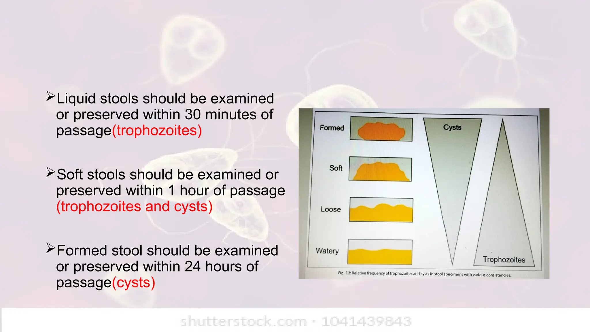
    Liquid stools shouldbe examined or preserved within 30 minutes of passage(trophozoites) Soft stools should be examined or preserved within 1 hour of passage (trophozoites and cysts) Formed stool should be examined or preserved within 24 hours of passage(cysts)
  • 6.
    Preservations of specimens 5or 10% formalin – preserves protozoal cysts, helminthic eggs and larvae Merthiolate Iodine Formalin-used both as stain and preservative Sodium acetate- Acetic acid formalin – fixative which gives better result when stained with iron haematoxylin Polyvinyl alcohol – preservative for protozoal cysts and trophozoites in stool.
  • 7.
    Macroscopic examination ofstool consistency Liquid stool – trophozoite (motile) Semisolid stool – cysts and trophozoites Formed stool – cyst forms Helminthic eggs and larvae are found in any type of stool. Presence of parasite Adult worm – ascaris lumbricoides, Ancylostoma duodenale, Trichuris trichiura, Enterobius vermicularis, intestinal flukes. Tapeworm segments. Blood and mucus Suggestive of amoebic dysentery
  • 8.
    Microscopic examination The salinewet mount is the standard preparation and is made by emulsifying a small quantity of stool in a drop of (0.85%) saline placed on a slide and applying a coverslip (22 mm × 22 mm) on top, avoiding air bubbles. The entire field under coverslip should be systematically examined with low power objective (10X) under low light intensity. Any suspicious object may then be examined with the high power objective.
  • 9.
    Wet mount Use Advantage Disadvantage Salinewet mount Preliminary identification of cysts Demonstrate helminthic eggs, motile trophozoites, larvae Can demonstrate viable, motile trophozoite Chromatoid body seen in cysts Saline stained cysts does not show nuclei, cytoplasm and glycogen material Lugol’s Iodine Further identification of protozoal cysts Iodine stained cysts show brown dot nuclei, yellowish cytoplasm and brown glycogen material Trophozoite killed with iodine so motile trophozoite not seen
  • 10.
    Parasites and theirdevelopmental stages found in stool Cysts/Trophozoites Eggs Larvae Adult worm Entamoeba histolytica Giardia lamblia Balantidium coli Sarcocystis spp. Cyclospora cayetanensis Cryptosporidium parvum Cestodes Taenia spp. Hymenolepsis nana Hymenolepsis diminuta Diphyllobothrium latum Nematodes Trichuris trichiura Enterobius vermicularis Ascaris lumbricoides Ancylostoma duodenale Necator americanus Strongyloides stercoralis Taenia solium Taenia saginata Diphyllobothrium latum Ascaris lumbricoides Enterobius vermicularis Trichinella spiralis Trematodes Schistosoma spp. Fasciolopsis buski Fasciola hepatica Clonorchis sinensis Opisthorchis spp.
  • 12.
    Bile stained eggs Ascarislumbricoides(fertilized egg) Trichuris trichiura Taenia species Non bile stained eggs Enterobius vermicularis Hymenolepsis nana Hymenolepsis diminuta Diphyllobothrium latum
  • 13.
    Permanent Stained Smears Thetwo methods - Wheatley’s trichrome stain and the iron-hematoxylin stain. Permanent stained smears are examined normally under oil immersion (100X) objective. Confirmation of the intestinal protozoan, both trophozoites and cysts, is the primary purpose of this technique.  Helminthic eggs and larvae take up too much stain and usually cannot be identified. Permanent smear can be prepared with both fresh and polyvinyl alcohol preserved stool specimen.
  • 14.
    Trichrome stain (Wheatley’smethod) The trichrome technique of Wheatley for stool specimens is a modification of Gomori’s original staining procedure for tissue. It is a quicker and simpler method, which produces uniformly well-stained smears of the intestinal protozoa, human cells, yeast cells, and artefact material in about 45 minutes or less. Helminth eggs and larvae can not be stained. Microsporidia spores can not be seen using the Wheatley Trichrome Stained smear.
  • 15.
    Trichrome stain IronHematoxylin stain Principle Uses chromotrope dyes to give contrast between cytoplasm, nuclei and chromatoid bodies Uses as a mordent with hematoxylin to give sharp nuclear detail Staining time Shorter, easier,quicker Longer, more labor - intensive Nucleus Moderate contrast Excellent contrast Cytoplasm Good differentiation of cytoplasm and chromatoid bodies Cytoplasm appears darker less contrast than nuclei Appearance Cytoplasm : blue/green Nuclei : red/purple chromatoid bodies: red Cytoplasm : Grey to black Nuclei : Dark black /blue Use Common for routine diagnosis of intestinal protozoa Used when precise nuclear morphology is required(research)
  • 16.
    Hot method (ModifiedZN stain) Cold method (Kinyon’s stain) Organisms detected Cryptosporidium, Isospora, Cyclospora Cryptosporidium, Isospora, Cyclospora Heating Recquired (carbol fuchsin applied with heat) Not recquired Decolorizer 5% sulphuric acid(mild) 1% sulphuric acid Appearance of oocysts Pink – red against blue background Pink – red against blue background Advantage Better penetration and contrast Simple, safe and avoids heating
  • 17.
    Concentration Methods When theparasites are scanty in stools, routine microscopic examination may fail to detect them. It is then necessary to selectively concentrate the protozoan cysts and helminth eggs and larvae. Concentration may be done using fresh or preserved feces.
  • 18.
    Technique Principle TypesAdvantage Disadvantage Floatation method The suspending fluids used are denser(has high specific gravity) than the parasitic forms, this helps in separation of helminthic eggs and protozoal cysts from faecal debris, parasite rise to surface. Saturated salt solution method Zinc sulphate floatation method Saturated magnesium sulphate solution method Sugar floatation method Both surface film and sediments can be examined for parasite Some helminthic eggs do not float in salt solution High specific gravity of the fluid may cause distortion in the morphology of eggs and cysts. Helminthic eggs which does not float in salt solution: operculated eggs of trematodes, eggs of Taenia species and unfertilised eggs of Ascaris lumbricoides, larvae of Strongyloides stercoralis
  • 19.
    Technique Principle TypesAdvantage Disadvantage Sedimentation method Suspending fluids used are lighter than the parasitic forms. So the parasite sink to the bottom so it can be recovered from the sediment. Sedimentation by gravity or by centrifugation. Formalin ether sedimentation method Simple gravity sedimentation method Formalin –Acetone sedimentation method Easy to perform More sensitive Morphology of eggs and cysts are preserved Formalin fixes the eggs, larvae and cysts so that they become non- infectious Sediment preparation contains more faecal debris Ether is extremely flammable and highly volatile
  • 20.
    Quantification of wormburden There are two methods Direct smear egg count Stoll’s method Direct smear method Two mg of faeces is mixed in a small drop of saline on a slide and cover the cover slip. Examined under low power of the microscope and count the eggs and calculate the number of eggs/gm of faeces.
  • 21.
    Stoll’s technique forcounting helminth egg 4 gm of stool and 56 mL of N/10 sodium hydroxide 0.075 mL on slid Multiply result in 200 Number in 1 gm
  • 22.
    NIH swab Eggs aredeposited in peri anal and perineal skin. The cellophane part is used for swabbing by rolling over the perianal area. Spread over glass slide and examined microscopically. This procedure should be repeated on three successive days.
  • 23.
    Scotch cellulose adhesivetape method The adhesive surface of the tape is then pressed against the perineal skin at several places. Then placed adhesive – side – down on the slide for examination. A drop of toluene or xylol may be placed between the tape and the slide to clear the preparation.
  • 24.
    Urine Examination Large volumeof urine samples should be allowed to settle for 1–2 hours.  About 50 mL of the bottom sediment of the sample is taken for centrifugation. The highly concentrated sediment after centrifugation is examined for direct wet mount microscopy. May show eggs of Schistosoma and Trichomonas vaginalis. Microfilaria may be detected from chylous urine in lymphatic filariasis.
  • 25.
    Examination of Blood Bloodexamination is the routine diagnostic method in malaria, filariasis, African trypanosomiasis, and babesiosis. Two methods Wet preparation Stained blood smear Parasites found in peripheral blood film Protozoa Nematodes Plasmodium spp Wuchereria bancrofti Babesia spp Brugia malayi Trypanosoma spp Loa loa Leishmania spp Mansonella spp
  • 26.
    Wet preparation A dropof anticoagulated blood is placed on a clean glass and cover slip is put over it. This preparation is examined microscopically for parasite such as Trypanosomes and Microfilariae.
  • 27.
    Stained blood smears Threetypes of blood smear Thin blood smear Thick blood smear Thin and thick blood smear on same slide
  • 28.
    Features Thin smearThick smear Preparation Small drop of blood spread into a thin film, fixed with methanol Large drop of blood spread thick, not fixed (RBC lysed during staining) Purpose Species identification Screening of presence of malaria parasite (more sensitive) Parasite density detection Detects parasite when parasitemia is moderate to high Detects parasites even at low density RBC morphology Preserved – useful for differentiating plasmodium species Destroyed (RBCs hemolysed) so species identification difficult. Advantage Provides detailed morphology of parasite and RBC Increased sensitivity by examining larger blood volume Disadvantage Less sensitive at low parasitemia Cannot accurately identify species alone
  • 29.
    Staining of bloodsmears Thick or thin smears are stained with Romanowsky’s stain. These stain include Giemsa stain Leishman’s stain Field’s stain Jaswant singh and Bhattacharjee stain Theses stains are combination of methylene blue and eosin.
  • 30.
    Quantitative buffy coat(QBC): This involves collection of the blood in a capillary tube coated internally with acridine orange stain, centrifugation and then examination of the buffy coat region under fluorescence microscopy. This is extremely useful for the detection of the malaria parasites and microfilariae
  • 31.
  • 32.
    Concentration of blood: Thisis useful for the detection of microfilariae from the blood specimen. Various concentration methods are: Sedimentation technique Gradient centrifugation Membrane filtration Knott concentration
  • 33.
    Types of buffycoat concentration methods Method procedure Gradient centrifugation Venous blood is collected in heparinised tube 4ml of Ficoll – hypaque solution is taken in a 15ml centrifuge tube and mixed with equal volume of blood Centrifuged Middle Ficoll – hypaque layer is examined for the Microfilariae. Membrane centrifugation 5ml heparinised blood is collected in syringe. Blood is filtered through the membranes. Microfilaria sticking to the surface is examined. Knott centrifugation 10ml of 2% formalin is collected in a 15ml centrifuge tube. 1ml of venous blood is collected aseptically and is add to the tube and mixed thoroughly Blood and formalin suspension is centrifuged at 200g for 2 mins Sediment is collected and then examined for microfilariae
  • 34.
    Diethylcarbamazine Provocation Test Oraladministration of diethylcarbamazine (DEC; 100 mg or 2 mg/kg of body weight) brings about mobilization of microfilariae into peripheral blood. After 30 mins, the capillary blood is collected by finger prick for demonstration of microfilariae by direct wet mount or staining. This is a great advantage for surveys. But the drug may cause febrile reactions, particularly in brugiasis.
  • 35.
    Sputum Examination Sputum isexamined for the detection of egg of paragonimus westermani. Parasites found in sputum P.Westermani eggs Rarely migrating larvae of ascaris E.Histolytica (trophozoites in case of pulmonary abscess) Rarely migrating larvae of strongyloides Pneumocystis jirovecii Rarely migrating larvae of Ancylostoma duodenale Rarely migrating larvae of necator americanus
  • 36.
    CSF examination Direct microscopicexamination of unstained and stained smears of CSF is useful for detection of Naegleria fowleri Acanthamoeba species Balamuthia mandrillaris Trypanosoma brucei
  • 37.
    Examination of aspirates Theaspirate from liver is useful in diagnosis of amoebic liver abscess and hydatid cyst. Duodenal aspirates may reveal trophozoites of Giardia lamblia.
  • 38.
    Duodenal Capsule Technique(Entero test) Entero test is a simple method of sampling duodenal contents.  The device is composed of a length of nylon yarn coiled inside a gelatine capsule.  The end of the yarn is affixed to the patient’s face. The capsule is then swallowed and the gelatin dissolves in the stomach. The weighted string is carried into the duodenum by peristalsis.  Bile-stained mucus is then retrieved after 3–4 hours and duodenal contents adherent to the yarn is scrapped off and examined under microscope. Usually 4–5 drops of material is obtained.  Entero test is used for detecting trophozoites of Giardia, larvae of Strongyloides, eggs of liver flukes, and oocysts of Isospora
  • 39.
    Examination of biopsy Musclebiopsy Cysticerci of Taenia solium Larvae of Trichinella spiralis Sarcocysts of sarcocystis lindemani Liver biopsy Entamoeba histolytica Leishmania donovani Echinococcus granulosus Brain biopsy Entamoeba histolytica Naegleria fowleri
  • 40.
    Culture Methods Many parasitescan now be grown in culture, but this has not become a routine diagnostic method in parasitic infections. It is sometimes employed for accurate identification of the parasite species. parasites Culture medium Entamoeba histolytica Boeck and Drbhohlav diphasic medium Balantidium coli balamuth’s medium Giardia lamblia Axenic culture Trichomonas vaginalis Trypticase serum media Acanthamoeba spp. Naegleria fowleri Agar plates Leishmania spp. Trypanosoma spp. NNN(Novy – MacNeal – Nicolle) medium Plasmodium spp. RPMI medium(Roswell Park Memorial Institute Medium)
  • 41.
    Animal inoculation Animal inoculationis not a routine diagnostic procedure in parasitic infection. It is useful in some parasites such as Toxoplasma gondii(intraperitoneal inoculation of mice) Leishmania donovani(intraperitoneal inoculation of hamsters) Trypanosoma species(intraperitoneal inoculation or tail vein inoculation in mice)
  • 42.
    Xenodiagnosis This method involvesthe diagnostic infection of a vector, in which the parasite multiplies and can be demonstrated. In Trypanosoma cruzi diagnosis may be established by letting the vector reduviid bug feed on suspected patients.  In 4–5 weeks, live flagellate forms can be seen in the feces of the bugs.
  • 43.
    Immunological Diagnosis Amoebiasis Invasive amoebiasis,particularly in liver abscess, serology is very useful. IHA is most widely employed. Titers of 1:256 or more are significant in cases of amoebic liver abscess and have prognostic value.  E. histolytica test was able to detect galactose lectin (galnac) antigen in almost all patients of amoebic liver abscess Antigen detection in parasitic disease Galactose lectin antigen Entamoeba histolytica Giardia- specific antigen 65 Giardia lamblia WKK and rk39 antigen Leishmania donovani HRP- 2 antigen Plasmodium falciparum Vivax specific LDH Plasmodium vivax 200 kDa Ag and OG43A antigen Wuchereria bancrofti
  • 44.
    Giardiasis ELISA and indirectimmunofluorescence test (IIF) have been developed for detection of Giardia. Commercially available ELISA (ProSpec T/Giardia) Kit detects Giardia- specific antigen 65 (GAS 65). The sensitivity of the test is 95% and specificity is 100%, when compared to conventional microscopy.
  • 45.
    Trypanosomiasis Serological tests usedto detect trypanosomiasis are IHA, indirect fluorescent antibody (IFA), and ELISA. Specific antibodies can be demonstrated by IFA and ELISA in CSF sample. Specific antibodies can be identified in the serum by these tests about 2–3 weeks after infection.
  • 46.
    Leishmaniasis  IHA, CounterImmuno Electrophoresis (CIEP), and DOT-ELISA are usually positive in Kala-azar.  Complement test using Witebsky Klingenstien, and Kuhn (WKK) antigen from the acid-fast Kedrowsky bacillus are relatively less sensitive. IFA test is positive very early in the disease, even before the appearance of symptoms and becomes negative within 6 months of cure. rK 39 micro ELISA test is a qualitative immuno chromatographic assay for detection of antibodies to Leishmania.
  • 47.
    Malaria IIF, ELISA, andIHA are sensitive and specific, but are not useful for diagnosis of acute malaria because antibodies persist for some years after cure. A negative test may, however help to exclude malaria.  Molecular assays such as antigen capture for detection of HRP-2 and pLDH have been applied for developing rapid dip-stick tests.
  • 48.
    Toxoplasmosis Serological tests offerthe most useful diagnostic method in toxoplasmosis.  The original Sabin-Feldman dye test, though very specific and sensitive, is no longer in use.  IIF, IHA, and CFT were other useful tests. The dye test remains positive for life, while CFT becomes negative soon after active infection. At present, ELISA is routinely used in Toxoplasma serology. It is very informative, as it provides titers of IgM and IgG antibodies separately for better interpretation of the results.
  • 49.
    Cryptosporidiosis • IFA andELISA using purified oocysts as antigens have been used to detect circulating antibodies specific to Cryptosporidium parvum . • but extensive cross-reactions limit their use in diagnosis.
  • 50.
    Trichinosis Bentonite flocculation slidetests and CFT become positive 3–4 weeks after infection.  IIF becomes positive even earlier.  ELISA is also available. Demonstration of seroconversion is diagnostic.
  • 51.
    Filariasis IHA and bentoniteflocculation tests with antigen from Dirofilaria immitis gives positive reaction in patients, and high titers in tropical pulmonary eosinophilia. But cross reactions are frequent. Immunochromatographic filariasis card test (ICT) is a new and rapid filarial antigen test that detects soluble Wuchereria bancrofti antigens in the serum of infected humans.
  • 52.
    Echinococcosis  Several serologicaltests have been developed using hydatid fluid or scolex antigens from hydatid cysts in sheep. IHA, IIF, CIEP, and ELISA are very sensitive. Cross-reactions occur with cysticercosis.
  • 53.
    Skin test Skin testDisease Casoni’s test Hydatid disease Montenegro test or leishmanin test Kala azar Frenkel’s test Toxoplasmosis Fairley’s test Schistosomiasis Bachman intradermal test Trichinellosis
  • 54.
    Molecular Methods DNA probeis a highly sensitive method for the diagnosis of malaria. It can detect even less than 10 parasite/µL of blood. Drug resistances in malaria are detected now by PCR techniques.  B1 gene of Toxoplasma gondii can be detected by PCR of the amniotic fluid in case of congenital toxoplasmosis. PCR have been developed for detection of filarial DNA from patients blood. Specific 17 kDa and 27 KDa sporozoite antigens are employed for epidemiological studies in cryptosporidiosis using western blot technique.
  • 55.
    Imaging technique X –ray – May show calcified cysts (hydatid cyst). USG – detects cystic lesions (hydatid cyst in liver). CT scan / MRI –May show Granulomatous Amoebic Encephalitis).
  • 56.
    Recent updates Kerala hasreported 80 cases and 21 deaths in the Primary Amoebic Meningoencephalitis (PAM) A rare but highly fatal brain infection caused by Naegleria fowleri.
  • 57.