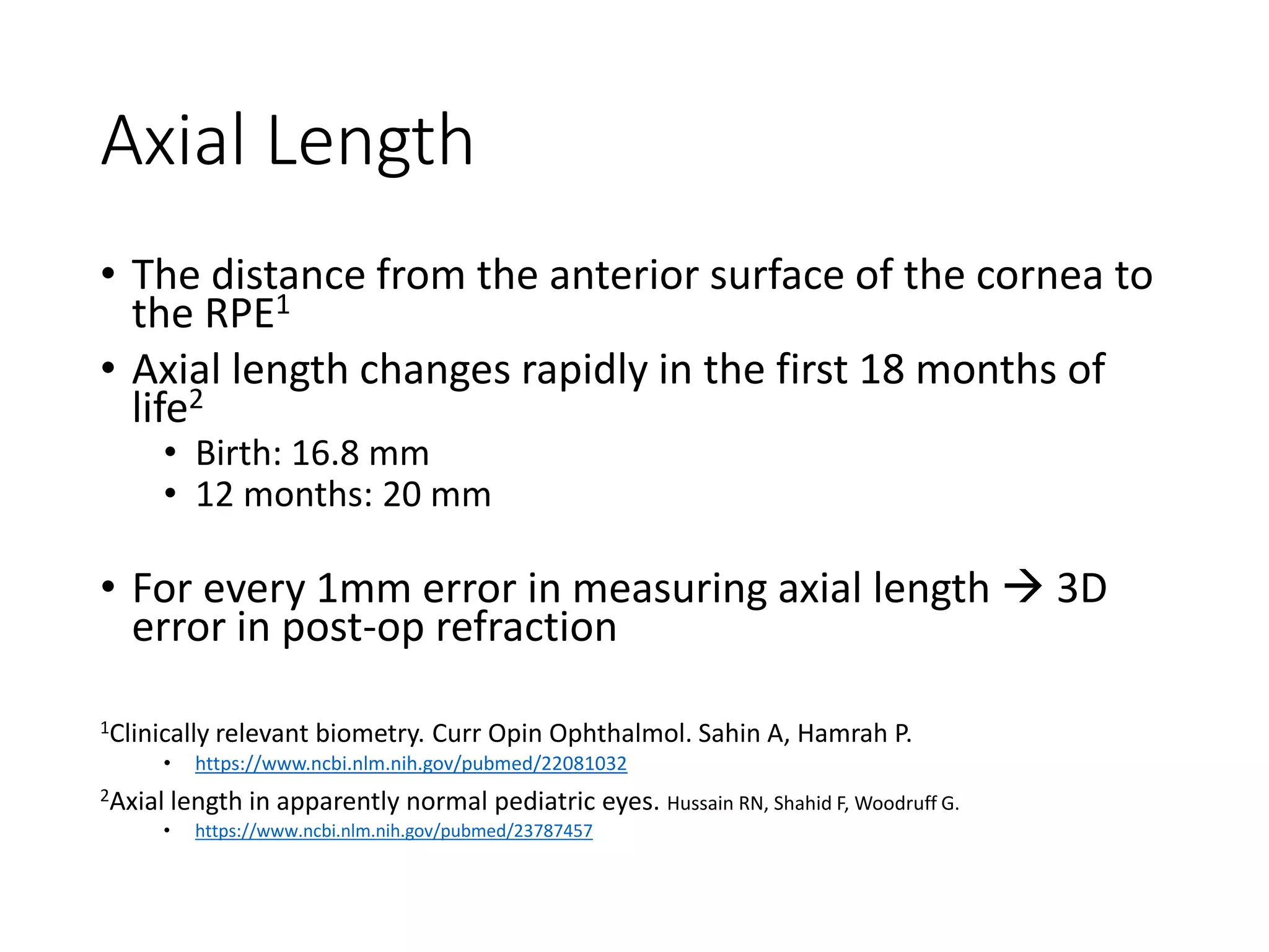
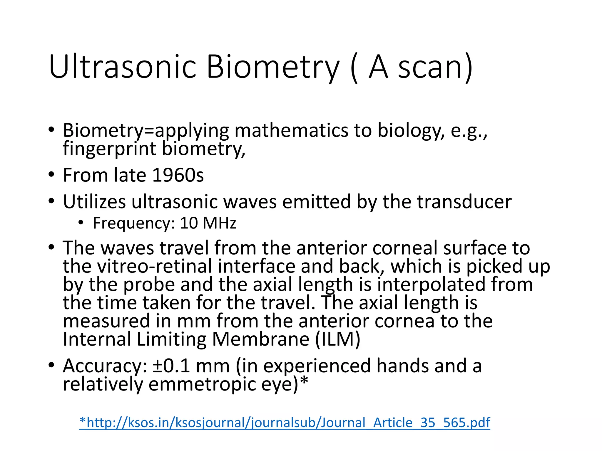
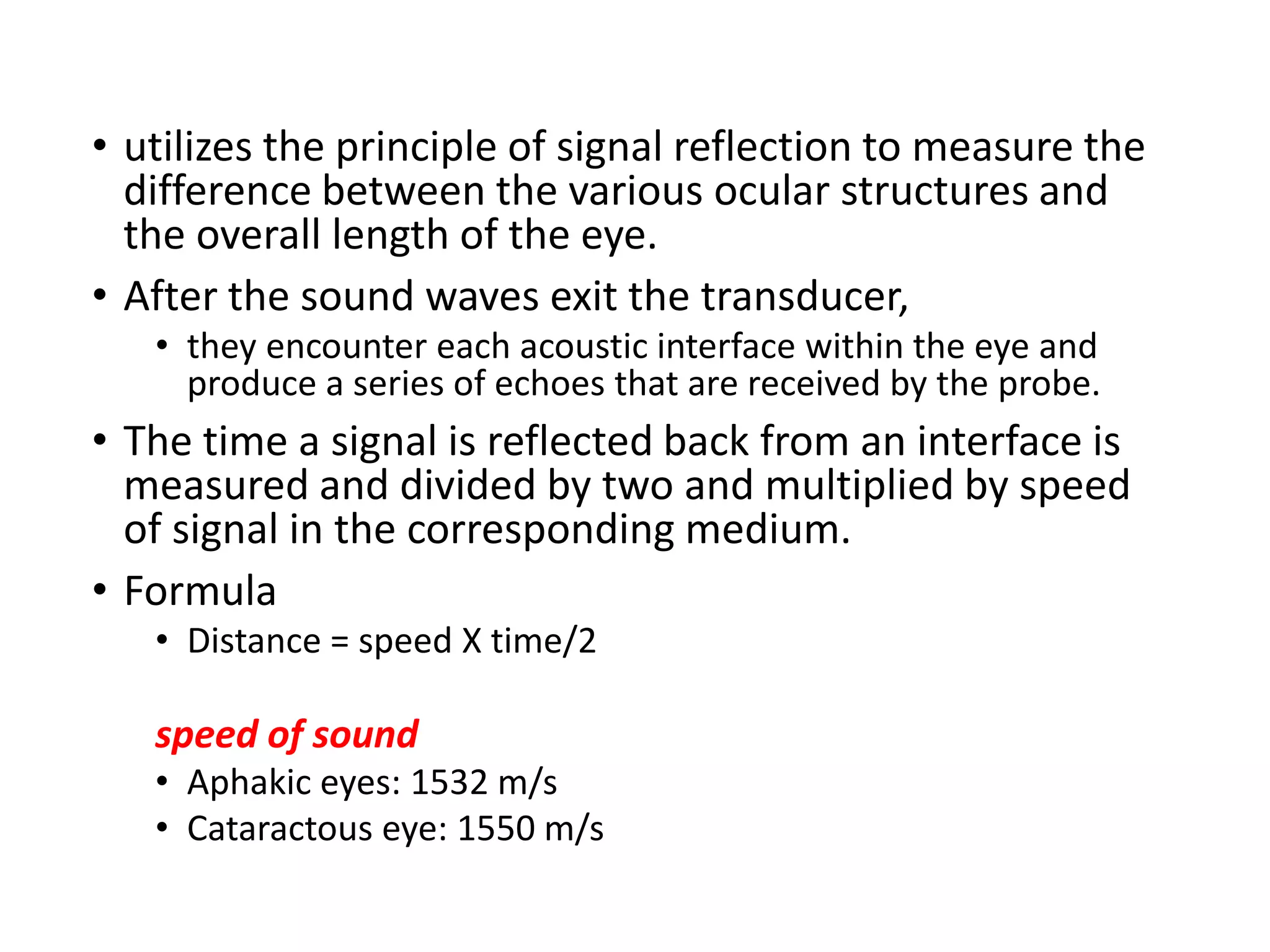
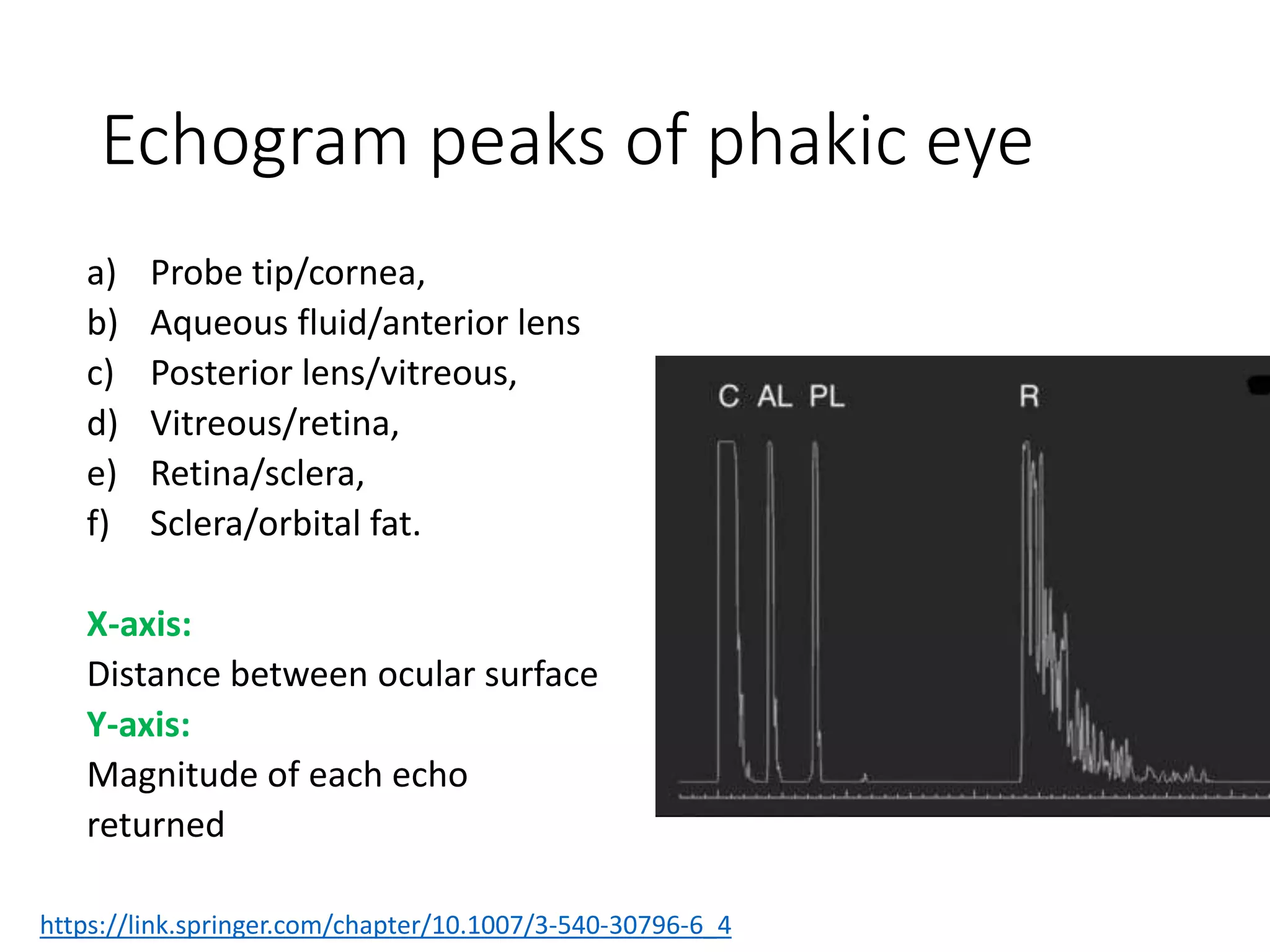
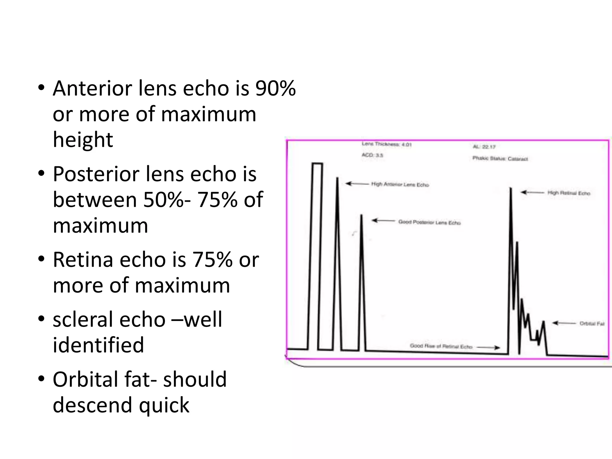
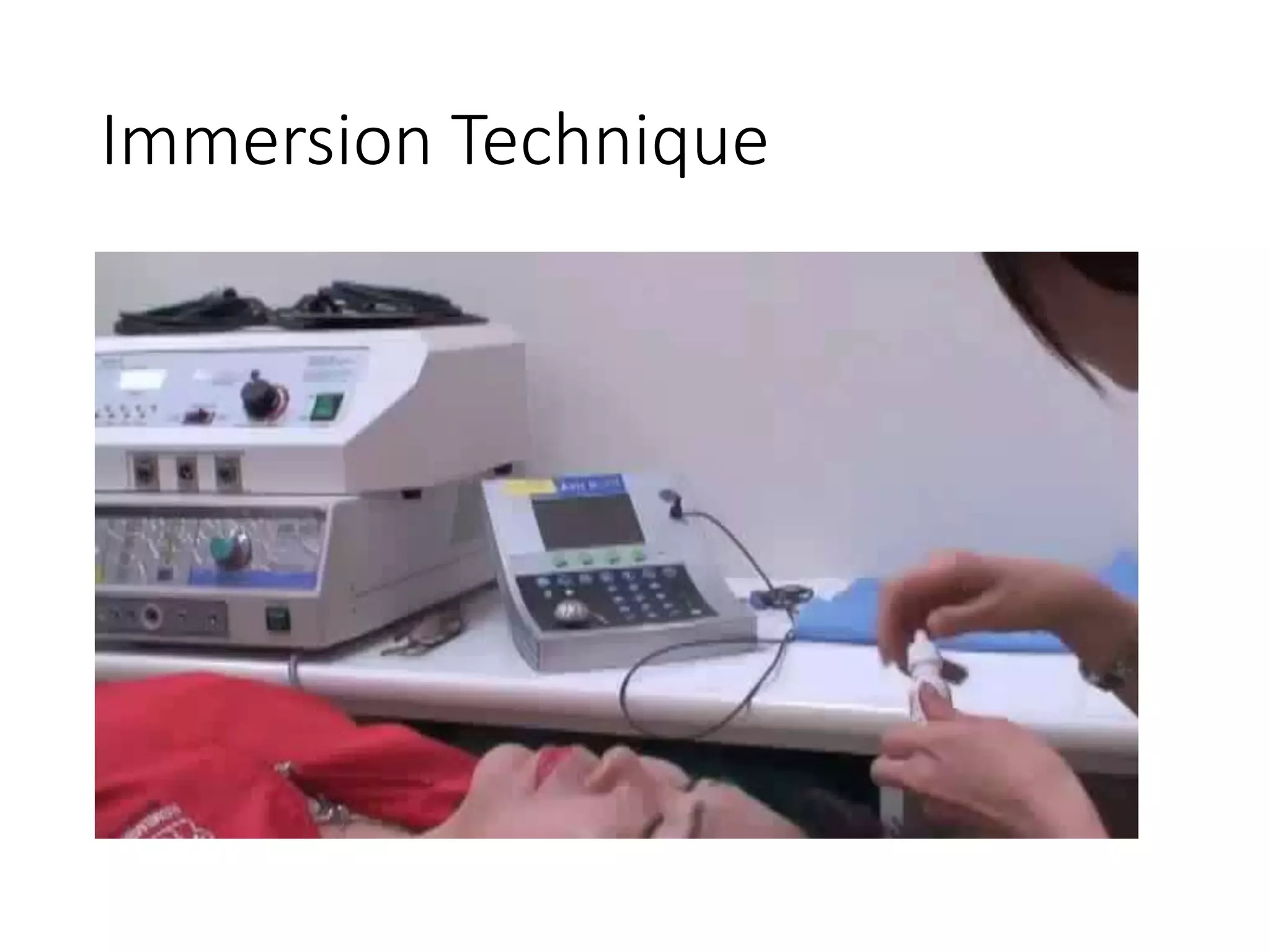
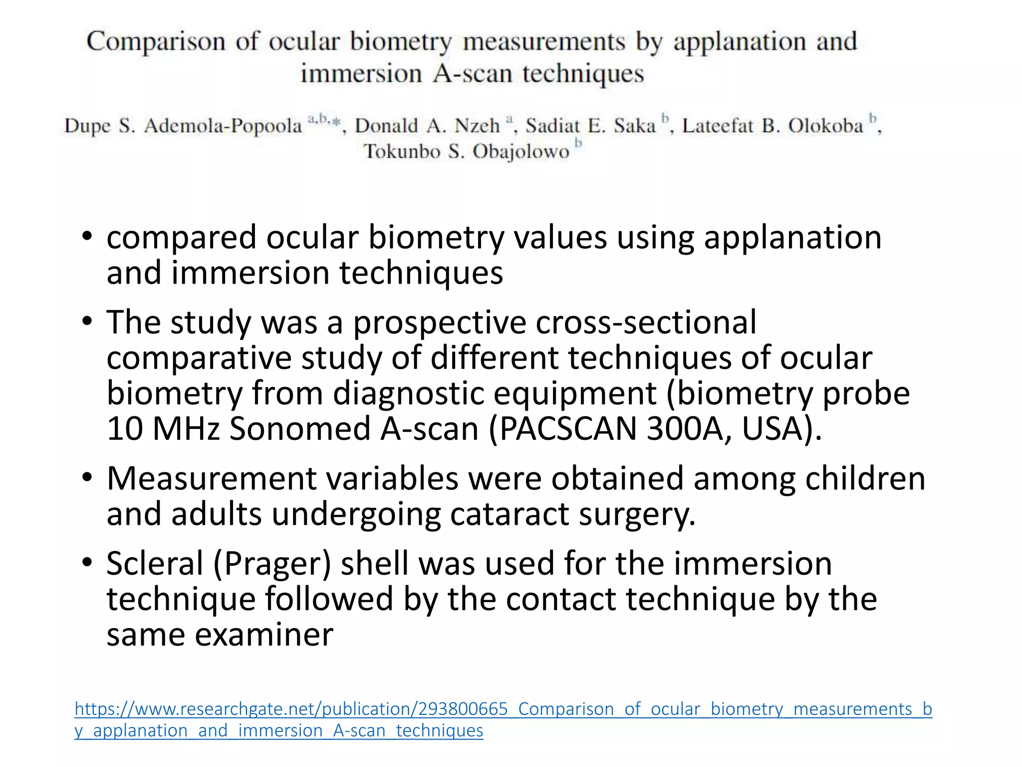
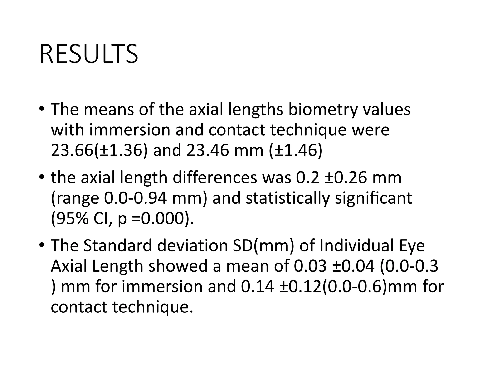
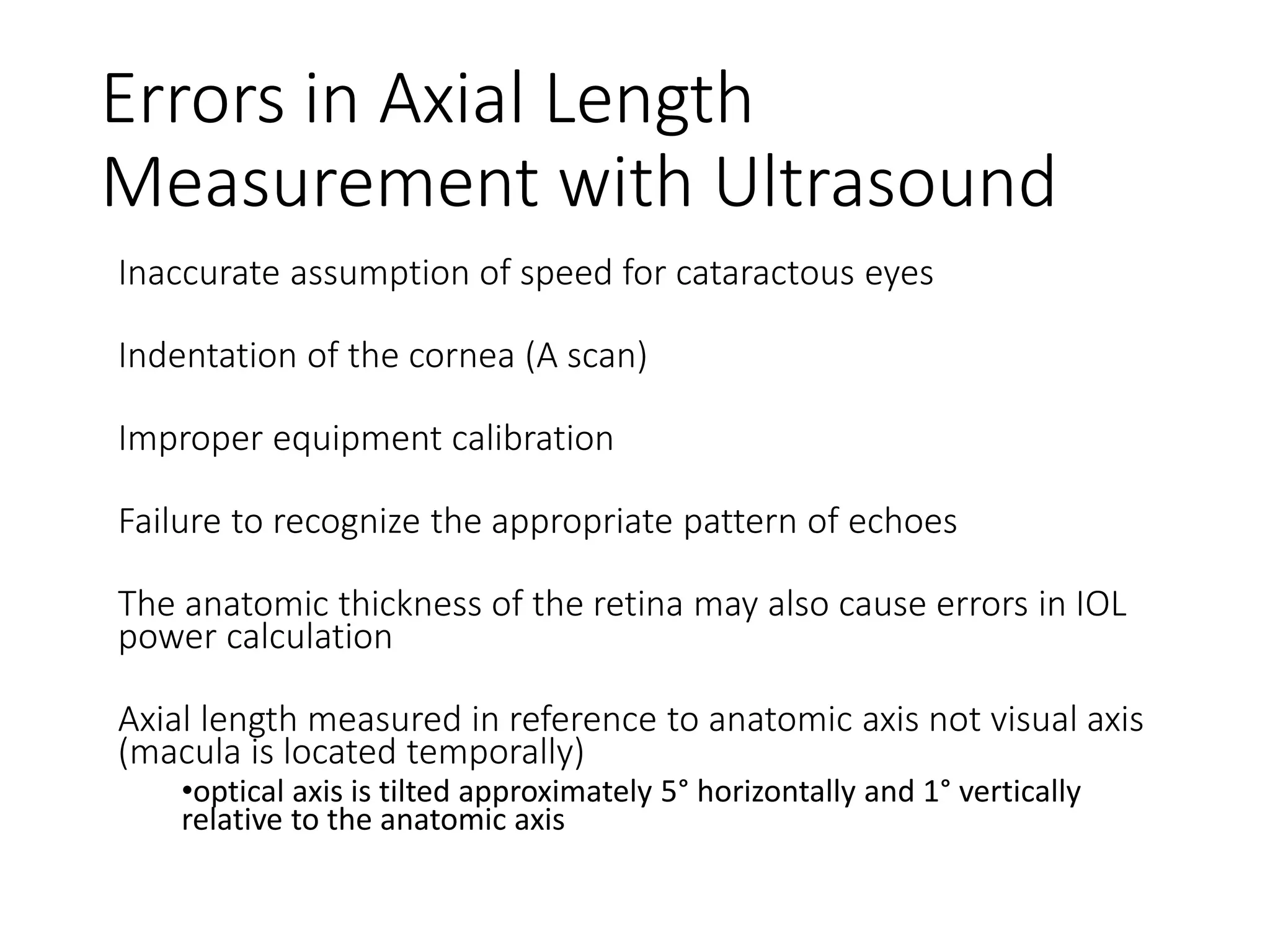
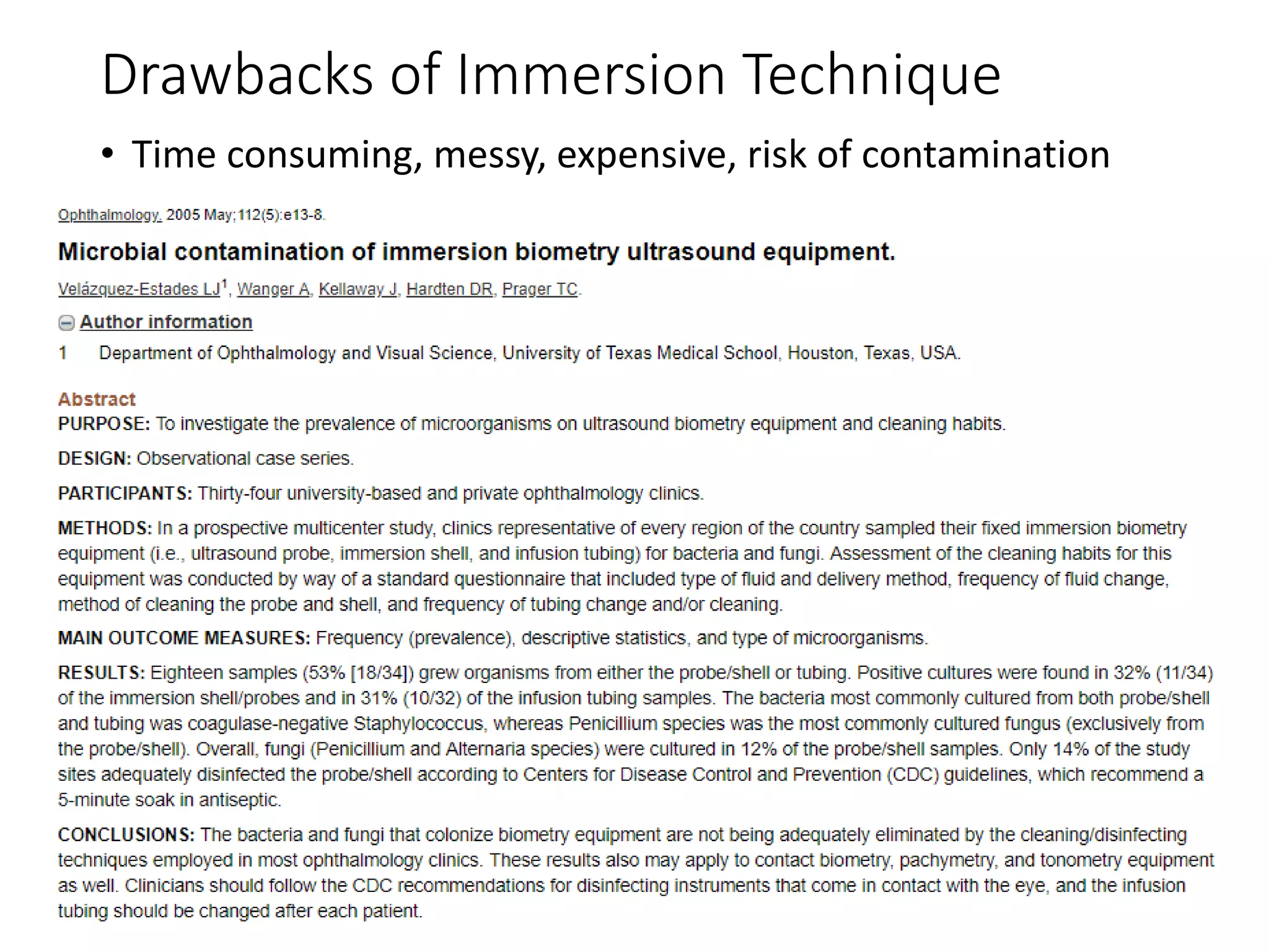
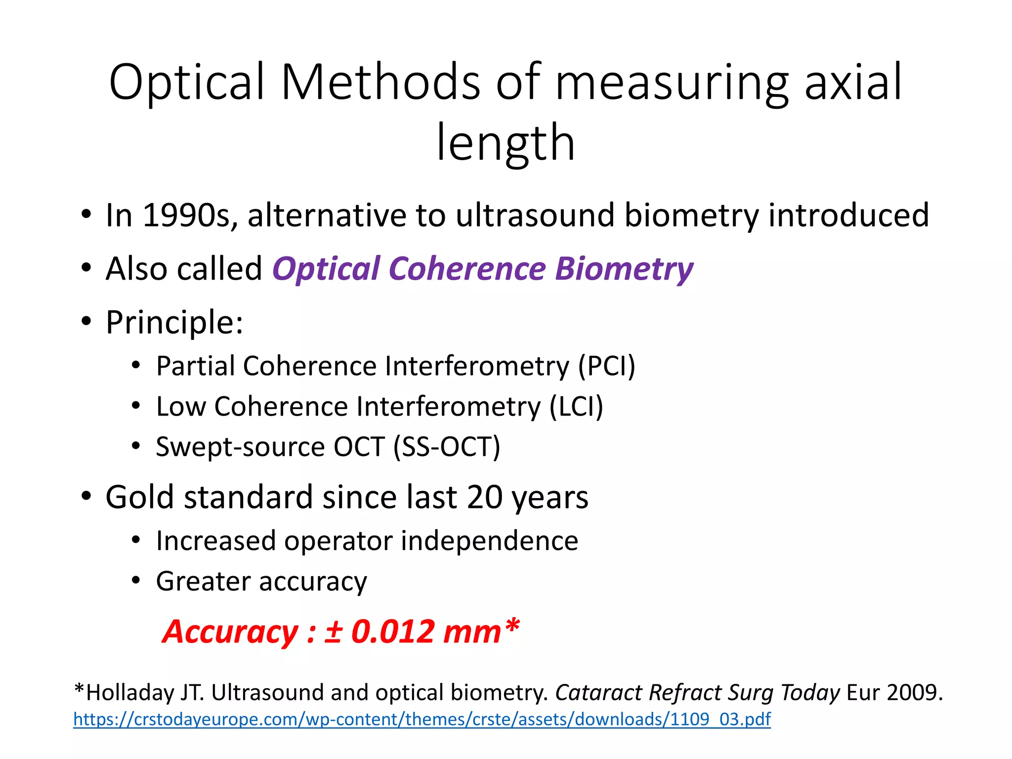
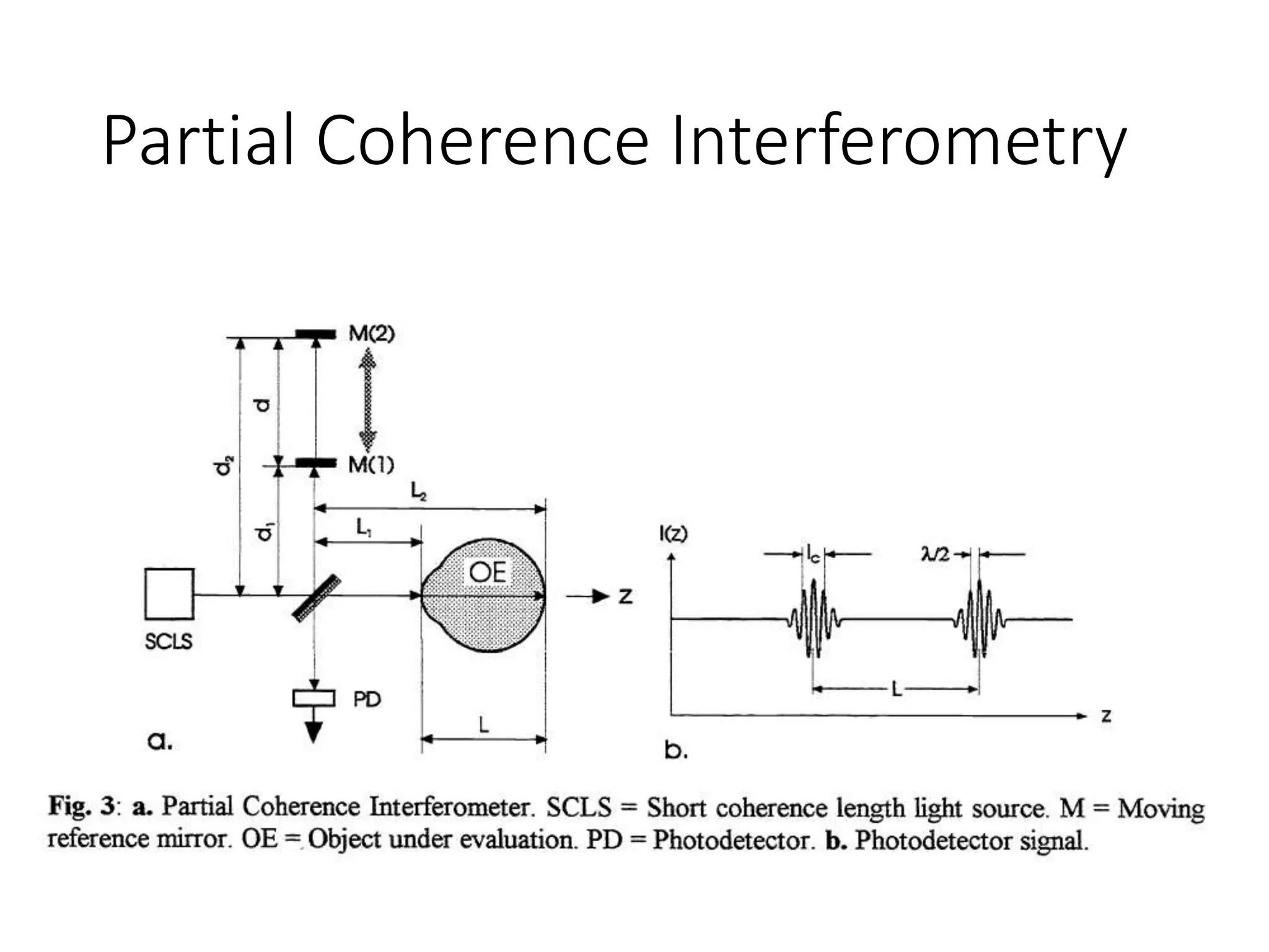
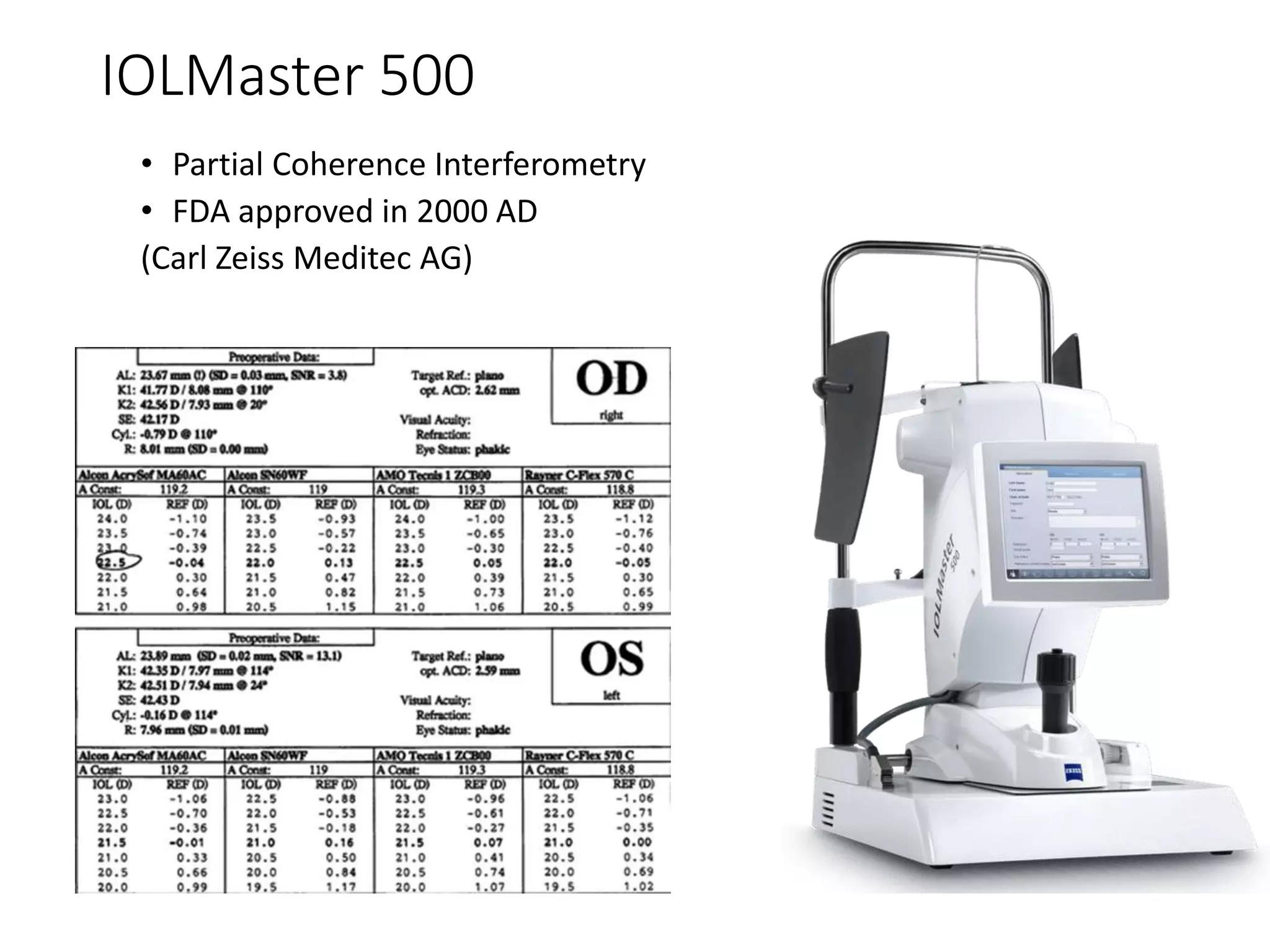
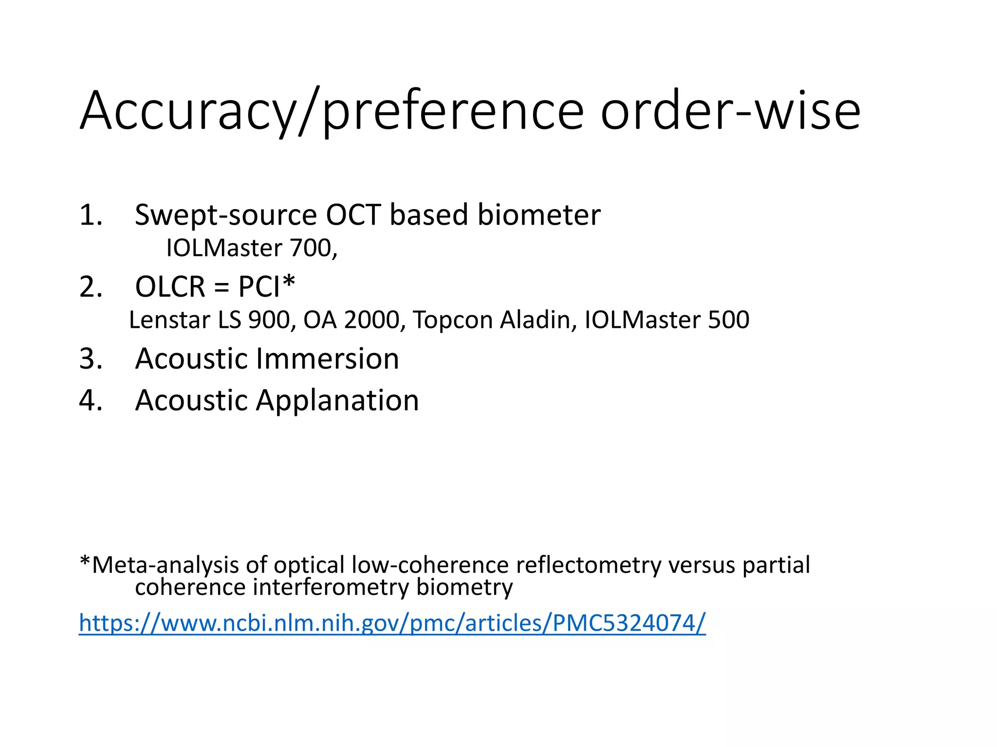
This document discusses various optical and non-optical methods of measuring axial length of the eye. It begins by defining axial length and noting its importance in intraocular lens power calculations. It then describes ultrasonic (A-scan) biometry, the historical standard, and optical biometry techniques like partial coherence interferometry used in devices like the IOLMaster 500. Key advantages of optical techniques are discussed as well as limitations of ultrasound. Details are provided on performing both immersion and non-immersion ultrasound techniques and interpreting the results.
Describes the basic of applanation tonometry, the factors affecting it and also how to perform the ideal tonometry. The slide are borrowed but it gives complete idea of mastering Applanation tonometry.
If the original owner of the slides has an objection i shall take down the ppt with due apologies.
Interventions to Reduce Myopia Progression in Children (Journal Club) (health...Bikash Sapkota
DIRECT DOWNLOAD LINK ❤❤https://healthkura.com/reduce-myopia/❤❤
Dear viewers Check Out my other piece of works at___ https://healthkura.com
Interventions to Reduce Myopia Progression in Children (Journal Club)
Objectives:
- To discuss about the different interventions to reduce myopia progression in children
- To determine the effectiveness of different interventions to slow down the progression of myopia in children
Interventions to Reduce Myopia Progression:
Environmental Considerations
- Time Spent Outdoors
- Near-Vision Activities
Spectacles & Contact Lenses
- Gas-Permeable Contact Lens Wear
- Bifocal & Multifocal Spectacles
- Soft Bifocal Contact Lenses
- Orthokeratology
Pharmacological Therapies
- Antimuscarinic Agents: Atropine & Pirenzepine
Under Correction of Myopia
To know Humphrey visual field analyser
To know about various types of perimetry
To identify field defect
To recognize that field defect is due to glaucoma or neurological lesion
To know that field defect is progressive or not
Interpretation of HVFA
introduction into Biometry and it's formulas.pptxQusaiAbusleem1
Biometry is the practice of applying mathematics to biology .
With regard to ophthalmology , there are several biometric systems used for making precise measurements of ocular structures : ultrasound (A- and B-scan , pachymeter) , low coherence interferometry (OCT) , laser interferometry (IOL Master , Lenstar)
These systems are used in measurement of axial length ,keratometry , pachymetry , and retinal thickness , among other things .
The term biometry , as it relates to ophthalmology , typically refers to preoperative measurements made for IOL calculations.
National Ocular Biometry Course (NOBC) 2015 An echoslide presentation Anis Suzanna Mohamad
This powerpoint presentation is basically about ocular biometry. Echo presentation is one of the method to deliver infomation that obtain from the course we attend to other staff in our Ophthalmology Department.
A scan biometry | How to Use A-scan? Types of A-Scan Biometry?Naeem Ahmad
A-SCAN BIOMETRY | What is A-Scan Biometry? How To Use It?
A-scan is the short form of amplitude scan.
This eye ultrasound gives details about the length of the eye.
A-Scan is an essential diagnostic tool used in ophthalmology.
The measurement of the eye’s axial length through an A-scan is necessary for placing an intraocular lens (IOL, artificial lens) during cataract surgery.
The total refractive power of the emmetropic eye is approximately 60D. Of this power, the cornea provides roughly 40D, and the crystalline lens 20 diopters.
When a cataract is removed, the lens is replaced by an artificial lens implant. By measuring both the length of the eye (A-scan Biometry) and the power of the cornea (keratometry).
It may also be used to assess vision abnormalities and other diseases involving the eye such as tumors.
A-scan techniques are based on the principles of ultrasonography. Sound travels in a wave pattern. For a sound to be heard by the human ear, the frequency must be between 20 and 20,000 Hz (20 kHz).
For an eye examination through A-scan, an ultrasound of frequency of around 10 MHz is used.
Specialized radiographic techniques /certified fixed orthodontic courses by I...Indian dental academy
The Indian Dental Academy is the Leader in continuing dental education , training dentists in all aspects of dentistry and offering a wide range of dental certified courses in different formats.
Indian dental academy provides dental crown & Bridge,rotary endodontics,fixed orthodontics,
Dental implants courses.for details pls visit www.indiandentalacademy.com ,or call
0091-9248678078
The Indian Dental Academy is the Leader in continuing dental education , training dentists in all aspects of dentistry and
offering a wide range of dental certified courses in different formats.for more details please visit
www.indiandentalacademy.com
Intraocular Lens (IOL) power calculation is a crucial step in cataract surgery and certain refractive surgeries like phakic IOL implantation. The goal is to determine the appropriate power of the IOL to be implanted into the eye, ensuring that the patient achieves their desired postoperative visual outcome. Several formulas and methods are available for IOL power calculation, and the choice of formula depends on various factors, including the patient's eye measurements and the surgeon's preference. Here, we describe the basic principles and some commonly used formulas.
Ocular Biometry:
Ocular biometry is the process of measuring various dimensions of the eye, primarily the axial length, corneal power, and anterior chamber depth. These measurements are essential for accurate IOL power calculation and achieving the desired post-surgical refractive outcome. Here are the key components of ocular biometry:
Axial Length: This measurement determines the overall length of the eye, from the cornea's front surface to the retina's back surface. Axial length is a critical factor in IOL power calculation because it helps determine the eye's focusing power.
Corneal Power: The cornea is the transparent front surface of the eye, and its curvature affects the eye's refractive power. Corneal power is typically measured using techniques like keratometry or corneal topography. It helps account for the eye's astigmatism and assists in selecting the appropriate IOL.
some basic notions on how they are measured is explored here.
This presentation is mainly focused on the clinical diagnosis and interpretation of oct macula.This is presented on 4th year optometry as topic presentation.
250+ High Frequency MCQs in Optometry and OphthalmologyRabindraAdhikary
The collection of high-ranked, top-rated high frequency multiple-choice questions suitable for any examination of optometry, ophthalmology and ophthalmic sciences with their answers for FREE. No Log in, No Pay!!
Contact lens care and maintenance
RGP care
Soft Contact lens care
Silicon Hydrogel Care
Contact Lens Disinfection
Thermal disinfection
Chemical Disinfection
Oxidative chemical disinfection
Troubleshooting bifocals and Market Availability in Nepal
Bifocals in Anisometropia
Prismatic Effect in Bifocal
Bifocal Prescription
Bifocals in High Astigmatism
Variables: Types and their Operational Definitions
Unit III: Problem identification formulation of research objectives and hypothesis (as part of M.Optom Curriculum of Pokhara University, Nepal)
Cholinergic agent: Autonomic Drugs
According to the M. Optom curriculum, we have prepared a concise presentation on Cholinergic or parasympathomimetic or cholinomimetic drugs
Multiple Choice Questions (MCQs) for Masters of Optometry Entrance Examinatio...RabindraAdhikary
Multiple Choice Questions (MCQs) for Masters of Optometry Entrance Examination, Pokhara University NEPAL
MCQs Optometry Nepal
Here we have included syllabus of entrance examinations for Master of Optometry in Pokhara University, entry requirements of candidate for the master of optometry course and multiple choice questions that appeared in the entrance examinations of 2019.
Prepared by: Rabindra Adhikary
for more MCQs:
http://ravinems.blogspot.com/2019/05/multiple-choice-questions-mcqs-for.html
Visual Implication in Diabetes Mellitus
These slides talk in detail about the visual implications of Diabetes Mellitus and how to address them systematically
Systemic Diseases and the Eye
Various systemic diseases affect the eye and it's functioning. Here we present those common systemic diseases that are responsible to cause effects in the eye.
What are the diseases that affect eye?
Eye is affected by the following diseases:
Systemic Hypertension (Increased blood pressure of the body)
Diabetes Melitus (Increased blood sugar level)
Systemic Lupus Erythromatosus (SLE)
AIDS and other Venereal Diseases like Syphilis
Sickle Cell Anemia,
Eales Disease and many more.
Look at the slides.
Ophthalmic Prisms: Prismatic Effects and DecentrationRabindraAdhikary
Ophthalmic Prisms: Prismatic Effects and Decentration
here we discuss about the ophthalmic prisms, the prismatic effects as caused by the decentration( moving the optical center away from the visual axis)
Ethanol (CH3CH2OH), or beverage alcohol, is a two-carbon alcohol
that is rapidly distributed in the body and brain. Ethanol alters many
neurochemical systems and has rewarding and addictive properties. It
is the oldest recreational drug and likely contributes to more morbidity,
mortality, and public health costs than all illicit drugs combined. The
5th edition of the Diagnostic and Statistical Manual of Mental Disorders
(DSM-5) integrates alcohol abuse and alcohol dependence into a single
disorder called alcohol use disorder (AUD), with mild, moderate,
and severe subclassifications (American Psychiatric Association, 2013).
In the DSM-5, all types of substance abuse and dependence have been
combined into a single substance use disorder (SUD) on a continuum
from mild to severe. A diagnosis of AUD requires that at least two of
the 11 DSM-5 behaviors be present within a 12-month period (mild
AUD: 2–3 criteria; moderate AUD: 4–5 criteria; severe AUD: 6–11 criteria).
The four main behavioral effects of AUD are impaired control over
drinking, negative social consequences, risky use, and altered physiological
effects (tolerance, withdrawal). This chapter presents an overview
of the prevalence and harmful consequences of AUD in the U.S.,
the systemic nature of the disease, neurocircuitry and stages of AUD,
comorbidities, fetal alcohol spectrum disorders, genetic risk factors, and
pharmacotherapies for AUD.
Report Back from SGO 2024: What’s the Latest in Cervical Cancer?bkling
Are you curious about what’s new in cervical cancer research or unsure what the findings mean? Join Dr. Emily Ko, a gynecologic oncologist at Penn Medicine, to learn about the latest updates from the Society of Gynecologic Oncology (SGO) 2024 Annual Meeting on Women’s Cancer. Dr. Ko will discuss what the research presented at the conference means for you and answer your questions about the new developments.
Couples presenting to the infertility clinic- Do they really have infertility...Sujoy Dasgupta
Dr Sujoy Dasgupta presented the study on "Couples presenting to the infertility clinic- Do they really have infertility? – The unexplored stories of non-consummation" in the 13th Congress of the Asia Pacific Initiative on Reproduction (ASPIRE 2024) at Manila on 24 May, 2024.
The prostate is an exocrine gland of the male mammalian reproductive system
It is a walnut-sized gland that forms part of the male reproductive system and is located in front of the rectum and just below the urinary bladder
Function is to store and secrete a clear, slightly alkaline fluid that constitutes 10-30% of the volume of the seminal fluid that along with the spermatozoa, constitutes semen
A healthy human prostate measures (4cm-vertical, by 3cm-horizontal, 2cm ant-post ).
It surrounds the urethra just below the urinary bladder. It has anterior, median, posterior and two lateral lobes
It’s work is regulated by androgens which are responsible for male sex characteristics
Generalised disease of the prostate due to hormonal derangement which leads to non malignant enlargement of the gland (increase in the number of epithelial cells and stromal tissue)to cause compression of the urethra leading to symptoms (LUTS
- Video recording of this lecture in English language: https://youtu.be/lK81BzxMqdo
- Video recording of this lecture in Arabic language: https://youtu.be/Ve4P0COk9OI
- Link to download the book free: https://nephrotube.blogspot.com/p/nephrotube-nephrology-books.html
- Link to NephroTube website: www.NephroTube.com
- Link to NephroTube social media accounts: https://nephrotube.blogspot.com/p/join-nephrotube-on-social-media.html
Explore natural remedies for syphilis treatment in Singapore. Discover alternative therapies, herbal remedies, and lifestyle changes that may complement conventional treatments. Learn about holistic approaches to managing syphilis symptoms and supporting overall health.
Title: Sense of Smell
Presenter: Dr. Faiza, Assistant Professor of Physiology
Qualifications:
MBBS (Best Graduate, AIMC Lahore)
FCPS Physiology
ICMT, CHPE, DHPE (STMU)
MPH (GC University, Faisalabad)
MBA (Virtual University of Pakistan)
Learning Objectives:
Describe the primary categories of smells and the concept of odor blindness.
Explain the structure and location of the olfactory membrane and mucosa, including the types and roles of cells involved in olfaction.
Describe the pathway and mechanisms of olfactory signal transmission from the olfactory receptors to the brain.
Illustrate the biochemical cascade triggered by odorant binding to olfactory receptors, including the role of G-proteins and second messengers in generating an action potential.
Identify different types of olfactory disorders such as anosmia, hyposmia, hyperosmia, and dysosmia, including their potential causes.
Key Topics:
Olfactory Genes:
3% of the human genome accounts for olfactory genes.
400 genes for odorant receptors.
Olfactory Membrane:
Located in the superior part of the nasal cavity.
Medially: Folds downward along the superior septum.
Laterally: Folds over the superior turbinate and upper surface of the middle turbinate.
Total surface area: 5-10 square centimeters.
Olfactory Mucosa:
Olfactory Cells: Bipolar nerve cells derived from the CNS (100 million), with 4-25 olfactory cilia per cell.
Sustentacular Cells: Produce mucus and maintain ionic and molecular environment.
Basal Cells: Replace worn-out olfactory cells with an average lifespan of 1-2 months.
Bowman’s Gland: Secretes mucus.
Stimulation of Olfactory Cells:
Odorant dissolves in mucus and attaches to receptors on olfactory cilia.
Involves a cascade effect through G-proteins and second messengers, leading to depolarization and action potential generation in the olfactory nerve.
Quality of a Good Odorant:
Small (3-20 Carbon atoms), volatile, water-soluble, and lipid-soluble.
Facilitated by odorant-binding proteins in mucus.
Membrane Potential and Action Potential:
Resting membrane potential: -55mV.
Action potential frequency in the olfactory nerve increases with odorant strength.
Adaptation Towards the Sense of Smell:
Rapid adaptation within the first second, with further slow adaptation.
Psychological adaptation greater than receptor adaptation, involving feedback inhibition from the central nervous system.
Primary Sensations of Smell:
Camphoraceous, Musky, Floral, Pepperminty, Ethereal, Pungent, Putrid.
Odor Detection Threshold:
Examples: Hydrogen sulfide (0.0005 ppm), Methyl-mercaptan (0.002 ppm).
Some toxic substances are odorless at lethal concentrations.
Characteristics of Smell:
Odor blindness for single substances due to lack of appropriate receptor protein.
Behavioral and emotional influences of smell.
Transmission of Olfactory Signals:
From olfactory cells to glomeruli in the olfactory bulb, involving lateral inhibition.
Primitive, less old, and new olfactory systems with different path
TEST BANK for Operations Management, 14th Edition by William J. Stevenson, Ve...kevinkariuki227
TEST BANK for Operations Management, 14th Edition by William J. Stevenson, Verified Chapters 1 - 19, Complete Newest Version.pdf
HOT NEW PRODUCT! BIG SALES FAST SHIPPING NOW FROM CHINA!! EU KU DB BK substit...GL Anaacs
Contact us if you are interested:
Email / Skype : kefaya1771@gmail.com
Threema: PXHY5PDH
New BATCH Ku !!! MUCH IN DEMAND FAST SALE EVERY BATCH HAPPY GOOD EFFECT BIG BATCH !
Contact me on Threema or skype to start big business!!
Hot-sale products:
NEW HOT EUTYLONE WHITE CRYSTAL!!
5cl-adba precursor (semi finished )
5cl-adba raw materials
ADBB precursor (semi finished )
ADBB raw materials
APVP powder
5fadb/4f-adb
Jwh018 / Jwh210
Eutylone crystal
Protonitazene (hydrochloride) CAS: 119276-01-6
Flubrotizolam CAS: 57801-95-3
Metonitazene CAS: 14680-51-4
Payment terms: Western Union,MoneyGram,Bitcoin or USDT.
Deliver Time: Usually 7-15days
Shipping method: FedEx, TNT, DHL,UPS etc.Our deliveries are 100% safe, fast, reliable and discreet.
Samples will be sent for your evaluation!If you are interested in, please contact me, let's talk details.
We specializes in exporting high quality Research chemical, medical intermediate, Pharmaceutical chemicals and so on. Products are exported to USA, Canada, France, Korea, Japan,Russia, Southeast Asia and other countries.
ARTIFICIAL INTELLIGENCE IN HEALTHCARE.pdfAnujkumaranit
Artificial intelligence (AI) refers to the simulation of human intelligence processes by machines, especially computer systems. It encompasses tasks such as learning, reasoning, problem-solving, perception, and language understanding. AI technologies are revolutionizing various fields, from healthcare to finance, by enabling machines to perform tasks that typically require human intelligence.
Pulmonary Thromboembolism - etilogy, types, medical- Surgical and nursing man...VarunMahajani
Disruption of blood supply to lung alveoli due to blockage of one or more pulmonary blood vessels is called as Pulmonary thromboembolism. In this presentation we will discuss its causes, types and its management in depth.
Evaluation of antidepressant activity of clitoris ternatea in animals
Optical and Non-optical Methods of Measuring Axial Length of Eye
1. Optical and Non-optical
Methods of Measuring Axial
Length
Rabindra Adhikary
M. Optom, 1st Batch
Tilganga Institute of Ophthalmology
Pokhara University
Facilitator:
Rupesh Poudel
14th August 2019
2. Axial Length
• The distance from the anterior surface of the cornea to
the RPE1
• Axial length changes rapidly in the first 18 months of
life2
• Birth: 16.8 mm
• 12 months: 20 mm
• For every 1mm error in measuring axial length 3D
error in post-op refraction
1Clinically relevant biometry. Curr Opin Ophthalmol. Sahin A, Hamrah P.
• https://www.ncbi.nlm.nih.gov/pubmed/22081032
2Axial length in apparently normal pediatric eyes. Hussain RN, Shahid F, Woodruff G.
• https://www.ncbi.nlm.nih.gov/pubmed/23787457
3. • Mean Axial Length of Normal adult Eye1
• 23.67 mm
• Mean Axial Length of Cataractous Eye2
• 23.65 mm
• Diurnal Fluctuation occurs in axial length by3
• 15-40μ
1. Axial length and optic disc size in normal eyes
C Oliveira, N Harizman, C A Girkin, A Xie, C Tello, J M Liebmann, and R Ritch
https://www.ncbi.nlm.nih.gov/pmc/articles/PMC1857588/
2. Biometry of 7,500 Cataractous Eyes
J. Hoffer, MD
https://www.sciencedirect.com/science/article/pii/S0002939414749177
3. Diurnal Axial Length Fluctuation in Human Eyes
Richard A. Stone; Graham E. Quinn; Ellie L. Francis; Gui-shuang Ying; D. Ian Flitcroft; Parag Parekh
https://iovs.arvojournals.org/article.aspx?articleid=2123886
4. Axial Length Comparison in different age groups
Age
(years)
Emmetropia Ametropia
Myopia Hypermetropia Astigmatism
Male Female Male Female Male Female Male Female
0-10 22.28 21.93 22.81 22.82 21.22 21.29 22.32 22.03
SD 0.50 0.67 1.10 1.68 0.21 0.31 0.24 0.32
11-20 23.23 23.42 24.07 24.18 21.97 21.71 23.13 23.06
SD 0.48 0.46 0.67 0.40 1.09 0.56 1.01 1.07
21-40 22.71 22.99 23.58 23.56 22.06 22.01 23.23 22.16
SD 1.21 0.91 1.65 2.53 0.90 1.28 2.26 1.39
>40 22.33 22.99 23.64 23.95 21.68 21.61 22.06 21.5
SD 0.49 0.71 1.69 1.72 0.35 0.47 0.49 0.26
Axial Length, Anterior Chamber Depth-A Study in Different Age Groups and Refractive Errors
Veena Bhardwaj and Gandhi Parth Rajeshbhai
https://www.ncbi.nlm.nih.gov/pmc/articles/PMC3843406/
5. Ultrasonic Biometry ( A scan)
• Biometry=applying mathematics to biology, e.g.,
fingerprint biometry,
• From late 1960s
• Utilizes ultrasonic waves emitted by the transducer
• Frequency: 10 MHz
• The waves travel from the anterior corneal surface to
the vitreo-retinal interface and back, which is picked up
by the probe and the axial length is interpolated from
the time taken for the travel. The axial length is
measured in mm from the anterior cornea to the
Internal Limiting Membrane (ILM)
• Accuracy: ±0.1 mm (in experienced hands and a
relatively emmetropic eye)*
*http://ksos.in/ksosjournal/journalsub/Journal_Article_35_565.pdf
6. • utilizes the principle of signal reflection to measure the
difference between the various ocular structures and
the overall length of the eye.
• After the sound waves exit the transducer,
• they encounter each acoustic interface within the eye and
produce a series of echoes that are received by the probe.
• The time a signal is reflected back from an interface is
measured and divided by two and multiplied by speed
of signal in the corresponding medium.
• Formula
• Distance = speed X time/2
speed of sound
• Aphakic eyes: 1532 m/s
• Cataractous eye: 1550 m/s
7. Echogram peaks of phakic eye
a) Probe tip/cornea,
b) Aqueous fluid/anterior lens
c) Posterior lens/vitreous,
d) Vitreous/retina,
e) Retina/sclera,
f) Sclera/orbital fat.
X-axis:
Distance between ocular surface
Y-axis:
Magnitude of each echo
returned
https://link.springer.com/chapter/10.1007/3-540-30796-6_4
8. • Anterior lens echo is 90%
or more of maximum
height
• Posterior lens echo is
between 50%- 75% of
maximum
• Retina echo is 75% or
more of maximum
• scleral echo –well
identified
• Orbital fat- should
descend quick
9. • The amplitude of the peak depends upon:
• Difference in the density at the acoustic interface
• Greater difference produce higher echoes
• The angle of incidence at the interface
• Perpendicular waves produces maximum height of spike
• Measurement of A scan:
• Spike Height
• Regularity
• Reflectivity
• Sound attenuation
10. Applanation technique
• the ultrasound probe
• in direct contact with the cornea., centrally at the
pupilary area
• Probe perpendicular
• Cornea to be anesthetized
• Inadvertent pressure may cause upto 0.3mm
corneal indentation
• Error of 1-1.5D so getting obsolete
11. • A number of readings are taken either manually or in
auto-mode
• Readings from improper spikes—removed
• Average taken in the value of axial length
• Accuracy: ± 0.10
• Inter-eye variation: ± 0.3 mm
Holladay JT. Ultrasound and optical biometry. Cataract Refract Surg Today
Eur 2009.
https://crstodayeurope.com/wp-content/themes/crste/assets/downloads/1109_03.pdf
12. Anatomic vs Optical AL
• Ultrasound measures
Anatomic Axial
Length
• Ant cornea – ILM
• Photoreceptors Layer
lies 200μm (160-400
μm) beyond ILM1
• Need to add 200μm to
get Optical Axial
Length(OAL)
• 200 μm ≈0.56D error2
• Optical Methods
measure OAL
2https://www.nature.com/articles/6700157
1https://www.ncbi.nlm.nih.gov/pubmed/25473345
13. Immersion technique
• Here a waterbath is placed on the cornea using a
scleral shell(eg. Praeger Shell). The hard tip
transducer is applied to the shell. The rest of the
procedure is similar to the contact method.
• If done properly, immersion technique gives very
accurate and reproducible results in all types of
cataracts.
14. Immersion Technique
• Unlike the applanation echogram, the immersion
technique produces an additional spike probe tip within
the coupling fluid.
• The patient lies supine, looking up at the ceiling and the
scleral shell is placed between the eyelids and centered
over the cornea.
• The scleral shell is then filled with coupling gel (40-60
mixture of Goniosol and Dacriose)
• The probe tip is placed into the solution.
• Align the ultrasound beam with the macula by having
the patient look at the probe tip fixation light, then
simply take your readings as usual.
17. RESULTS
• The means of the axial lengths biometry values
with immersion and contact technique were
23.66(±1.36) and 23.46 mm (±1.46)
• the axial length differences was 0.2 ±0.26 mm
(range 0.0-0.94 mm) and statistically significant
(95% CI, p =0.000).
• The Standard deviation SD(mm) of Individual Eye
Axial Length showed a mean of 0.03 ±0.04 (0.0-0.3
) mm for immersion and 0.14 ±0.12(0.0-0.6)mm for
contact technique.
18. Errors in Axial Length
Measurement with Ultrasound
Inaccurate assumption of speed for cataractous eyes
Indentation of the cornea (A scan)
Improper equipment calibration
Failure to recognize the appropriate pattern of echoes
The anatomic thickness of the retina may also cause errors in IOL
power calculation
Axial length measured in reference to anatomic axis not visual axis
(macula is located temporally)
•optical axis is tilted approximately 5° horizontally and 1° vertically
relative to the anatomic axis
19. Drawbacks of Immersion Technique
• Time consuming, messy, expensive, risk of contamination
20. • Recommendations: The shell and probe should be
soaked in alcohol or hydrogen peroxide for at least
5 minutes. The immersion shell should be allowed
to dry completely and flushed with balanced saline
solution (BSS).
21. Optical Methods of measuring axial
length
• In 1990s, alternative to ultrasound biometry introduced
• Also called Optical Coherence Biometry
• Principle:
• Partial Coherence Interferometry (PCI)
• Low Coherence Interferometry (LCI)
• Swept-source OCT (SS-OCT)
• Gold standard since last 20 years
• Increased operator independence
• Greater accuracy
Accuracy : ± 0.012 mm*
*Holladay JT. Ultrasound and optical biometry. Cataract Refract Surg Today Eur 2009.
https://crstodayeurope.com/wp-content/themes/crste/assets/downloads/1109_03.pdf
23. Interferometer
• Two-beam interferometers (4 types)
• The Fizeau Intearferometer
• The Michelson Interferometer
• used in optical biometers
• The Mach-Zehnder Interferometer
• The Sagnac interferometer
Interferometers. P . Hariharan Division of Applied Physics CSIRO Sydney , Australia
http://www.photonics.intec.ugent.be/education/IVPV/res_handbook/v2ch21.pdf
24. M=0,1,2,3,…
L1 changes continuously, L2 is fixed
Therefore this instrument allows you to observe distance changes
of the order of the wavelength of light.
28. IOLMaster 500
• Partial Coherence Interferometry
• FDA approved in 2000 AD
(Carl Zeiss Meditec AG)
29. IOLMaster 500
• First optical biometer introduced in 1999
• 780nm laser diode infrared light is used
• Measures keratometry analyzing the anterior corneal
curvature at 6 reference points at approx. 2.3mm optical
zone
• The ACD is measured using slit-lamp illumination and is
defined as a distance from the corneal epithelium and to
the anterior lens surface
• WTW is obtained by analyzing the image of the iris using an
infrared light source (wavelength 880 nm)
• All measurements are performed simultaneously.
• IOLMaster 500 is currently considered as a gold-standard
biometer.
https://www.ncbi.nlm.nih.gov/pubmed/9780097
30. ALM
• Switching to ALM mode will
automatically change the
magnification ratio: a smaller section
of the eye becomes visible with the
reflection of the alignment light and
a vertical line (1, Fig. 27).
• The patient should look at the red
fixation point in the center. A cross-
hair (3, Fig. 27) with a circle in the
middle will appear on the display.
31. • Ask the patient if he or she sees the fixation
point. If the patient fails to fixate properly, the
visual axis will not be correctly recognized, which
may result in measuring errors.
• poor visual acuity/high ametropia (> 4 D)?
• measure through the spectacles. If the procedure is
followed correctly, no measuring errors will be
produced.
• Measurements should not be taken while a patient is
wearing contact lenses, as it will result in measuring
errors.
32. • IOLMaster requires 5 measurements (max. 20 per
eye in a day)
• If a reading deviate from another by more than
0.05 mm, it will be displayed in red and the
message Multiple peaks will appear
• *Error in the display field denotes readings with an
SNR smaller than 1.6.
• The following considerations should be made:
• AL < 22mm (Wang-Koch Axial Length mod + Holladay1)
• AL > 25mm (Hoffer Q or Holladay 2 )
*https://www.ncbi.nlm.nih.gov/pubmed/21183100
33. • If the AL difference between RE and LE exceeds 0.3
mm, a message appears to check the readings once
again.
34. IOLMaster: Modes and Features
• Axial Length
• Corneal Curvature
• White-to-White (optional)
• Anterior Chamber Depth
• IOL Power Calculation
• Phakic (Anterior and Posterior Chamber) IOL Power
Calculation (optional)
• Lens Constant Personalization
• Post Refractive Surgery Corneal Power Calculation
(optional)
• Data Export (optional)
35. Limitation of IOLMaster 500
• To get the measurement, fixation is required.
• So, in dense cataract (VA<3/60) it is not possible to carry
out the biometry
• inaccurate measurements may result in cases of
other media opacities
• corneal opacity
• vitreous hemorrhage
• Nystagmus also may give rise to inaccurate readings
In these cases Ultrasound Biometry
36. Measurement Range of
IOLMaster 500
Axial Length 14-40
Corneal Radii 5-10
ACD 1.5-6.5
White to white 8-16
Resolution Range
AL/CR/W-W 0.01 mm
Formula SRK II , SRK/T, Holladay, Hoffer Q, Haigis
37. 3 Advantages of optical method
over ultrasound method
1. Measures length along the visual axis
• cornea to fovea
2. Measures true distance that we want
• optical AL
3. Uses light instead of Sound
• The shorter the wavelength the more precise is the
measurement
38. • Lenstar LS 900 Optical Biometer
(Haag Streit)
• Optical Principle: OLCR
• Using a 820 µm super-luminescent diode as light
source
• allows the measurement of the AL, CCT, LT and
ACD
• The retinal thickness can also be determined
• requires subjective alignment of a cursor
39. • also uses 950 µm light to assess by image analysis
central corneal curvature using two rings of
diameter 1.65 mm and 2.30 mm of 16 light spot
each
• WTW and PS are obtained by fitting the best circle
with the lowest error square to the detected edge
40. NIDEK AL Scan Optical Biometer
• Optical Principle: PCI
• uses an 830 nm infrared laser diode.
• measures keratometry (K) at 36 measurement
points in two circles with diameters of 2.4 mm and
3.3 mm
• ACD and CCT are measured with an incorporated
Scheimpflug camera with a 470 nm monochromatic
light
• WTW and PS are obtained by analysing the image
of the iris edge
41. Oculus Pentacam AXL
• Introduced in 2015
• PCI
• Scheimpflug camera which rotates around the
eye
• AS tomography, ACD, CCT and WTW
measurements, corneal topography, anterior and
posterior corneal surface and spherical
aberrations AL measurement is added
• Calculation of toric IOLs
• based on the total corneal refractive power taking into
account the influence of the posterior corneal surface.
42. Others
• Topcon Aladin Biometer
• Principle: OLCI
• Super-luminescent diode of 830 nm
• AL: 15-38 mm
• Built-in full Placido topography
• Point & shoot acquisition: all necessary measurements are
taken in under 5 seconds
• OA 2000 Optical Biometer
(Tomey Corp)
• Principle: Swept-source OCT
• Tomey Fourier domain A-scan technology
measure almost all cases of dense cataract
• Cornea curvature: 5.0 ~ 11 mm
• AL: 14-40 mm
43. • PURPOSE:
• To investigate the relationship between optical biometry and
applanation ultrasound measurement of the axial length of
the eye.
• MATERIALS AND METHODS:
• This prospective study enrolled 55 (68 eyes) consecutive
patients scheduled for cataract surgery at Dhahran Eye
Specialist Hospital Every eye underwent two measurements
each with an optical biometer and with applanation
ultrasound. Only patients with cataract and no other
ophthalmic pathology or disease were enrolled. Comparison,
correlation and repeatability of axial length with both devices
were analyzed. Agreement between devices was evaluated. A
regression formula to convert measurements between
devices was investigated. A p value less than 0.05 was
statistically significant.
https://www.ncbi.nlm.nih.gov/pubmed/25473345
44. • RESULTS:
• There was strong repeatability (99.4%) and agreement
(r = 0.987) between both devices (p < 0.001); the
difference between devices was mainly in short eyes
(p = 0.031).
• CONCLUSION:
• Optical biometry and applanation ultrasound
measurements of axial length correlate well. However,
optical biometry is preferable in short eyes.
45. • Comparison of optical biometry and applanation ultrasound measurements of the axial length of
the eye, Fouad R. Nakhli, COMT
• https://www.ncbi.nlm.nih.gov/pubmed/25473345
47. IOLMaster 700
• Swept Source Biometry
• Also called spectral-domain OCT
• Swept-source OCT scans sequentially through a series of
wavelengths to enhance the interference pattern, which
is then decoded with Fourier transformation into an A-
scan trace.
• Multiple adjacent A-scans can be combined to form a B-
scan image. Advantages of swept-source OCT include a
deeper range of imaging into the eye, less sensitivity
reduction with depth, and faster scanning speeds.
• Highly repeatable and accurate measurement upto date
• Full-eye length tomography
• Unusual eye geometires including lens decentration or tilt
48. • Performs a 1-mm central retinal scan to ensure
safety.
• Makes 2,000 scans/sec, Test time ≈ 50 sec
• The software’s telecentric keratometry provides
accurate corneal readings
• with its 950-nm light source, it penetrates the
sclera as well.
• Incredibly robust B-scan of almost any eye, with the
exception of white mature cataracts.
49. • uses OCT in 6 scan lines at 0, 300, 600, 900, 1200,
and 1500 to measure AL, ACD, lens thickness, and
CCT
• The K readings are calculated by analyzing the
anterior corneal curvature at 18 reference points in
hexagonal patterns at approximately 1.5, 2.4, and
3.2 mm optical zones
https://www.ncbi.nlm.nih.gov/pubmed/27531293
50.
51. • Purpose To compare the measurements and failure
rates obtained with a new swept source optical
coherence tomography (OCT)-based biometry to
IOLMaster 500.
• Design Observational cross-sectional study and
evaluation of a new diagnostic technology.
• Methods 188 eyes of 101 subjects were included in the
study. Measurements of axial length (AL), anterior
chamber depth (ACD), corneal power (K1 and K2) and
the measurement failure rate with the new Zeiss
IOLMaster 700 were compared with those obtained
with the IOLMaster 500.
https://bjo.bmj.com/content/100/9/1201
52. • The differences between both methods were
assessed using the paired samples t test, and their
correlation was evaluated by intraclass correlation
coefficient (ICC).
• Results:
• The agreements between two devices were outstanding
regarding
• AL (ICC=1.0)
• ACD (ICC=0.920)
• K1 (ICC=0.992) and
• K2 (ICC=0.989)
53. • IOLMaster 700 was able to measure ACD AL, K1
and K2 in all eyes within high-quality SD limits of
the manufacturer
• IOLMaster 500 was able to measure ACD in 175
eyes
• Measurement not possible in 13 eyes
• ALM was not possible in 17 eyes with IOLMaster
500
• Nine of these eyes had posterior subcapsular cataracts
and eight had dense nuclear cataracts
54. conclusion
• Although the agreement between the two devices
was excellent, the IOLMaster 700 was more
effective in obtaining biometric measurements in
eyes with posterior sub-capsular and dense nuclear
cataracts.
55. • The purpose of this study is to compare the
predictive accuracy of intraocular lens (IOL)
calculations made with partial coherence
interferometry (PCI, IOLMaster, version 5) and
swept-source optical coherence tomography (SS-
OCT, Argos). Axial length (AL), mean keratometry
value (K), and anterior chamber depth (ACD) were
obtained using PCI and SS-OCT optical biometers.
Intraocular lens (IOL) power calculations were
made using the Barret-Universal II, Haigis, Hoffer Q,
SRK/T, and T2 formulas and compared the
predictive accuracy between biometers.
56. • In 153 eyes (153 patients), axial length
measurements made with PCI (24.65 ± 2.35 mm)
and SS-OCT (24.62 ± 2.29 mm) were significantly
different (P < 0.001). Corneal power (P = 0.97) and
anterior chamber depth (P = 0.51) were not
significantly different between biometer
• Conclusion:
• predictive accuracy of PCI and SS-OCT were nearly the
same. However, in medium-long eyes, the predictive
accuracy of SS-OCT for IOL calculations was higher.
https://www.ncbi.nlm.nih.gov/pmc/articles/PMC6137182/
57. • Conclusion:
• Two biometers showed high repeatability and relatively
good agreements. The swept-source optical biometer
demonstrated better repeatability, penetration, and an
overall lower prediction error.
• Both the IOLMaster 700 and Galilei G6 showed good
repeatability, although the IOLMaster 700 showed
better repeatability than the Galilei G6.
• The proportion of eyes with an absolute prediction error
within 0.5 D was 85.0% for the IOLMaster 700 and was
80.0% for the Galilei G6 based on the SRK/T formula
https://www.ncbi.nlm.nih.gov/pubmed/29375908
58. • estimate the repeatability of biometric parameters
obtained with a new swept-source biometer and to
compare the agreement with that of partial
coherence interferometry (PCI) and optical low-
coherence reflectometry (OLCR).
• Swept-source biometry showed high repeatability
performance for all biometric parameters.
59. Accuracy/preference order-wise
1. Swept-source OCT based biometer
IOLMaster 700,
2. OLCR = PCI*
Lenstar LS 900, OA 2000, Topcon Aladin, IOLMaster 500
3. Acoustic Immersion
4. Acoustic Applanation
*Meta-analysis of optical low-coherence reflectometry versus partial
coherence interferometry biometry
https://www.ncbi.nlm.nih.gov/pmc/articles/PMC5324074/
60. • In non-optical method
• Axial length can also be measured by Imaging
technologies like CT scan and more precisely by MRI
• While these techniques are employed only when
patients are severely emaciated and are not done to
calculate axial length for IOL.
• They are done instead to find the extent of tumors (eg
retinoblastoma) or mass that are life threatening.
61. • Good Bye…Till I will come with the next Topics:
1. IOL Calculation
• Old and new formulas
2. Pearls of getting best Biometry Outcomes