This document discusses different types of otitis media including acute otitis media, chronic otitis media, and serous otitis media. It covers the etiology, risk factors, clinical manifestations, diagnostic evaluations, and management for each type. The main types are acute bacterial infection of the middle ear (acute otitis media), chronic infection with tissue damage (chronic otitis media), and non-infectious fluid accumulation (serous otitis media). Diagnosis involves examination, tests to check for fluid/infection, and treatment involves antibiotics, drainage procedures, and addressing underlying causes.
Otitis media is a group of inflammatory diseases of the middle ear. The two main types are acute otitis media (AOM) and otitis media with effusion (OME). AOM is an infection of rapid onset that usually presents with ear pain.
Ototoxicity is, quite simply, ear poisoning (oto = ear, toxicity = poisoning), which results from exposure to drugs or chemicals that damage the inner ear or the vestibulo-cochlear nerve (the nerve sending balance and hearing information from the inner ear to the brain).
this contain detailed information about introduction, definition, causes, risk factor,treatment, medical and surgical management, nursing care given to the patient ,patient teaching.
What specific questions you will ask to reach the diagnosis?
Give the differential diagnosis?
Give management plan of your diagnosis?
What complications can develop?
Write the treatment of your diagnosis?
This is an insidious condition characterized by accumulation of nonpurulent effusion in the middle ear cleft.
The effusion is mostly viscid and thick but sometimes it is thin and serous.
This condition is commonly seen in the school going children.
it is also known as;
Secretory otitis media.
Mucoid otitis media.
Glue ear.
inflammation of the ear, usually distinguished as otitis externa (of the passage of the outer ear), otitis media (of the middle ear), and otitis interna (of the inner ear; labyrinthitis).
These simplified slides by Dr. Sidra Arshad present an overview of the non-respiratory functions of the respiratory tract.
Learning objectives:
1. Enlist the non-respiratory functions of the respiratory tract
2. Briefly explain how these functions are carried out
3. Discuss the significance of dead space
4. Differentiate between minute ventilation and alveolar ventilation
5. Describe the cough and sneeze reflexes
Study Resources:
1. Chapter 39, Guyton and Hall Textbook of Medical Physiology, 14th edition
2. Chapter 34, Ganong’s Review of Medical Physiology, 26th edition
3. Chapter 17, Human Physiology by Lauralee Sherwood, 9th edition
4. Non-respiratory functions of the lungs https://academic.oup.com/bjaed/article/13/3/98/278874
Pulmonary Thromboembolism - etilogy, types, medical- Surgical and nursing man...VarunMahajani
Disruption of blood supply to lung alveoli due to blockage of one or more pulmonary blood vessels is called as Pulmonary thromboembolism. In this presentation we will discuss its causes, types and its management in depth.
HOT NEW PRODUCT! BIG SALES FAST SHIPPING NOW FROM CHINA!! EU KU DB BK substit...GL Anaacs
Contact us if you are interested:
Email / Skype : kefaya1771@gmail.com
Threema: PXHY5PDH
New BATCH Ku !!! MUCH IN DEMAND FAST SALE EVERY BATCH HAPPY GOOD EFFECT BIG BATCH !
Contact me on Threema or skype to start big business!!
Hot-sale products:
NEW HOT EUTYLONE WHITE CRYSTAL!!
5cl-adba precursor (semi finished )
5cl-adba raw materials
ADBB precursor (semi finished )
ADBB raw materials
APVP powder
5fadb/4f-adb
Jwh018 / Jwh210
Eutylone crystal
Protonitazene (hydrochloride) CAS: 119276-01-6
Flubrotizolam CAS: 57801-95-3
Metonitazene CAS: 14680-51-4
Payment terms: Western Union,MoneyGram,Bitcoin or USDT.
Deliver Time: Usually 7-15days
Shipping method: FedEx, TNT, DHL,UPS etc.Our deliveries are 100% safe, fast, reliable and discreet.
Samples will be sent for your evaluation!If you are interested in, please contact me, let's talk details.
We specializes in exporting high quality Research chemical, medical intermediate, Pharmaceutical chemicals and so on. Products are exported to USA, Canada, France, Korea, Japan,Russia, Southeast Asia and other countries.
Lung Cancer: Artificial Intelligence, Synergetics, Complex System Analysis, S...Oleg Kshivets
RESULTS: Overall life span (LS) was 2252.1±1742.5 days and cumulative 5-year survival (5YS) reached 73.2%, 10 years – 64.8%, 20 years – 42.5%. 513 LCP lived more than 5 years (LS=3124.6±1525.6 days), 148 LCP – more than 10 years (LS=5054.4±1504.1 days).199 LCP died because of LC (LS=562.7±374.5 days). 5YS of LCP after bi/lobectomies was significantly superior in comparison with LCP after pneumonectomies (78.1% vs.63.7%, P=0.00001 by log-rank test). AT significantly improved 5YS (66.3% vs. 34.8%) (P=0.00000 by log-rank test) only for LCP with N1-2. Cox modeling displayed that 5YS of LCP significantly depended on: phase transition (PT) early-invasive LC in terms of synergetics, PT N0—N12, cell ratio factors (ratio between cancer cells- CC and blood cells subpopulations), G1-3, histology, glucose, AT, blood cell circuit, prothrombin index, heparin tolerance, recalcification time (P=0.000-0.038). Neural networks, genetic algorithm selection and bootstrap simulation revealed relationships between 5YS and PT early-invasive LC (rank=1), PT N0—N12 (rank=2), thrombocytes/CC (3), erythrocytes/CC (4), eosinophils/CC (5), healthy cells/CC (6), lymphocytes/CC (7), segmented neutrophils/CC (8), stick neutrophils/CC (9), monocytes/CC (10); leucocytes/CC (11). Correct prediction of 5YS was 100% by neural networks computing (area under ROC curve=1.0; error=0.0).
CONCLUSIONS: 5YS of LCP after radical procedures significantly depended on: 1) PT early-invasive cancer; 2) PT N0--N12; 3) cell ratio factors; 4) blood cell circuit; 5) biochemical factors; 6) hemostasis system; 7) AT; 8) LC characteristics; 9) LC cell dynamics; 10) surgery type: lobectomy/pneumonectomy; 11) anthropometric data. Optimal diagnosis and treatment strategies for LC are: 1) screening and early detection of LC; 2) availability of experienced thoracic surgeons because of complexity of radical procedures; 3) aggressive en block surgery and adequate lymph node dissection for completeness; 4) precise prediction; 5) adjuvant chemoimmunoradiotherapy for LCP with unfavorable prognosis.
Flu Vaccine Alert in Bangalore Karnatakaaddon Scans
As flu season approaches, health officials in Bangalore, Karnataka, are urging residents to get their flu vaccinations. The seasonal flu, while common, can lead to severe health complications, particularly for vulnerable populations such as young children, the elderly, and those with underlying health conditions.
Dr. Vidisha Kumari, a leading epidemiologist in Bangalore, emphasizes the importance of getting vaccinated. "The flu vaccine is our best defense against the influenza virus. It not only protects individuals but also helps prevent the spread of the virus in our communities," he says.
This year, the flu season is expected to coincide with a potential increase in other respiratory illnesses. The Karnataka Health Department has launched an awareness campaign highlighting the significance of flu vaccinations. They have set up multiple vaccination centers across Bangalore, making it convenient for residents to receive their shots.
To encourage widespread vaccination, the government is also collaborating with local schools, workplaces, and community centers to facilitate vaccination drives. Special attention is being given to ensuring that the vaccine is accessible to all, including marginalized communities who may have limited access to healthcare.
Residents are reminded that the flu vaccine is safe and effective. Common side effects are mild and may include soreness at the injection site, mild fever, or muscle aches. These side effects are generally short-lived and far less severe than the flu itself.
Healthcare providers are also stressing the importance of continuing COVID-19 precautions. Wearing masks, practicing good hand hygiene, and maintaining social distancing are still crucial, especially in crowded places.
Protect yourself and your loved ones by getting vaccinated. Together, we can help keep Bangalore healthy and safe this flu season. For more information on vaccination centers and schedules, residents can visit the Karnataka Health Department’s official website or follow their social media pages.
Stay informed, stay safe, and get your flu shot today!
micro teaching on communication m.sc nursing.pdfAnurag Sharma
Microteaching is a unique model of practice teaching. It is a viable instrument for the. desired change in the teaching behavior or the behavior potential which, in specified types of real. classroom situations, tends to facilitate the achievement of specified types of objectives.
MANAGEMENT OF ATRIOVENTRICULAR CONDUCTION BLOCK.pdfJim Jacob Roy
Cardiac conduction defects can occur due to various causes.
Atrioventricular conduction blocks ( AV blocks ) are classified into 3 types.
This document describes the acute management of AV block.
ARTIFICIAL INTELLIGENCE IN HEALTHCARE.pdfAnujkumaranit
Artificial intelligence (AI) refers to the simulation of human intelligence processes by machines, especially computer systems. It encompasses tasks such as learning, reasoning, problem-solving, perception, and language understanding. AI technologies are revolutionizing various fields, from healthcare to finance, by enabling machines to perform tasks that typically require human intelligence.
Title: Sense of Taste
Presenter: Dr. Faiza, Assistant Professor of Physiology
Qualifications:
MBBS (Best Graduate, AIMC Lahore)
FCPS Physiology
ICMT, CHPE, DHPE (STMU)
MPH (GC University, Faisalabad)
MBA (Virtual University of Pakistan)
Learning Objectives:
Describe the structure and function of taste buds.
Describe the relationship between the taste threshold and taste index of common substances.
Explain the chemical basis and signal transduction of taste perception for each type of primary taste sensation.
Recognize different abnormalities of taste perception and their causes.
Key Topics:
Significance of Taste Sensation:
Differentiation between pleasant and harmful food
Influence on behavior
Selection of food based on metabolic needs
Receptors of Taste:
Taste buds on the tongue
Influence of sense of smell, texture of food, and pain stimulation (e.g., by pepper)
Primary and Secondary Taste Sensations:
Primary taste sensations: Sweet, Sour, Salty, Bitter, Umami
Chemical basis and signal transduction mechanisms for each taste
Taste Threshold and Index:
Taste threshold values for Sweet (sucrose), Salty (NaCl), Sour (HCl), and Bitter (Quinine)
Taste index relationship: Inversely proportional to taste threshold
Taste Blindness:
Inability to taste certain substances, particularly thiourea compounds
Example: Phenylthiocarbamide
Structure and Function of Taste Buds:
Composition: Epithelial cells, Sustentacular/Supporting cells, Taste cells, Basal cells
Features: Taste pores, Taste hairs/microvilli, and Taste nerve fibers
Location of Taste Buds:
Found in papillae of the tongue (Fungiform, Circumvallate, Foliate)
Also present on the palate, tonsillar pillars, epiglottis, and proximal esophagus
Mechanism of Taste Stimulation:
Interaction of taste substances with receptors on microvilli
Signal transduction pathways for Umami, Sweet, Bitter, Sour, and Salty tastes
Taste Sensitivity and Adaptation:
Decrease in sensitivity with age
Rapid adaptation of taste sensation
Role of Saliva in Taste:
Dissolution of tastants to reach receptors
Washing away the stimulus
Taste Preferences and Aversions:
Mechanisms behind taste preference and aversion
Influence of receptors and neural pathways
Impact of Sensory Nerve Damage:
Degeneration of taste buds if the sensory nerve fiber is cut
Abnormalities of Taste Detection:
Conditions: Ageusia, Hypogeusia, Dysgeusia (parageusia)
Causes: Nerve damage, neurological disorders, infections, poor oral hygiene, adverse drug effects, deficiencies, aging, tobacco use, altered neurotransmitter levels
Neurotransmitters and Taste Threshold:
Effects of serotonin (5-HT) and norepinephrine (NE) on taste sensitivity
Supertasters:
25% of the population with heightened sensitivity to taste, especially bitterness
Increased number of fungiform papillae
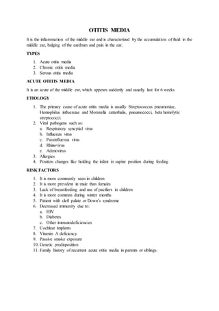
1. OTITIS MEDIA
It is the inflammation of the middle ear and is characterized by the accumulation of fluid in the
middle ear, bulging of the eardrum and pain in the ear.
TYPES
1. Acute otitis media
2. Chronic otitis media
3. Serous otitis media
ACUTE OTITIS MEDIA
It is an acute of the middle ear, which appears suddenly and usually last for 6 weeks
ETIOLOGY
1. The primary cause of acute otitis media is usually Streptococcus pneumoniae,
Hemophilus influenzae and Moraxella catarrhalis, pneumococci, beta hemolytic
streptococci.
2. Viral pathogens such as:
a. Respiratory syncytial virus
b. Influenza virus
c. Parainfluenza virus
d. Rhinovirus
e. Adenovirus
3. Allergies
4. Position changes like holding the infant in supine position during feeding
RISK FACTORS
1. It is more commonly seen in children
2. It is more prevalent in male than females
3. Lack of breastfeeding and use of pacifiers in children
4. It is more common during winter months
5. Patient with cleft palate or Down’s syndrome
6. Decreased immunity due to:
a. HIV
b. Diabetes
c. Other immunodeficiencies
7. Cochlear implants
8. Vitamin A deficiency
9. Passive smoke exposure
10. Genetic predisposition
11. Family history of recurrent acute otitis media in parents or siblings.
2. PATHOPHYSIOLOGY
CLINICAL MANIFESTATIONS
1. Otalgia: severe deep throbbing pain
2. Local engorgement of the blood vessels
3. Swelling of the mucous membrane lining
4. Serous exudates develop
5. Sensation of fullness
6. Fever
7. Tinnitus may occur
8. Drainage/ discharge from the ear
9. Exudate may cause the eardrum to rupture
10. Hearing loss
In Children
1. Young children with otitis media may be irritable, fussy or have problems in feeding
or sleeping
2. Older children exhibit the pain and fullness in the ear
3. Fever
4. Signs of upper respiratory infection, such as runny or stuffy nose or a cough
5. Rupture of eardrum
6. The pus then drains from the middle ear into the ear canal
DIAGNOSTIC EVALUATION
1. History collection
2. Physical examination
3. 3. Tympanocentesis is used to detect presence of middle ear fluid. Ear drainage swab is
sent to laboratory for culture in order to identify pathogens
4. Computed tomography of the temporal bones may identify mastoiditis, epidural
abscess, sigmoid sinus thrombophlebitis, meningitis, brain abscess, subdural abscess,
ossicular disease.
5. Magnetic resonance imaging is helpful to confirm fluid collections in the middle ear
MANAGEMENT
Medical management
1. Administer analgesics and antipyretic to the patient.
2. Provide complete bed rest to the patient
3. Administer antibiotics to control infection
4. Administration of nasal vasoconstrictors to open blocked Eustachian tubes and
application of dry heat etc.
SURGICAL MANAGEMENT
1. Myringotomy and Tympanotomy
An incision in the tympanic membrane known as myringotomy or tympanotomy, may
be performed to permit fluid that has collected in the middle ear to drain
NURSING MANAGEMENT
1. Aspirate the fluid and send for culture following tympanotomy
2. Place cotton loosely in the outer ear to collect drainage
3. Change the cotton when it becomes moist to lessen the danger of secondary infection
4. Discharge may be infectious, so wash hands after changing cotton plugs or cleaning
the ear
5. Monitor vital signs
6. Antibiotics should be continued for several days even though symptoms have
subsided
7. Assess pain level, administer prescribed analgesics and divert the patient
COMPLICATIONS
1. Hearing loss
2. Tympanic membrane rupture
3. Cholesteatoma (cyst like mass in middle ear)
4. Tympanosclerosis
5. Mastoditis
6. Labyrinthitis
7. Facial paralysis
8. Cholesterol granuloma
4. CHRONIC OTITIS MEDIA
It is a chronic inflammation of the middle ear with tissue damage
a. It is characterized by chronic purulent discharge from the middle ear. It is the result of
repeated episodes of acute otitis media causing irreversible tissue pathology, and a
persistent perforation of the tympanic membrane.
ETIOLOGY
It occurs as a result of inadequate treatment of acute otitis media. It is caused by:
1. Streptococcus-group A beta hemolytic streptococci
2. Staphylococcus
3. Proteus
4. Pseudomonas organisms are the most common.
RISK FACTORS
1. Bacteria more often affect children with chronic suppurative otitis media
2. Immunocompromised persons are at high risk
PATHOPHYSIOLOGY
CLINICAL MANIFESTATIONS
1. Deafness
2. Pain less or dull ache: pain occurs occasionally
3. Dizziness
4. Odorless or foul-smelling ear discharge
5. Tenderness of mastoid
6. Fever, post-auricular erythema and edema
7. Cholesteatoma (it is an ingrowth of skin of the external layer of the eardrum into the
middle ear)
5. 8. Decreased or absent tympanic membrane mobility(tympanosclerosis)
9. Conductive hearing loss.
DIAGNOSTIC EVALUATION
1. History collection
2. Ear examination, audiometric test to check for hearing loss, otoscopy to check tympanic
membrane
3. Culture and sensitivity test of ear discharge
4. X-ray to check mastoid
5. Computed tomography of the temporal bones may identify infection and cholesteatoma
6. Magnetic Resonance Imaging is helpful to confirm fluid collections in the middle ear.
MANAGEMENT
Medical Management
1. Local treatment consists of careful suctioning of the ear under microscopic guidance
2. Instillation of antibiotic and steroid drops or application of antibiotic powder is used to
treat a purulent discharge ear drops containing neomycin, tobramycin, ciprofloxacin are
instilled into middle ear.
3. Systemic antibiotics are usually not prescribed except in case of acute infection or when
mastoditis develops. IV antibiotics like ampicillin, sulbactam and cefuroxime.
NURSING MANAGEMENT
1. Explain the procedure to client and family members
2. Aspirate the fluid and send for culture following tympanotomy
3. Place cotton loosely in the outer ear to collect drainage
4. Change the cotton when it becomes moist to lessen the danger of secondary infection
5. Discharge may be infectious, so wash hands after changing cotton plugs or cleaning the
ear
6. Onitor vital signs
7. Antibiotics should be continued for several days even though symptoms have subsided
8. Assess pain level, administer prescribed analgesics and divert the patient
COMPLICATIONS
1. Hearing loss
2. Tympanic membrane rupture
3. Labyrinthitis
4. Temporal abscess
5. Meningitis
6. Intracranial abscess
6. SEROUS OTITIS MEDIA
It is also called otitis media with effusion or non-suppurative otitis media.
In this type of otitis media, no affected fluid accumulates in the middle ear. This condition is
found in primarily in children.
ETIOLOGY
1. Viral upper respiratory infection
2. Residual otitis media, inadequate treatment of acute suppurative otitis media
3. Allergy
a. Allergic rhinitis infection
b. Chronic sinus infection
4. Enlarged lymphoid tissue: adenoidal tissue growth
5. Pressure injury caused by an inability to equalize presume between environment and
middle ear.
PATHOPHYSIOLOGY
7. CLINICAL MANIFESTATIONS
1. Many patients are asymptomatic
2. Sensation of fullness in affected ear
3. Popping, cracking, bubbling or clicking sounds with swallowing and jaw movement
4. Hearing an echo while speaking
5. Having a vague feeling of top heaviness and tympanic membrane retraction
6. Slide conductive hearing loss ranging from 15 to 35 Db
7. Budging tympanic membrane without any redness on otoscopic examination.
DIAGNOSTIC EVALUATION
1. History collection
2. Physical examination
3. Audiometric studies, Rinne test, Weber test, whisper test to check hearing
4. Imaging tests may be done.
MANAGEMENT
1. Inflation of Eustachian tube several times per day using Valsalva maneuver may be the
only treatment required.
2. Nasopharyngeal decongestant therapy may be helpful
3. If medical management fails, then myringotomy and aspiration of middle ear fluid may
be needed.
4. Treat the underlying cause:
a. Treatment of allergies
b. Adenoidectomy for hypertrophied adenoids
c. Adequate treatment of upper respiratory infections and otitis media.
NURSING MANAGEMENT
1. Instruct the patient to perform Valsalva’s maneuver several times daily to maintain
Eustachian tube patency
2. Instruct patient to get prompt treatment of otitis media to prevent further complications
3. Instruct patient about medication, their correct administration
4. If a nasopharyngeal decongestant is prescribed teach correct instillation.
5. Instruct parents not to feed their infant in a supine position or put him/her to bed with
a feeding bottle.
COMPLIATIONS
1. Hearing loss
2. Speech impairment