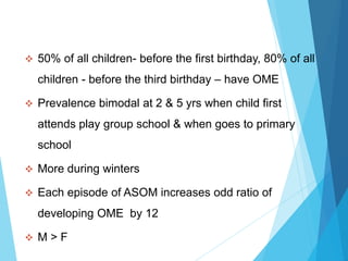
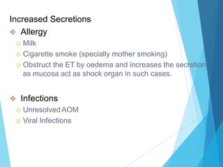
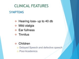
3. DEFINITION
Chronic accumulation of non purulent serous or
mucoid fluid within middle ear cleft.
Time that fluid has to be present for the condition to be
chronic is usually taken as 12 weeks.
Mostly affects children
Insidious onset
Sterile in nature.
Behind an intact and retracted TM.
With conductive hearing loss
4. 50% of all children- before the first birthday, 80% of all
children - before the third birthday – have OME
Prevalence bimodal at 2 & 5 yrs when child first
attends play group school & when goes to primary
school
More during winters
Each episode of ASOM increases odd ratio of
developing OME by 12
M > F
5. AETIOLOGY
ET Malfunctioning.
Obstruction
Adenoid hyperplasia
Tumours (nasopharyngeal carcinoma)
Palatal defects
Palatal paralysis
Hyperbaric oxygen therapy
Oedema during radiation therapy
Spread of Infection
Chronic adenoiditis
Chronic rhino-sinusitis
Chronic tonsillitis
High prevalence in HIV patients
6. Increased Secretions
Allergy
Milk
Cigarette smoke (specially mother smoking)
Obstruct the ET by oedema and increases the secretion
as mucosa act as shock organ in such cases.
Infections
Unresolved AOM
Viral Infections
7. PATHOGENESIS
Eustachian tube dysfunction
Failure of aeration
Failure of drainage
Increased secretion in ME
Due to Increase in secretory glands
Spontaneous resolution if
Drainage via ET restablished
Perforation of the tympanic membrane ocure
If both continue OME
8. CLINICAL FEATURES
Hearing loss- up to 40 db
Mild otalgia
Ear fullness
Tinnitus
Children
Delayed Speech and defective speech
PoorAcademics
SYMPTOMS
9. Clinical Features: Signs
Conductive hearing Loss - TFT
Otoscopy
-Signs of TM retraction.
-Loss of light reflex
-Colour – Yellow/Grey/ Blue
-Thin blood vessel along handle of malleus ant periphery of tm
Signs of Effusion
Air Bubbles
Fluid Levels
10. Pneumatic Otoscopy
Used to assess the mobility and position of TM
Observe TM movement by
Increasing Pressure in EAM – Siegel’s speculum
Increasing pressure in ME - Valsalva namoeuver
TM may be Mobile/ Partially Mobile/ Immobile
11. Evaluation
TFT-
Rinne - negative
Weber- lateralized towards more diseased ear.
ABC –normal both ear
Audiometry
Pure ToneAudiometry
Bilateral Conductive Hearng Loss
20 – 40dB
Sometimes SNHL due to fluid pressing round window
12. Tympanometry (sensitivity 96 %)
Assess compliance of TM
Mobility of TM on increase/ decrease of pressure in EAM
Graphic representation
4 patterns
A/As/Ad/B/C
In OME – B & C
Reduced compliance with flat curve with a shift to
negative side.
14. Radiology
Xray Skull Lateral view
Adenoid Hyperplasia
Xray Mastoid Schuller’s View
Clouding of air cell
MRI
Absence of fluid does not imply an absence of OME, as
one-third of patients in MRI study had fluid in mastoid, but
not in the mesotympanum (Kew et al)
Nasal and nasopharyngeal examination.
16. Anti-allergy measures
Antihistamines
Nasal Steroids spray
Surface tension lowering agent
N acetyl cyteine 30 mg tds X 15 days beneficial
Antibiotics (no long term benefit, can be used for initial 2 wks)
Middle Ear Aeration
Valsalva Maneuver
17. Surgical Treatment
Myringotomy
Myringotomy with ventillation tubes (improves hearing by 12
dB)
Adenoidectomy (improves hearing by 8 dB)
Tonsillectomy
Cortical Matoidectomy (in failure of ventilation tube cases)
18. Ventilation tubes
Also known as grommet
Inserted through radial incision in AS or AI quadrant of
TM.
Longer a tube stays in situ longer it can be potentially
benefit
On other hand, longer a tube is in situ the greater the
chance of complications like-
Infection
Granulation tissue
Permanent perforation
Thinning of TM with possible retraction
19. TYPES
Shepard Armstrong
Reuter Bobbin
Goodie t tube
Silicone T tube
Shah tube
FUNCTION
Ventilate the middle ear
Drain the middle ear
Improve the hearing
Epithelium will revert back to normal
20. Complications of ventilation tubes
Intra op
Displacement into middle ear
Damage to ossicles
Early post op
Blockage
Granulation around tube
Ear infection
Otorrhoea
Late post op
Permanent perforation
Tympanosclerosis
TM atrophy & retraction