Downloaded 164 times
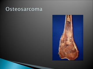


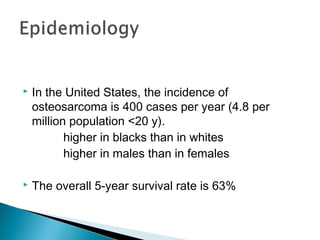
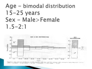
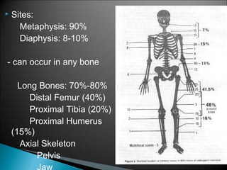
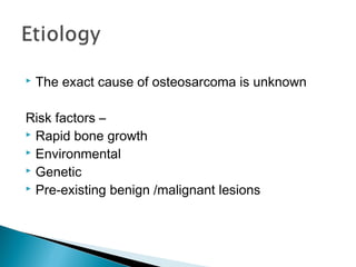
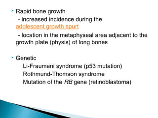
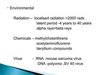
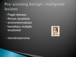
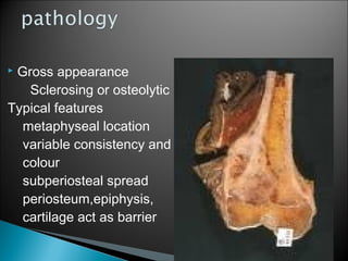
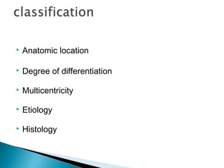
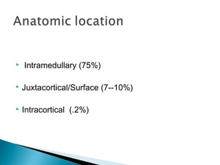
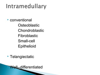
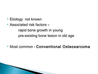
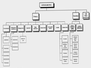


Osteosarcoma is a malignant bone tumor characterized by the production of osteoid bone. It is the second most common primary bone malignancy and occurs most often in the metaphysis of long bones in adolescents and young adults. The tumor may be conventional, presenting as osteoblastic, chondroblastic or fibroblastic subtypes, or may be a rare telangiectatic or well-differentiated variant. While the exact cause is unknown, risk factors include rapid bone growth, genetic conditions, radiation exposure and pre-existing bone diseases.


![Osteosarcoma[2]](https://cdn.slidesharecdn.com/ss_thumbnails/osteosarcoma2-130423123803-phpapp01-thumbnail.jpg?width=640&height=640&fit=bounds)


