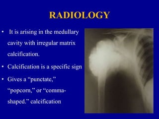
Moderator :Director & HOD
PROF.DR.K.PRAKASAM M.S.Ortho,D.Ortho,DSC (HON)
PRESENTOR:DR.THOUSEEF.A.MAJEED
M S Ortho PG
VMKVMCH Salem
CHONDROsARCOMA
• It is a malignant bone tumour of cartilageneous
origin
• 9% of all primary malignancies of bone
• Primary chondrosarcoma - Between 40 and 60 years
• Secondary chondrosarcoma-Between 25 and 45
years
Classification
• PRIMARY CHONDROSARCOMA
• SECONDARY CHONDROSARCOMA
PRIMARY CHONDROSARCOMA
• Central (medullary)
• Intra cortical
• Clear cell
• Mesenchymal
• Dedifferentiated
SECONDARY CHONDROSARCOMA (from
pre existing lesions)
• Multiple enchondromas & Exostosis
• Chondroblastoma
• Irradiation induced
• Fibrous dysplasia
Location
Types according to the site
• CENTRAL
• PERIPHERAL (surface type)
COMMON SITES
• Pelvis
• Proximal femur
• Proximal humerus.
• Ribs
• Rarely occur in the hand
• Incidence is higher among males
CLINICAL FEATURES
• A palpable mass with
increasing pain
• Hard in consistency with
lobulated surface
• Continuous with the bone
• slow growing .
• Mechanical restriction of joint
movements.
Secondary chondrosarcomas
• Arise at the site of a pre-existing benign cartilage
lesion.
• Most frequently in the enchondromas and
multiple hereditary exostoses.
INCIDENCE –
• 5% for patients with multiple hereditary exostoses
• Approximately 1% for patients with solitary
osteochondromas
RADIOLOGY
• It is arising in the medullary
cavity with irregular matrix
calcification.
• Calcification is a specific sign
• Gives a “punctate,”
“popcorn,” or “comma-
shaped.” calcification
• The size of the cartilaginous cap of an
osteochondroma, is to be evaluated with
Computerised Tomography (CT-Scan) or Magnetic
resonance imaging (MRI).
• It is important in evaluating the possibility of a
secondary chondrosarcoma.
HISTOLOGY
CONVENTIONAL CHONDROSARCOMA
• Composed of malignant cells
with abundant cartilaginous
matrix.
• If malignant osteoid is present
 the diagnosis should be
chondroblastic osteosarcoma
Diagnosing Factors that favor the malignant
transformation
• Hypercellularity
• Plump nuclei
• More than occasional binucleate cells
• Entrapment of bony trabeculae.
Histological subtypes
• Clear cell chondrosarcoma
• Mesenchymal chondrosarcoma
• Dedifferentiated chondrosarcoma
Clear cell chondrosarcoma
• Low-grade malignancy.
• Round cells with abundant
clear cytoplasm
• Distinct cytoplasmic
borders with a background
of cartilaginous matrix.
• Multinucleated giant cells
are usually apparent.
• Clear cell Chondrosarcoma has a strong tendency to
arise in an epiphysis
• Benign radiographic features and can be confused with
chondroblastoma or giant cell tumor.
Mesenchymal chondrosarcoma
• High-grade tumor
• Small round blue cells
• Islands of benign-appearing
cartilage.
• The cellular portions have a
“Hemangiopericytomatous” pattern
of growth with “staghorn-like”
vessels.
Dedifferentiated chondrosarcoma
High-grade sarcoma
• ( Commonly osteosarcoma followed
in frequency by fibrosarcoma and
malignant fibrous histiocytoma)
• The radiography shows aggressive
radiolucent area
Teatment
Low-grade chondrosarcoma
• Extended curettage with the use of intraoperative
adjuvant treatments.
High-grade chondrosarcoma
• Wide or radical resection
• Amputation.
• The local recurrence rate after intraoperative
tumor contamination is high.
• For lesions in an expendable locations,
Primary wide resection without a biopsy may
be indicated
• It is to decrease the chance of tumor
contamination.
• After wide resection  local recurrence is
less than 10%
• Pulmonary metastases Treated with
surgical resection if possible.
• Chemotherapy has no role in the treatment of
conventional chondrosarcoma
• Radiotherapy is used only as a palliative measure
for surgically inaccessible lesions.
Inaccessible areas include
• Tumour arising from floor of pelvis
Vertebrae &spinous process
PROGNOSIS
The prognosis depends on
• Size
• Grade
• Location of the lesion.
Low-grade lesions
• Greater than 90% 10-year survival rate
High-grade conventional chondrosarcoma
• 20% to 40% 10-year survival rate
Chondrosarcoma

Chondrosarcoma

  • 1.
    Moderator :Director &HOD PROF.DR.K.PRAKASAM M.S.Ortho,D.Ortho,DSC (HON) PRESENTOR:DR.THOUSEEF.A.MAJEED M S Ortho PG VMKVMCH Salem CHONDROsARCOMA
  • 2.
    • It isa malignant bone tumour of cartilageneous origin
  • 3.
    • 9% ofall primary malignancies of bone • Primary chondrosarcoma - Between 40 and 60 years • Secondary chondrosarcoma-Between 25 and 45 years
  • 4.
  • 5.
    PRIMARY CHONDROSARCOMA • Central(medullary) • Intra cortical • Clear cell • Mesenchymal • Dedifferentiated
  • 6.
    SECONDARY CHONDROSARCOMA (from preexisting lesions) • Multiple enchondromas & Exostosis • Chondroblastoma • Irradiation induced • Fibrous dysplasia
  • 7.
    Location Types according tothe site • CENTRAL • PERIPHERAL (surface type)
  • 8.
    COMMON SITES • Pelvis •Proximal femur • Proximal humerus. • Ribs • Rarely occur in the hand • Incidence is higher among males
  • 9.
    CLINICAL FEATURES • Apalpable mass with increasing pain • Hard in consistency with lobulated surface • Continuous with the bone • slow growing . • Mechanical restriction of joint movements.
  • 10.
    Secondary chondrosarcomas • Ariseat the site of a pre-existing benign cartilage lesion. • Most frequently in the enchondromas and multiple hereditary exostoses.
  • 11.
    INCIDENCE – • 5%for patients with multiple hereditary exostoses • Approximately 1% for patients with solitary osteochondromas
  • 12.
    RADIOLOGY • It isarising in the medullary cavity with irregular matrix calcification. • Calcification is a specific sign • Gives a “punctate,” “popcorn,” or “comma- shaped.” calcification
  • 14.
    • The sizeof the cartilaginous cap of an osteochondroma, is to be evaluated with Computerised Tomography (CT-Scan) or Magnetic resonance imaging (MRI). • It is important in evaluating the possibility of a secondary chondrosarcoma.
  • 15.
  • 16.
    CONVENTIONAL CHONDROSARCOMA • Composedof malignant cells with abundant cartilaginous matrix. • If malignant osteoid is present  the diagnosis should be chondroblastic osteosarcoma
  • 17.
    Diagnosing Factors thatfavor the malignant transformation • Hypercellularity • Plump nuclei • More than occasional binucleate cells • Entrapment of bony trabeculae.
  • 18.
    Histological subtypes • Clearcell chondrosarcoma • Mesenchymal chondrosarcoma • Dedifferentiated chondrosarcoma
  • 19.
    Clear cell chondrosarcoma •Low-grade malignancy. • Round cells with abundant clear cytoplasm • Distinct cytoplasmic borders with a background of cartilaginous matrix. • Multinucleated giant cells are usually apparent.
  • 20.
    • Clear cellChondrosarcoma has a strong tendency to arise in an epiphysis • Benign radiographic features and can be confused with chondroblastoma or giant cell tumor.
  • 21.
    Mesenchymal chondrosarcoma • High-gradetumor • Small round blue cells • Islands of benign-appearing cartilage. • The cellular portions have a “Hemangiopericytomatous” pattern of growth with “staghorn-like” vessels.
  • 22.
    Dedifferentiated chondrosarcoma High-grade sarcoma •( Commonly osteosarcoma followed in frequency by fibrosarcoma and malignant fibrous histiocytoma) • The radiography shows aggressive radiolucent area
  • 23.
    Teatment Low-grade chondrosarcoma • Extendedcurettage with the use of intraoperative adjuvant treatments.
  • 24.
    High-grade chondrosarcoma • Wideor radical resection • Amputation.
  • 25.
    • The localrecurrence rate after intraoperative tumor contamination is high. • For lesions in an expendable locations, Primary wide resection without a biopsy may be indicated • It is to decrease the chance of tumor contamination.
  • 26.
    • After wideresection  local recurrence is less than 10% • Pulmonary metastases Treated with surgical resection if possible.
  • 27.
    • Chemotherapy hasno role in the treatment of conventional chondrosarcoma • Radiotherapy is used only as a palliative measure for surgically inaccessible lesions. Inaccessible areas include • Tumour arising from floor of pelvis Vertebrae &spinous process
  • 28.
    PROGNOSIS The prognosis dependson • Size • Grade • Location of the lesion.
  • 29.
    Low-grade lesions • Greaterthan 90% 10-year survival rate High-grade conventional chondrosarcoma • 20% to 40% 10-year survival rate