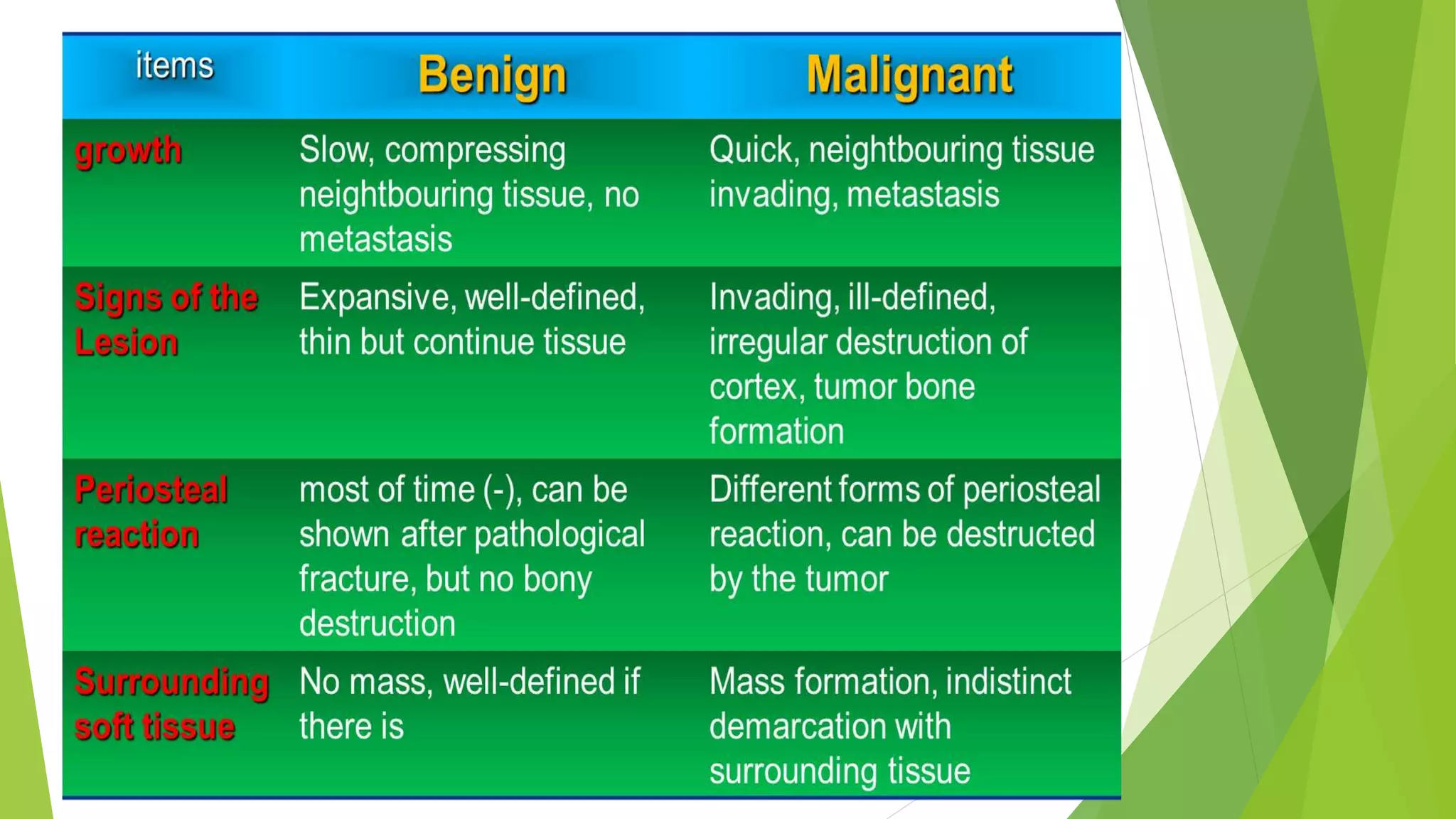
Bone tumors an tumor like lesions
BY DR ALIYA ZAMAN
 Most bone tumors develop during the first decades of life
 Propensity to originate in the long bones of the
extremities.
 Most osteosarcomas occur during adolescence, with half
arising around the knee, either in the distal femur or
proximal tibia.
 In contrast, chondrosarcomas tend to develop during mid-
to late adulthood and involve the trunk, limb girdles, and
proximal long bones.
 Most bone tumors arise without any prior known cause.
 Benign lesions are frequently asymptomatic and are
detected as incidental findings
Osteoma
 Osteomas are benign lesions of bone
 They are most commonly encountered in the head and neck,
including the paranasal sinuses
 but can occur elsewhere as well
 they are typically seen in middle age.
 usually solitary and present as localized, slowly growing, hard,
exophytic masses on the bone surface.
 Multiple lesions are a feature of Gardner syndrome
 Histologically, osteomas are a bland mixture of woven and lamellar
bone.
 Although they may cause local mechanical problems (e.g., obstruction
of a sinus cavity) and cosmetic deformities, they are not invasive and
do not undergo malignant transformation.
Osteoid osteoma and osteoblastoma
 Both have identical histological features but differ in size
,site of origin and symptoms.
 Osteoid osteomas arise most often in the proximal femur
and tibia, and are by definition less than 2 cm, whereas
osteoblastomas are larger.
 Localized pain is an almost universal complaint with
osteoid osteomas, and is usually relieved by aspirin.
 Osteoblastomas arise most often in the vertebral column;
they also cause pain, although it is often more difficult to
localize and is not responsive to aspirin.
 Osteoid osteoma treated by radiofrequency ablation
osteoblastoma curetted by excised en bloc.
 Malignant transformation is rare
Morphology
 Grossly,
 Both lesions are round-to-oval masses of hemorrhagic gritty tan
tissue.
 A rim of sclerotic bone is present at the edge of both types of
tumors; however, it is much more conspicuous in osteoid
osteomas.
 Microscopically,
 Both neoplasms are composed of interlacing trabeculae of
woven bone surrounded by osteoblasts.
 The intervening stroma is loose, vascular connective
tissue containing variable numbers of giant cells.
 Small osteolytic lesion (up to 1.5 cm) with or without central
calcification.
 Surrounded by a prominent zone of reactive sclerosis due to a
periosteal and endosteal reaction, which may obscure the central
nidus.
 In juxta-articular localisation, the reactive sclerosis may be absent.
osteosarcoma Osteosarcoma is a bone-producing malignant
mesenchymal tumor.
 Osteosarcoma is the most common primary malignant
tumor of bone,
 Accounting for approximately 20% of primary bone
cancers
 2000 cases are diagnosed annually in the United States.
 Osteosarcomas occur in all age groups but some 75% of
patients are younger than age 20, with a second peak
occuring in the elderly,
 usually with other conditions, including Paget disease,
bone infarcts, and prior irradiation.
 Men are more commonly affected than women (1.6 : 1)
 Although any bone can be involved, most tumors arise in the
metaphyseal region of the long bones of the extremities, with
 almost 60% occurring about the knee,
 15% around the hip, 10% at the shoulder,
 and 8% in the jaw.
 Several subtypes of osteosarcoma are recognized on the basis of the
site of involvement within the bone (e.g.,
medullary vs cortical),
degree of differentiation,
solitary vs multicentric,
presence of underlying disease, and
histologic variants;
 THE MOST COMMON TYPE OF OSTEOSARCOMA IS PRIMARY, SOLITARY,
INTRAMEDULLARY, AND POORLY DIFFERENTIATED, PRODUCING A
PREDOMINANTLY BONY MATRIX.
PATHOGENESIS
 70% has genetic mutaion
 RB ,,germline mutation cause 1000 folds
increased risk of osteosarcoma and in sporadic
cases 70% shows presence of RB mutation.
 TP53 mutation
 Patient with LI-Fraumeni syndrome
 INK4alpha ,,it encode two tumor suppressor gene
 MDM2 and CDK4 ,,,which inhibit p53 and RB
function
Morphology
GROSSLY
 osteosarcomas are gritty, gray-white tumors, often
exhibiting hemorrhage and cystic degeneration.
 Tumors frequently destroy the surrounding cortices
and produce soft tissue masses.
 They spread extensively in the medullary canal,
infiltrating and replacing the marrow, but only
infrequently penetrating the epiphyseal plate or
entering the joint space
MICROSCOPICALLY
 Tumor cells vary in size and shape, and frequently have large
hyperchromatic nuclei; bizarre tumor giant cells are common,
as are mitoses.
 The production of mineralized or unmineralized bone
(osteoid) by malignant cells is essential for diagnosis of
osteosarcoma.
 The neoplastic bone is typically coarse and ragged but can also
be deposited in broad sheets.
 Cartilage and fibrous tissue can also be present in varying
amounts.
 When malignant cartilage is abundant, the tumor is called a
chondroblastic osteosarcoma.
 Vascular invasion is common, as is spontaneous tumor necrosis.
Clinical course
 Osteosarcomas typically present as painful enlarging masses,
although a pathologic fracture can be the first symptom.
 Radiographs usually show a large, destructive, mixed lytic and
blastic mass with indistinct infiltrating margins.
 The tumor frequently breaks through the cortex and lifts the
periosteum, resulting in reactive periosteal bone formation.
 A triangular shadow on x-ray between the cortex and raised
periosteum (Codman triangle) is characteristic of
osteosarcomas. Osteosarcomas typically spread
hematogenously; at the time of diagnosis, approximately 10% to
20% of patients have demonstrable pulmonary metastases.
Pulmonary metastasis is very common
10-20% cases
 5 yrs survival rate is 60-70%
 90% of patient who died due to osteosarcoma has
lungs metastasis
 Other metastatic organs are bones,brain,and
elsewhere
 With mets survival rate is <20%
 Secondary osteosarcomas occur in an older age
group than do primary osteosarcomas. They most
commonly develop in the setting of Paget disease
or previous radiation exposure. Secondary
osteosarcomas are highly aggressive tumors that
do not respond well to therapy
CARTILAGE FORMING
TUMORS
OSTEOCHNODROMA
CHOMDROMAS
CHOMDROSARCOMA
 These neoplasms produce hyaline or myxoid
cartilage
 fibrocartilage and elastic cartilage are rare
components
 Benign cartilage tumors are much more common
than malignant ones.
OSTEOCHONDROMA
 Also called exostoses
 Relatively common benign cartilage-capped outgrowths
attached by a bony stalk to the underlying skeleton.
 Solitary osteochondromas are usually first diagnosed in
late adolescence and early adulthood
 male-to-female ratio of 3 : 1
 Multiple osteochondromas become apparent during
childhood, occurring as multiple hereditary exostosis, an
autosomal dominant disorder.
 Inactivation of both copies of the EXT gene in
chondrocytes is implicated in both sporadic and hereditary
osteochondromas.
 Osteochondromas develop only in bones of
endochondral origin arising at the metaphysis
near the growth plate of long tubular bones,
especially about the knee
 Occasionally they develop from bones of the
pelvis, scapula, and ribs, and in these sites are
frequently sessile.
 Rarely, exostoses involve the short tubular bones
of hands and feet.
MORPHOLOGY
 Osteochondromas vary from 1-20cm in size.
 The cap is benign hyaline cartilage, resembling
disorganized growth plate undergoing endochondral
ossification.
 Newly formed bone forms the inner portion of the head
and stalk,
 with the stalk cortex merging with the cortex of the host
bone.
CLINICAL FEATURES
 Osteochondromas are slow-growing masses
 that are painful when they impinge on a nerve or if the
stalk is fractured.
 In many cases, they are detected only incidentally.
 In multiple hereditary exostosis, deformity of the
underlying bone suggests an associated disturbance in
epiphyseal growth.
 Osteochondromas rarely progress to chondrosarcoma or
other sarcoma
 patients with the hereditary syndrome are at increased
risk of malignant transformation.
 Chondromas are benign tumors of hyaline cartilage.
 When they arise within the medulla, they are termed
ENCHONDROMAS
 when on the bone surface they are called juxtacortical
chondromas.
 Enchondromas are usually diagnosed in persons between
ages 20 and 50
 they are typically solitary and located in the metaphyseal
region of tubular bones,
 the favored sites being the short tubular bones of the
hands and feet.
Ollier disease is characterized by multiple chondromas
preferentially involving one side of the body, and
Maffucci syndrome is characterized by multiple chondromas
associated with benign soft tissue angiomas.
MORPHOLOGY
 Enchondromas are gray-blue, translucent nodules
 usually smaller than 3 cm.
Microscopically
 well-circumscribed hyaline matrix and cytologically
benign chondrocytes.
 At the periphery, there is endochondral ossification, while
the center frequently calcifies and dies.
 In the hereditary multiple chondromatoses, the islands of
cartilage exhibit greater cellularity and atypia, making
them difficult to distinguish from chondrosarcoma.
CLINICAL FEATURES
 Most enchondromas are detected as incidental findings;
 occasionally they are painful or cause pathologic fractures.
 On x-ray, the unmineralized nodules of cartilage produce well-
circumscribed oval lucencies surrounded by thin rims of radiodense
bone (O-ring sign).
 Calcified matrix exhibits irregular opacities.
 The growth potential of chondromas is limited, and most remain
stable, although they can recur if incompletely excised.
 Solitary chondromas rarely undergo malignant transformation
 those associated with enchondromatoses are at increased risk.
Maffucci syndrome is associated with an increased risk of
developing other types of malignancies, including ovarian carcinomas and
brain gliomas.
CHONDROSARCOMA
 It comprise a variety of tumors sharing the ability to
produce neoplastic cartilage
 They are subclassified according to site (e.g.,
intramedullary vs juxtacortical), and histologic variants.
 Occur roughly half as frequently as osteosarcomas
 Most patients are age 40 or older
 Men affected twice as frequently as women.
MORPHOLOGY
 Conventional chondrosarcomas arise within the medullary cavity of
the bone to form an expansile glistening mass that often erodes the
cortex
 translucent firm, bluish gray lesion,
with smooth, shiny cut surfaces.
 They exhibit malignant hyaline and myxoid cartilage.
In myxoid chondrosarcomas
 the tumors are viscous and gelatinous
 matrix oozes from the cut surface
 Spotty calcifications are typically present
 central necrosis can create cystic spaces
 The adjacent cortex is thickened or eroded
 Tumor grade is determined by
cellularity,
cytologic atypia, and
mitotic activity
 Low-grade tumors resemble normal cartilage.
 Higher grade lesions contain pleomorphic chondrocytes
with frequent mitotic figures.
 Multinucleate cells are present with lacunae containing
two or more chondrocytes.
 Approximately 10% of patients with conventional low-grade
chondrosarcomas have a second high-grade poorly differentiated
component (dedifferentiated chondrosarcomas) that include foci of
fibro- or osteosarcomas.
 Other histologic variants include clear-cell and mesenchymal
chondrosarcomas.
CLINICAL FEATURES
 Commonly arise in the pelvis, shoulder, and ribs
 Chondrosarcomas rarely involve the distal extremities.
 They typically present as painful, progressively enlarging masses.
 A slowly growing low-grade tumor causes reactive thickening of the
cortex
 The more radiolucent the tumor the greater the likelihood that it is
high grade.
 Conventional chondrosarcomas are indolent and low-grade,
 with a 5-year survival rate of 80% to 90% (vs 43% for grade 3 tumors)
 Grade 1 tumors rarely metastasize
 whereas 70% of the grade 3 tumors disseminate.
 Size is another prognostic feature, with tumors larger than
10 cm being significantly more aggressive than smaller
tumors.
 Chondrosarcomas metastasize hematogenously,
preferentially to the lungs and skeleton.
 Conventional chondrosarcomas are treated with wide
surgical excision
 Chemotherapy is added for the mesenchymal and
dedifferentiated variants because of their aggressive
clinical course.
Tumors of unknown origin
Eving sarcoma family tumors
Giant cell tmors
Aneurysmal bone cyst
EWING SARCOMA and Primitive
Neuroectodermal Tumor
 Primary malignant small round-cell tumors of bone and
soft tissue.
 Because they share an identical chromosome
translocation, they should be viewed as the same tumor,
differing only in degree of differentiation.
 PNETs demonstrate neural differentiation whereas Ewing
sarcomas are undifferentiated by traditional marker
analysis.
 These two malignancies account for 6% to 10% of
primary malignant bone tumors.
 Youngest age of presentation
 80% of diagnosed cases have age younger than 20
 Most patients are 10 to 15 years old
 Boys are affected slightly more frequently than
girls
 Whites are more affected
 Translocation that causes fusion of the EWS gene
on 22q12 with a member of the ETS family of
transcription factors.
 Approximately 95% of patients with Ewing tumor
have t(11;22) (q24;q12) or t(21;22) (q22;q12).
Morphology
 Ewing sarcoma and PNETs arise in the medullary cavity
 invade the cortex and periosteum to produce a soft tissue
mass.
 Gross---The tumor is tan-white, frequently with
hemorrhage and necrosis.
 Histologically----
 It is composed of sheets of uniform small, round cells
that are slightly larger than lymphocytes with few mitoses
and little intervening stroma .
 The cells have scant glycogen-rich cytoplasm.
 The presence of Homer-Wright rosettes (tumor cells
circled about a central fibrillary space) indicates neural
differentiation.
Clinical features
 Ewing sarcoma and PNETs typically present as painful
enlarging masses in the diaphyses of long tubular bones
(especially the femur) and the pelvic flat bones.
 Some patients have systemic signs and symptoms,
including fever, elevated erythrocyte sedimentation rate,
anemia, and leukocytosis that can mimic infection.
 X-rays show a destructive lytic tumor with infiltrative
margins and extension into surrounding soft tissues.
 There is a characteristic periosteal reaction depositing
bone in an onionskin fashion.
 Treatment includes chemotherapy and surgical excision
with or without radiation.
 The 5-year survival is currently 75%, with 50% achieving
GIANT-CELL TUMOR OF BONE
Osteoclastoma
 GCT is relatively uncommon
 it is benign but locally aggressive
 usually arising in individuals in their 20s to 40s.
 It is suggested that the giant cell component is
likely a reactive macrophage population and the
mononuclear cells are neoplastic.
 The latter show complex cytogenetic
abnormalities.
Morphology
 Tumors are large and red-brown with frequent cystic
degeneration.
 They are composed of uniform oval mononuclear cells
with frequent mitoses, with scattered osteoclast-type
giant cells containing 100 or more nuclei
 Necrosis, hemorrhage, and reactive bone formation are
also commonly present.
CLINICAL FEATURES
 Although almost any bone can be involved, the majority of
GCTs arise in the epiphysis of long bones around the knee
(distal femur and proximal tibia), frequently causing
arthritis-like symptoms.
 Occasionally, GCTs present as pathologic fractures.
 Most are solitary.
 Radiographically, GCTs are large, purely lytic, and
eccentric
 The overlying cortex is frequently destroyed, producing a
bulging soft tissue mass with a thin shell of reactive bone.
 Although GCTs are histologically benign, roughly half recur
after simple curettage
 4% metastasize to the lungs.
Aneurysmal bone cyst (ABC)
 ABC is tumor characterized by multioculated blood-filled
cystic spaces.
 Uncommon
 Expanding osteolytic lesion of blood-filled spaces of
variable size separated by connective tissue septa with
osteoclast giant cells and variable reactive bone
 Usually ages 1-20 years
 No gender preference
 Benign but grows rapidly
 Simple curettage is followed by recurrence in 20%
 rarely transforms to osteosarcoma
 Sites: metaphysis of posterior vertebrae (often multiple),
flat bones, shaft of long bones; rarely within wall of major
artery or in soft tissue
 May also be secondary to trauma or arise in preexisting
bone lesion (giant cell tumor, chondroblastoma, fibrous
dysplasia)
 Eccentric expansion of bone, cortical erosion and
destruction, small peripheral area of periosteal bone
formation
GROSS
 Spongy, hemorrhagic mass covered by thin shell of reactive bone
 Small amount of tissue compared to large size of lesion on Xray
MICROSCOPICALLY
 Large cystic spaces filled with blood and separated by fibrous septa,
alternating with solid areas
 Cysts and septa lined by fibroblasts, myofibroblasts and histiocytes
but not endothelium
 Clusters of osteoclast-like multinucleated giant cells with loose
spindly stroma to cellular stroma, reactive woven bone, degenerated
calcifying fibromyxoid tissue
 Variable mitotic figures and hemosiderin
 No malignant osteoid, no atypia
Bone tumour
Bone tumour
Bone tumour
Bone tumour

Bone tumour

  • 1.
    Bone tumors antumor like lesions BY DR ALIYA ZAMAN
  • 2.
     Most bonetumors develop during the first decades of life  Propensity to originate in the long bones of the extremities.  Most osteosarcomas occur during adolescence, with half arising around the knee, either in the distal femur or proximal tibia.  In contrast, chondrosarcomas tend to develop during mid- to late adulthood and involve the trunk, limb girdles, and proximal long bones.  Most bone tumors arise without any prior known cause.  Benign lesions are frequently asymptomatic and are detected as incidental findings
  • 7.
    Osteoma  Osteomas arebenign lesions of bone  They are most commonly encountered in the head and neck, including the paranasal sinuses  but can occur elsewhere as well  they are typically seen in middle age.  usually solitary and present as localized, slowly growing, hard, exophytic masses on the bone surface.  Multiple lesions are a feature of Gardner syndrome  Histologically, osteomas are a bland mixture of woven and lamellar bone.  Although they may cause local mechanical problems (e.g., obstruction of a sinus cavity) and cosmetic deformities, they are not invasive and do not undergo malignant transformation.
  • 9.
    Osteoid osteoma andosteoblastoma  Both have identical histological features but differ in size ,site of origin and symptoms.  Osteoid osteomas arise most often in the proximal femur and tibia, and are by definition less than 2 cm, whereas osteoblastomas are larger.  Localized pain is an almost universal complaint with osteoid osteomas, and is usually relieved by aspirin.  Osteoblastomas arise most often in the vertebral column; they also cause pain, although it is often more difficult to localize and is not responsive to aspirin.  Osteoid osteoma treated by radiofrequency ablation osteoblastoma curetted by excised en bloc.  Malignant transformation is rare
  • 11.
    Morphology  Grossly,  Bothlesions are round-to-oval masses of hemorrhagic gritty tan tissue.  A rim of sclerotic bone is present at the edge of both types of tumors; however, it is much more conspicuous in osteoid osteomas.  Microscopically,  Both neoplasms are composed of interlacing trabeculae of woven bone surrounded by osteoblasts.  The intervening stroma is loose, vascular connective tissue containing variable numbers of giant cells.
  • 12.
     Small osteolyticlesion (up to 1.5 cm) with or without central calcification.  Surrounded by a prominent zone of reactive sclerosis due to a periosteal and endosteal reaction, which may obscure the central nidus.  In juxta-articular localisation, the reactive sclerosis may be absent.
  • 16.
    osteosarcoma Osteosarcoma isa bone-producing malignant mesenchymal tumor.  Osteosarcoma is the most common primary malignant tumor of bone,  Accounting for approximately 20% of primary bone cancers  2000 cases are diagnosed annually in the United States.  Osteosarcomas occur in all age groups but some 75% of patients are younger than age 20, with a second peak occuring in the elderly,  usually with other conditions, including Paget disease, bone infarcts, and prior irradiation.  Men are more commonly affected than women (1.6 : 1)
  • 17.
     Although anybone can be involved, most tumors arise in the metaphyseal region of the long bones of the extremities, with  almost 60% occurring about the knee,  15% around the hip, 10% at the shoulder,  and 8% in the jaw.  Several subtypes of osteosarcoma are recognized on the basis of the site of involvement within the bone (e.g., medullary vs cortical), degree of differentiation, solitary vs multicentric, presence of underlying disease, and histologic variants;  THE MOST COMMON TYPE OF OSTEOSARCOMA IS PRIMARY, SOLITARY, INTRAMEDULLARY, AND POORLY DIFFERENTIATED, PRODUCING A PREDOMINANTLY BONY MATRIX.
  • 19.
    PATHOGENESIS  70% hasgenetic mutaion  RB ,,germline mutation cause 1000 folds increased risk of osteosarcoma and in sporadic cases 70% shows presence of RB mutation.  TP53 mutation  Patient with LI-Fraumeni syndrome  INK4alpha ,,it encode two tumor suppressor gene  MDM2 and CDK4 ,,,which inhibit p53 and RB function
  • 20.
    Morphology GROSSLY  osteosarcomas aregritty, gray-white tumors, often exhibiting hemorrhage and cystic degeneration.  Tumors frequently destroy the surrounding cortices and produce soft tissue masses.  They spread extensively in the medullary canal, infiltrating and replacing the marrow, but only infrequently penetrating the epiphyseal plate or entering the joint space
  • 25.
    MICROSCOPICALLY  Tumor cellsvary in size and shape, and frequently have large hyperchromatic nuclei; bizarre tumor giant cells are common, as are mitoses.  The production of mineralized or unmineralized bone (osteoid) by malignant cells is essential for diagnosis of osteosarcoma.  The neoplastic bone is typically coarse and ragged but can also be deposited in broad sheets.  Cartilage and fibrous tissue can also be present in varying amounts.  When malignant cartilage is abundant, the tumor is called a chondroblastic osteosarcoma.  Vascular invasion is common, as is spontaneous tumor necrosis.
  • 27.
    Clinical course  Osteosarcomastypically present as painful enlarging masses, although a pathologic fracture can be the first symptom.  Radiographs usually show a large, destructive, mixed lytic and blastic mass with indistinct infiltrating margins.  The tumor frequently breaks through the cortex and lifts the periosteum, resulting in reactive periosteal bone formation.  A triangular shadow on x-ray between the cortex and raised periosteum (Codman triangle) is characteristic of osteosarcomas. Osteosarcomas typically spread hematogenously; at the time of diagnosis, approximately 10% to 20% of patients have demonstrable pulmonary metastases.
  • 29.
    Pulmonary metastasis isvery common 10-20% cases
  • 31.
     5 yrssurvival rate is 60-70%  90% of patient who died due to osteosarcoma has lungs metastasis  Other metastatic organs are bones,brain,and elsewhere  With mets survival rate is <20%  Secondary osteosarcomas occur in an older age group than do primary osteosarcomas. They most commonly develop in the setting of Paget disease or previous radiation exposure. Secondary osteosarcomas are highly aggressive tumors that do not respond well to therapy
  • 32.
    CARTILAGE FORMING TUMORS OSTEOCHNODROMA CHOMDROMAS CHOMDROSARCOMA  Theseneoplasms produce hyaline or myxoid cartilage  fibrocartilage and elastic cartilage are rare components  Benign cartilage tumors are much more common than malignant ones.
  • 33.
    OSTEOCHONDROMA  Also calledexostoses  Relatively common benign cartilage-capped outgrowths attached by a bony stalk to the underlying skeleton.  Solitary osteochondromas are usually first diagnosed in late adolescence and early adulthood  male-to-female ratio of 3 : 1  Multiple osteochondromas become apparent during childhood, occurring as multiple hereditary exostosis, an autosomal dominant disorder.  Inactivation of both copies of the EXT gene in chondrocytes is implicated in both sporadic and hereditary osteochondromas.
  • 34.
     Osteochondromas developonly in bones of endochondral origin arising at the metaphysis near the growth plate of long tubular bones, especially about the knee  Occasionally they develop from bones of the pelvis, scapula, and ribs, and in these sites are frequently sessile.  Rarely, exostoses involve the short tubular bones of hands and feet.
  • 35.
    MORPHOLOGY  Osteochondromas varyfrom 1-20cm in size.  The cap is benign hyaline cartilage, resembling disorganized growth plate undergoing endochondral ossification.  Newly formed bone forms the inner portion of the head and stalk,  with the stalk cortex merging with the cortex of the host bone.
  • 40.
    CLINICAL FEATURES  Osteochondromasare slow-growing masses  that are painful when they impinge on a nerve or if the stalk is fractured.  In many cases, they are detected only incidentally.  In multiple hereditary exostosis, deformity of the underlying bone suggests an associated disturbance in epiphyseal growth.  Osteochondromas rarely progress to chondrosarcoma or other sarcoma  patients with the hereditary syndrome are at increased risk of malignant transformation.
  • 41.
     Chondromas arebenign tumors of hyaline cartilage.  When they arise within the medulla, they are termed ENCHONDROMAS  when on the bone surface they are called juxtacortical chondromas.  Enchondromas are usually diagnosed in persons between ages 20 and 50  they are typically solitary and located in the metaphyseal region of tubular bones,  the favored sites being the short tubular bones of the hands and feet. Ollier disease is characterized by multiple chondromas preferentially involving one side of the body, and Maffucci syndrome is characterized by multiple chondromas associated with benign soft tissue angiomas.
  • 42.
    MORPHOLOGY  Enchondromas aregray-blue, translucent nodules  usually smaller than 3 cm. Microscopically  well-circumscribed hyaline matrix and cytologically benign chondrocytes.  At the periphery, there is endochondral ossification, while the center frequently calcifies and dies.  In the hereditary multiple chondromatoses, the islands of cartilage exhibit greater cellularity and atypia, making them difficult to distinguish from chondrosarcoma.
  • 45.
    CLINICAL FEATURES  Mostenchondromas are detected as incidental findings;  occasionally they are painful or cause pathologic fractures.  On x-ray, the unmineralized nodules of cartilage produce well- circumscribed oval lucencies surrounded by thin rims of radiodense bone (O-ring sign).  Calcified matrix exhibits irregular opacities.  The growth potential of chondromas is limited, and most remain stable, although they can recur if incompletely excised.  Solitary chondromas rarely undergo malignant transformation  those associated with enchondromatoses are at increased risk. Maffucci syndrome is associated with an increased risk of developing other types of malignancies, including ovarian carcinomas and brain gliomas.
  • 46.
    CHONDROSARCOMA  It comprisea variety of tumors sharing the ability to produce neoplastic cartilage  They are subclassified according to site (e.g., intramedullary vs juxtacortical), and histologic variants.  Occur roughly half as frequently as osteosarcomas  Most patients are age 40 or older  Men affected twice as frequently as women.
  • 47.
    MORPHOLOGY  Conventional chondrosarcomasarise within the medullary cavity of the bone to form an expansile glistening mass that often erodes the cortex  translucent firm, bluish gray lesion, with smooth, shiny cut surfaces.
  • 48.
     They exhibitmalignant hyaline and myxoid cartilage. In myxoid chondrosarcomas  the tumors are viscous and gelatinous  matrix oozes from the cut surface  Spotty calcifications are typically present  central necrosis can create cystic spaces  The adjacent cortex is thickened or eroded
  • 51.
     Tumor gradeis determined by cellularity, cytologic atypia, and mitotic activity  Low-grade tumors resemble normal cartilage.  Higher grade lesions contain pleomorphic chondrocytes with frequent mitotic figures.  Multinucleate cells are present with lacunae containing two or more chondrocytes.
  • 57.
     Approximately 10%of patients with conventional low-grade chondrosarcomas have a second high-grade poorly differentiated component (dedifferentiated chondrosarcomas) that include foci of fibro- or osteosarcomas.  Other histologic variants include clear-cell and mesenchymal chondrosarcomas.
  • 59.
    CLINICAL FEATURES  Commonlyarise in the pelvis, shoulder, and ribs  Chondrosarcomas rarely involve the distal extremities.  They typically present as painful, progressively enlarging masses.  A slowly growing low-grade tumor causes reactive thickening of the cortex  The more radiolucent the tumor the greater the likelihood that it is high grade.  Conventional chondrosarcomas are indolent and low-grade,  with a 5-year survival rate of 80% to 90% (vs 43% for grade 3 tumors)  Grade 1 tumors rarely metastasize  whereas 70% of the grade 3 tumors disseminate.
  • 60.
     Size isanother prognostic feature, with tumors larger than 10 cm being significantly more aggressive than smaller tumors.  Chondrosarcomas metastasize hematogenously, preferentially to the lungs and skeleton.  Conventional chondrosarcomas are treated with wide surgical excision  Chemotherapy is added for the mesenchymal and dedifferentiated variants because of their aggressive clinical course.
  • 61.
    Tumors of unknownorigin Eving sarcoma family tumors Giant cell tmors Aneurysmal bone cyst
  • 62.
    EWING SARCOMA andPrimitive Neuroectodermal Tumor  Primary malignant small round-cell tumors of bone and soft tissue.  Because they share an identical chromosome translocation, they should be viewed as the same tumor, differing only in degree of differentiation.  PNETs demonstrate neural differentiation whereas Ewing sarcomas are undifferentiated by traditional marker analysis.
  • 63.
     These twomalignancies account for 6% to 10% of primary malignant bone tumors.  Youngest age of presentation  80% of diagnosed cases have age younger than 20  Most patients are 10 to 15 years old  Boys are affected slightly more frequently than girls  Whites are more affected  Translocation that causes fusion of the EWS gene on 22q12 with a member of the ETS family of transcription factors.  Approximately 95% of patients with Ewing tumor have t(11;22) (q24;q12) or t(21;22) (q22;q12).
  • 64.
    Morphology  Ewing sarcomaand PNETs arise in the medullary cavity  invade the cortex and periosteum to produce a soft tissue mass.  Gross---The tumor is tan-white, frequently with hemorrhage and necrosis.  Histologically----  It is composed of sheets of uniform small, round cells that are slightly larger than lymphocytes with few mitoses and little intervening stroma .  The cells have scant glycogen-rich cytoplasm.  The presence of Homer-Wright rosettes (tumor cells circled about a central fibrillary space) indicates neural differentiation.
  • 68.
    Clinical features  Ewingsarcoma and PNETs typically present as painful enlarging masses in the diaphyses of long tubular bones (especially the femur) and the pelvic flat bones.  Some patients have systemic signs and symptoms, including fever, elevated erythrocyte sedimentation rate, anemia, and leukocytosis that can mimic infection.  X-rays show a destructive lytic tumor with infiltrative margins and extension into surrounding soft tissues.  There is a characteristic periosteal reaction depositing bone in an onionskin fashion.  Treatment includes chemotherapy and surgical excision with or without radiation.  The 5-year survival is currently 75%, with 50% achieving
  • 70.
    GIANT-CELL TUMOR OFBONE Osteoclastoma  GCT is relatively uncommon  it is benign but locally aggressive  usually arising in individuals in their 20s to 40s.  It is suggested that the giant cell component is likely a reactive macrophage population and the mononuclear cells are neoplastic.  The latter show complex cytogenetic abnormalities.
  • 71.
    Morphology  Tumors arelarge and red-brown with frequent cystic degeneration.  They are composed of uniform oval mononuclear cells with frequent mitoses, with scattered osteoclast-type giant cells containing 100 or more nuclei  Necrosis, hemorrhage, and reactive bone formation are also commonly present.
  • 75.
    CLINICAL FEATURES  Althoughalmost any bone can be involved, the majority of GCTs arise in the epiphysis of long bones around the knee (distal femur and proximal tibia), frequently causing arthritis-like symptoms.  Occasionally, GCTs present as pathologic fractures.  Most are solitary.  Radiographically, GCTs are large, purely lytic, and eccentric  The overlying cortex is frequently destroyed, producing a bulging soft tissue mass with a thin shell of reactive bone.  Although GCTs are histologically benign, roughly half recur after simple curettage  4% metastasize to the lungs.
  • 77.
    Aneurysmal bone cyst(ABC)  ABC is tumor characterized by multioculated blood-filled cystic spaces.  Uncommon  Expanding osteolytic lesion of blood-filled spaces of variable size separated by connective tissue septa with osteoclast giant cells and variable reactive bone  Usually ages 1-20 years  No gender preference  Benign but grows rapidly  Simple curettage is followed by recurrence in 20%  rarely transforms to osteosarcoma
  • 78.
     Sites: metaphysisof posterior vertebrae (often multiple), flat bones, shaft of long bones; rarely within wall of major artery or in soft tissue  May also be secondary to trauma or arise in preexisting bone lesion (giant cell tumor, chondroblastoma, fibrous dysplasia)  Eccentric expansion of bone, cortical erosion and destruction, small peripheral area of periosteal bone formation
  • 79.
    GROSS  Spongy, hemorrhagicmass covered by thin shell of reactive bone  Small amount of tissue compared to large size of lesion on Xray MICROSCOPICALLY  Large cystic spaces filled with blood and separated by fibrous septa, alternating with solid areas  Cysts and septa lined by fibroblasts, myofibroblasts and histiocytes but not endothelium  Clusters of osteoclast-like multinucleated giant cells with loose spindly stroma to cellular stroma, reactive woven bone, degenerated calcifying fibromyxoid tissue  Variable mitotic figures and hemosiderin  No malignant osteoid, no atypia