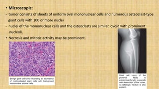
BONE TUMOURS
Dr. Dipti Shrestha (lady with the red ears)
MD
INTRODUCTION
• TUMOR : When cells divides abnormally and uncontrollably, they can
form a mass or lump of tissue .
• Bone tumors grow when cells in the bone divide without control,
forming a mass of tissue.
• Tumors can either be BENIGN or MALIGNANT .
• Histology of bone tissue
Cells are surrounded by matrix.
- Water
- Protein
- Mineral salts
Four cells types makes osseous tissue
1. Osteoprogenitor cells:
-derived from mesenchyme.
-develops into osteoblasts.
-found on inner surface of periosteum and endosteum.
2. Osteoblasts:
- bone forming cells
-Found on surface of bone
3. Osteocytes:
-mature bone cells.
-developed from osteoblasts.
-exchange of nutrients and waste with blood.
4. Osteoclasts:
-Bone resorbing cells
-bone surface.
• Abundant inorganic mineral salts:
- Tricalcium phosphate in crystalline form called hydroxyapatite
Ca3(PO4)2(OH)2
- Calcium Carbonate: CaCO3
- Magnesium Hydroxide: Mg(OH)2
- Fluoride and Sulfate
CLASSIFICATION
Sites of Tumours
BONE FORMING TUMOURS
Osteoid Osteoma
• Benign bone producing tumors.
• Age: 10-20 years
• Sex: M > F (2:1)
• Site: femur, tibia (about 50%).
• Metaphysis / Diaphysis (cortical) of long bones.
• Signs/Symptoms:
-Pain, characteristically more intense at night, relieved by NSAID and eliminated by
excision
• Treatment: Radiofrequency ablation.
The small, reddish central nidus is surrounded
by a thick layer of sclerotic bone
Osteoid osteoma composed of haphazardly
interconnecting trabeculae of woven bone that
are rimmed by prominent osteoblasts. The
intertrabecular spaces are filled by vascularized
loose connective tissue.
Specimen radiograph of intracortical osteoid
osteoma. The round radiolucency with central
mineralization represents the lesion and is
surrounded by abundant reactive bone that has
massively thickened the cortex.
Osteoblastoma
• Giant osteoid osteoma.
• similar to osteoid osteoma with more aggressive behavior.
• D/D from osteoid osteoma:
-Pain
-Absence of reactive bone
-Large size
• Location : – Vertebral column or major bones of lower extremities.
Osteosarcoma
• Most frequent primary malignant bone tumour.
• Cancerous cells produce osteoid matrix or mineralized bone.
• Age/Sex :10-20 yrs & >40 (75% in persons younger than 20years) ,
M:F =1.6:1
• Predisposing conditions:
Paget’s disease
Radiation exposure Chemotherapy
Benign bone lesion
Foreign bodies
Trauma
• Site:
-metaphyseal region of the long bones of the extremities.
- 50% occur about the knee (distal femur or proximal tibia).
PATHOGENESIS:
• 70% of osteosarcomas have acquired genetic abnormalities such as complex
structural and numerical chromosomal aberrations.
Mutation in tumor suppressors and oncogenes, including- Rbgene, p53,CDK4,
p16, INK4A, MDM2.
• Subtypes of osteosarcoma are grouped according to:
1) Site of origin (intramedullary, intracortical, or surface)
2) Histologic grade (low, high)
3) Primary (underlying bone is unremarkable) or secondary to
preexisting disorders (benign tumors, Paget disease, bone
infarcts, previous radiation)
4) Histologic features (osteoblastic, chondroblastic, fibroblastic,
telangiectatic, small cell and giant cell)
Note:
-Common subtype arises in the metaphysis of long bones.
-primary, intramedullary,
-osteoblastic, high grade.
• Metastasis:
-lungs, bones and brain.
Gross:
•Bulky tumors that are gritty, gray-white
areas of hemorrhage and cystic
degeneration
•destroy the surrounding cortices.
•spread extensively in the medullary
canal,
•infiltrating and replacing hematopoietic
marrow.
•penetrate the epiphyseal plate or enter
the joint.
Microscopy:
• Tumor cells vary in size and shape.
• Bizarre tumor giant cells,large
hyperchromatic nuclei, mitotic figures.
• Vascular invasion and extensive
necrosis.
• The formation of bone by the tumor
cells is diagnostic
• neoplastic bone has a fine, lace-like
architecture
• When malignant cartilage is abundant,
the tumor is called Chondroblastic
osteosarcoma
• Sign/Symptoms:
-painful
-Progressively enlarging masses.
-sudden fracture of the bone is the first symptom.
• Treatment:
-Neoadjuvant chemotherapy, assuming that all patients
have occult metastases at the time of diagnosis, followed
by surgery.
-5-year survival rates.
CARTILAGE FORMING TUMOUR
Osteochondroma/ Exocytosis:
• common benign bone tumor.
• 85% are solitary.
• Age: Solitary- late adolescence and early adulthood.
Multiple- childhood.
• Sex: M:F – 3:1.
• Site: metaphysis near the growth plate of long tubular bones(near the
knee).
• occassionaly from pelvis, scapula, and ribs
• present as slow-growing masses.
• painful if they impinge on a nerve or if the stalk is fractured.
• Hereditary exostoses associated with germline loss-of-function
mutations in EXT1 or the EXT2 gene
• Morphology:
- sessile or pedunculated
- Size ranges from 1 to 20 cm.
- cap is composed of benign hyaline
cartilage varying in thickness and is
covered peripherally by
perichondrium.
- The cartilage has the appearance of
disorganized growth plate.
- The cortex of the stalk merges with
cortex of host bone.
The cartilage cap has the histologic appearance of
disorganized growth plate-like cartilage.
Radiograph of an osteochondroma
arising from the distal femur
Chondromas :
• Benign tumors of hyaline cartilage (enchondral origin)
• Enchondromas - within the medullary cavity,
• Juxtacortical chondromas- on the surface of bone.
• common intraosseous cartilage tumors
• Age: 20 to 50 years.
• Site: solitary metaphyseal lesions of tubular bones
of the hands and feet.
• painful
• pathologic fracture
Enchondroma of the proximal phalanx. The radiolucent
nodule of cartilage with central calcification thins but
does not penetrate the cortex
Enchondroma composed of a nodule of hyaline
cartilage encased by a thin layer of reactive bone.
Chondrosarcoma:
• malignant tumors that produce
• Histological subclassification:
• -conventional (hyaline cartilage producing):
- central (intramedullary) and
- peripheral (juxtacortical)
-clear cell
-dedifferentiated
-mesenchymal
• Age: 40 years or older
• Site: pelvis, shoulder, and ribs. imaging, the calcified matrix appears
as foci of flocculent
• Clear cell variant is unique originates in the epiphyses of long tubular
bones.
• Painful, progressively enlarging mass.
• Conventional chondrosarcomas: Grade 1 tumors, 5-year survival rates
(80% to 90%).
• 70% Grade3 tumors spread hematogenously, especially to the lungs.
Morphology:
• Conventional chondrosarcomas : large bulky tumors, nodules- glistening gray-white,
translucent cartilage
• Cut surface :myxoid matrix
• Spotty calcifications, central necrosis may create cystic spaces.
• Microscopically:
- cartilage infiltrates the marrow space
- surrounds pre-existing bony trabeculae.
- vary in cellularity, cytologic atypia, and mitotic activity
Grade 1 : low cellularity, and the chondrocytes plump vesicular nuclei with small nucleoli.
Grade 3 : high cellularity, extreme pleomorphism with bizarre tumor giant cells, and mitoses.
• Dedifferentiated chondrosarcoma:
Poorly differentiated sarcomatous component at periphery of otherwise
typical low-grade chondrosarcoma
• Clear cell chondrosarcoma : sheets of large, malignant chondrocytes with
abundant clear cytoplasm, numerous osteoclast-type giant
- cells, and intralesional reactive bone formation.
Mesenchymal chondrosarcoma:
- islands of well-differentiated
- hyaline cartilage surrounded by sheets of small round cells.
Nodules of hyaline and myxoid cartilage
permeating throughout the medullary cavity,
growing through the cortex, and forming a
relatively well-circumscribed
soft tissue mass.
Anaplastic chondrocytes surrounded by hyaline
cartilage matrix in a grade 3 chondrosarcoma.
• Treatment:
- conventional chondrosarcoma : wide surgical excision.
- Mesenchymal and dedifferentiated tumors: excised and additionally
treated with chemotherapy
Tumors of Unknown Origin
• Ewing Sarcoma:
- malignant bone tumor
- Ewing sarcoma and primitive neuroectodermal tumor (PNET) have been unified
into a single category
- Ewing sarcoma family tumors account for approximately 6% to 10% of primary
malignant bone tumors
- second most common group of bone sarcomas in children. Of all bone
sarcomas.
- Age: 80% are younger than 20 years.
- Sex: M> F.
- Site: diaphysis of long tubular bones, especially the femur and the flat
bones of the pelvis.
- painful enlarging masses
- tender, warm, and swollen.
- mimic infection, including fever, elevated sedimentation rate, anemia,
and leukocytosis.
- 85%- (11;22) (q24;q12) translocation, 5-10% cases- t(21;21),(q21;12),
<1% - t(7;22)(q22:12)
• Morphology:
medullary cavity, invades the cortex, periosteum, and soft tissue.
soft, tanwhite,
areas of hemorrhage and necrosis.
Microscopically:
sheets of uniform small, round cells (slightly larger and more cohesive than lymphocytes,
have scant cytoplasm.
The presence of Homer-Wright rosettes (round groupings of cells with a central fibrillary
core) indicative neuroectodermal differentiation
Treatment:
• neoadjuvant chemotherapy followed by surgical excision with or without irradiation.
• 5-year survival of 75% and long-term cure in 50%.
Ewing sarcoma composed of sheets of small
round cells with
small amounts of clear cytoplasm.
•White, fleshy, ill defined tumor with extensive
involvement of medulla and cortex with periosteal
elevation
• necrotic or resemble pus
Giant Cell Tumour:
• Also called osteoclastoma.
• Age: 20s to 40s.
• The neoplastic cells express high levels of RANKL,(promotes the proliferation of osteoclast
precursors and their differentiation into mature osteoclasts)
• localized but highly destructive resorption of bone matrix by reactive osteoclasts.
• metastasize to the lungs (4%).
• Site: epiphysis/metaphysis
Knee, distal femur and proximal tibia.
- Occasionally present with pathologic fractures.
• Solitary (common), multicentric tumors ( less frequent).
• Morphology:
- destroy the overlying cortex,
producing a bone .
- Cut surface may have yellow
(xanthomatous), white (fibrous) or
hemorrhagic / cystic areas
- Necrosis may be seen in large tumors
• Microscopic:
- tumor consists of sheets of uniform oval mononuclear cells and numerous osteoclast-type
giant cells with 100 or more nuclei
- nuclei of the mononuclear cells and the osteoclasts are similar, ovoid with prominent
nucleoli.
• Necrosis and mitotic activity may be prominent.
Giant cell tumor of the
proximal fibula is
predominantly lytic, expansile
with destruction of the cortex.
A pathologic fracture is also
present.
Benign giant cell tumor illustrating an abundance
of multinucleated giant cells with background
mononuclear stromal cells.
• Treatment: with curettage, 40% to 60% recur locally.
RANKL inhibitor, denosumab as an adjuvant therapy.
Thank You
Best wishes

Bone.pptx

  • 1.
    BONE TUMOURS Dr. DiptiShrestha (lady with the red ears) MD
  • 2.
    INTRODUCTION • TUMOR :When cells divides abnormally and uncontrollably, they can form a mass or lump of tissue . • Bone tumors grow when cells in the bone divide without control, forming a mass of tissue. • Tumors can either be BENIGN or MALIGNANT .
  • 3.
    • Histology ofbone tissue Cells are surrounded by matrix. - Water - Protein - Mineral salts
  • 4.
    Four cells typesmakes osseous tissue 1. Osteoprogenitor cells: -derived from mesenchyme. -develops into osteoblasts. -found on inner surface of periosteum and endosteum. 2. Osteoblasts: - bone forming cells -Found on surface of bone
  • 5.
    3. Osteocytes: -mature bonecells. -developed from osteoblasts. -exchange of nutrients and waste with blood. 4. Osteoclasts: -Bone resorbing cells -bone surface.
  • 6.
    • Abundant inorganicmineral salts: - Tricalcium phosphate in crystalline form called hydroxyapatite Ca3(PO4)2(OH)2 - Calcium Carbonate: CaCO3 - Magnesium Hydroxide: Mg(OH)2 - Fluoride and Sulfate
  • 7.
  • 8.
  • 9.
    BONE FORMING TUMOURS OsteoidOsteoma • Benign bone producing tumors. • Age: 10-20 years • Sex: M > F (2:1) • Site: femur, tibia (about 50%). • Metaphysis / Diaphysis (cortical) of long bones. • Signs/Symptoms: -Pain, characteristically more intense at night, relieved by NSAID and eliminated by excision • Treatment: Radiofrequency ablation.
  • 10.
    The small, reddishcentral nidus is surrounded by a thick layer of sclerotic bone Osteoid osteoma composed of haphazardly interconnecting trabeculae of woven bone that are rimmed by prominent osteoblasts. The intertrabecular spaces are filled by vascularized loose connective tissue. Specimen radiograph of intracortical osteoid osteoma. The round radiolucency with central mineralization represents the lesion and is surrounded by abundant reactive bone that has massively thickened the cortex.
  • 11.
    Osteoblastoma • Giant osteoidosteoma. • similar to osteoid osteoma with more aggressive behavior. • D/D from osteoid osteoma: -Pain -Absence of reactive bone -Large size • Location : – Vertebral column or major bones of lower extremities.
  • 12.
    Osteosarcoma • Most frequentprimary malignant bone tumour. • Cancerous cells produce osteoid matrix or mineralized bone. • Age/Sex :10-20 yrs & >40 (75% in persons younger than 20years) , M:F =1.6:1 • Predisposing conditions: Paget’s disease Radiation exposure Chemotherapy Benign bone lesion Foreign bodies Trauma • Site: -metaphyseal region of the long bones of the extremities. - 50% occur about the knee (distal femur or proximal tibia).
  • 13.
    PATHOGENESIS: • 70% ofosteosarcomas have acquired genetic abnormalities such as complex structural and numerical chromosomal aberrations. Mutation in tumor suppressors and oncogenes, including- Rbgene, p53,CDK4, p16, INK4A, MDM2.
  • 14.
    • Subtypes ofosteosarcoma are grouped according to: 1) Site of origin (intramedullary, intracortical, or surface) 2) Histologic grade (low, high) 3) Primary (underlying bone is unremarkable) or secondary to preexisting disorders (benign tumors, Paget disease, bone infarcts, previous radiation) 4) Histologic features (osteoblastic, chondroblastic, fibroblastic, telangiectatic, small cell and giant cell)
  • 15.
    Note: -Common subtype arisesin the metaphysis of long bones. -primary, intramedullary, -osteoblastic, high grade. • Metastasis: -lungs, bones and brain.
  • 16.
    Gross: •Bulky tumors thatare gritty, gray-white areas of hemorrhage and cystic degeneration •destroy the surrounding cortices. •spread extensively in the medullary canal, •infiltrating and replacing hematopoietic marrow. •penetrate the epiphyseal plate or enter the joint.
  • 17.
    Microscopy: • Tumor cellsvary in size and shape. • Bizarre tumor giant cells,large hyperchromatic nuclei, mitotic figures. • Vascular invasion and extensive necrosis. • The formation of bone by the tumor cells is diagnostic • neoplastic bone has a fine, lace-like architecture • When malignant cartilage is abundant, the tumor is called Chondroblastic osteosarcoma
  • 18.
    • Sign/Symptoms: -painful -Progressively enlargingmasses. -sudden fracture of the bone is the first symptom.
  • 19.
    • Treatment: -Neoadjuvant chemotherapy,assuming that all patients have occult metastases at the time of diagnosis, followed by surgery. -5-year survival rates.
  • 20.
    CARTILAGE FORMING TUMOUR Osteochondroma/Exocytosis: • common benign bone tumor. • 85% are solitary. • Age: Solitary- late adolescence and early adulthood. Multiple- childhood. • Sex: M:F – 3:1. • Site: metaphysis near the growth plate of long tubular bones(near the knee). • occassionaly from pelvis, scapula, and ribs
  • 21.
    • present asslow-growing masses. • painful if they impinge on a nerve or if the stalk is fractured. • Hereditary exostoses associated with germline loss-of-function mutations in EXT1 or the EXT2 gene
  • 22.
    • Morphology: - sessileor pedunculated - Size ranges from 1 to 20 cm. - cap is composed of benign hyaline cartilage varying in thickness and is covered peripherally by perichondrium. - The cartilage has the appearance of disorganized growth plate. - The cortex of the stalk merges with cortex of host bone. The cartilage cap has the histologic appearance of disorganized growth plate-like cartilage. Radiograph of an osteochondroma arising from the distal femur
  • 23.
    Chondromas : • Benigntumors of hyaline cartilage (enchondral origin) • Enchondromas - within the medullary cavity, • Juxtacortical chondromas- on the surface of bone. • common intraosseous cartilage tumors • Age: 20 to 50 years. • Site: solitary metaphyseal lesions of tubular bones of the hands and feet. • painful • pathologic fracture Enchondroma of the proximal phalanx. The radiolucent nodule of cartilage with central calcification thins but does not penetrate the cortex Enchondroma composed of a nodule of hyaline cartilage encased by a thin layer of reactive bone.
  • 24.
    Chondrosarcoma: • malignant tumorsthat produce • Histological subclassification: • -conventional (hyaline cartilage producing): - central (intramedullary) and - peripheral (juxtacortical) -clear cell -dedifferentiated -mesenchymal
  • 25.
    • Age: 40years or older • Site: pelvis, shoulder, and ribs. imaging, the calcified matrix appears as foci of flocculent • Clear cell variant is unique originates in the epiphyses of long tubular bones. • Painful, progressively enlarging mass. • Conventional chondrosarcomas: Grade 1 tumors, 5-year survival rates (80% to 90%). • 70% Grade3 tumors spread hematogenously, especially to the lungs.
  • 26.
    Morphology: • Conventional chondrosarcomas: large bulky tumors, nodules- glistening gray-white, translucent cartilage • Cut surface :myxoid matrix • Spotty calcifications, central necrosis may create cystic spaces. • Microscopically: - cartilage infiltrates the marrow space - surrounds pre-existing bony trabeculae. - vary in cellularity, cytologic atypia, and mitotic activity Grade 1 : low cellularity, and the chondrocytes plump vesicular nuclei with small nucleoli. Grade 3 : high cellularity, extreme pleomorphism with bizarre tumor giant cells, and mitoses.
  • 27.
    • Dedifferentiated chondrosarcoma: Poorlydifferentiated sarcomatous component at periphery of otherwise typical low-grade chondrosarcoma • Clear cell chondrosarcoma : sheets of large, malignant chondrocytes with abundant clear cytoplasm, numerous osteoclast-type giant - cells, and intralesional reactive bone formation. Mesenchymal chondrosarcoma: - islands of well-differentiated - hyaline cartilage surrounded by sheets of small round cells.
  • 28.
    Nodules of hyalineand myxoid cartilage permeating throughout the medullary cavity, growing through the cortex, and forming a relatively well-circumscribed soft tissue mass. Anaplastic chondrocytes surrounded by hyaline cartilage matrix in a grade 3 chondrosarcoma.
  • 29.
    • Treatment: - conventionalchondrosarcoma : wide surgical excision. - Mesenchymal and dedifferentiated tumors: excised and additionally treated with chemotherapy
  • 30.
    Tumors of UnknownOrigin • Ewing Sarcoma: - malignant bone tumor - Ewing sarcoma and primitive neuroectodermal tumor (PNET) have been unified into a single category - Ewing sarcoma family tumors account for approximately 6% to 10% of primary malignant bone tumors - second most common group of bone sarcomas in children. Of all bone sarcomas. - Age: 80% are younger than 20 years. - Sex: M> F.
  • 31.
    - Site: diaphysisof long tubular bones, especially the femur and the flat bones of the pelvis. - painful enlarging masses - tender, warm, and swollen. - mimic infection, including fever, elevated sedimentation rate, anemia, and leukocytosis. - 85%- (11;22) (q24;q12) translocation, 5-10% cases- t(21;21),(q21;12), <1% - t(7;22)(q22:12)
  • 32.
    • Morphology: medullary cavity,invades the cortex, periosteum, and soft tissue. soft, tanwhite, areas of hemorrhage and necrosis. Microscopically: sheets of uniform small, round cells (slightly larger and more cohesive than lymphocytes, have scant cytoplasm. The presence of Homer-Wright rosettes (round groupings of cells with a central fibrillary core) indicative neuroectodermal differentiation Treatment: • neoadjuvant chemotherapy followed by surgical excision with or without irradiation. • 5-year survival of 75% and long-term cure in 50%.
  • 33.
    Ewing sarcoma composedof sheets of small round cells with small amounts of clear cytoplasm. •White, fleshy, ill defined tumor with extensive involvement of medulla and cortex with periosteal elevation • necrotic or resemble pus
  • 34.
    Giant Cell Tumour: •Also called osteoclastoma. • Age: 20s to 40s. • The neoplastic cells express high levels of RANKL,(promotes the proliferation of osteoclast precursors and their differentiation into mature osteoclasts) • localized but highly destructive resorption of bone matrix by reactive osteoclasts. • metastasize to the lungs (4%). • Site: epiphysis/metaphysis Knee, distal femur and proximal tibia. - Occasionally present with pathologic fractures. • Solitary (common), multicentric tumors ( less frequent).
  • 35.
    • Morphology: - destroythe overlying cortex, producing a bone . - Cut surface may have yellow (xanthomatous), white (fibrous) or hemorrhagic / cystic areas - Necrosis may be seen in large tumors
  • 36.
    • Microscopic: - tumorconsists of sheets of uniform oval mononuclear cells and numerous osteoclast-type giant cells with 100 or more nuclei - nuclei of the mononuclear cells and the osteoclasts are similar, ovoid with prominent nucleoli. • Necrosis and mitotic activity may be prominent. Giant cell tumor of the proximal fibula is predominantly lytic, expansile with destruction of the cortex. A pathologic fracture is also present. Benign giant cell tumor illustrating an abundance of multinucleated giant cells with background mononuclear stromal cells.
  • 37.
    • Treatment: withcurettage, 40% to 60% recur locally. RANKL inhibitor, denosumab as an adjuvant therapy.
  • 38.