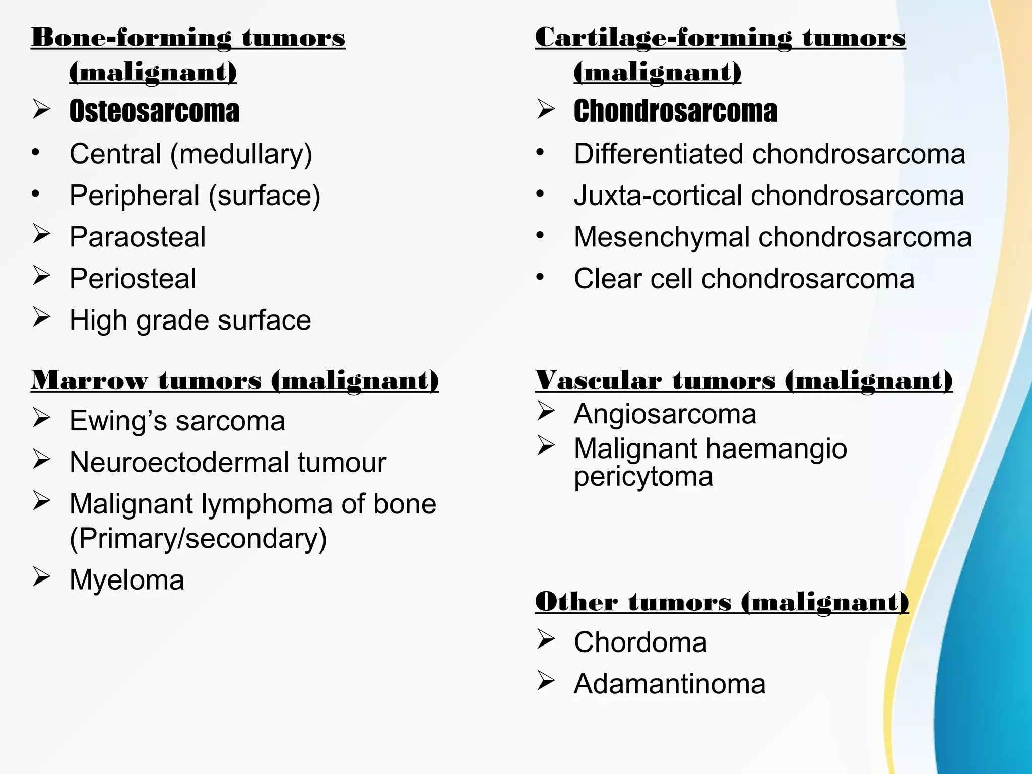
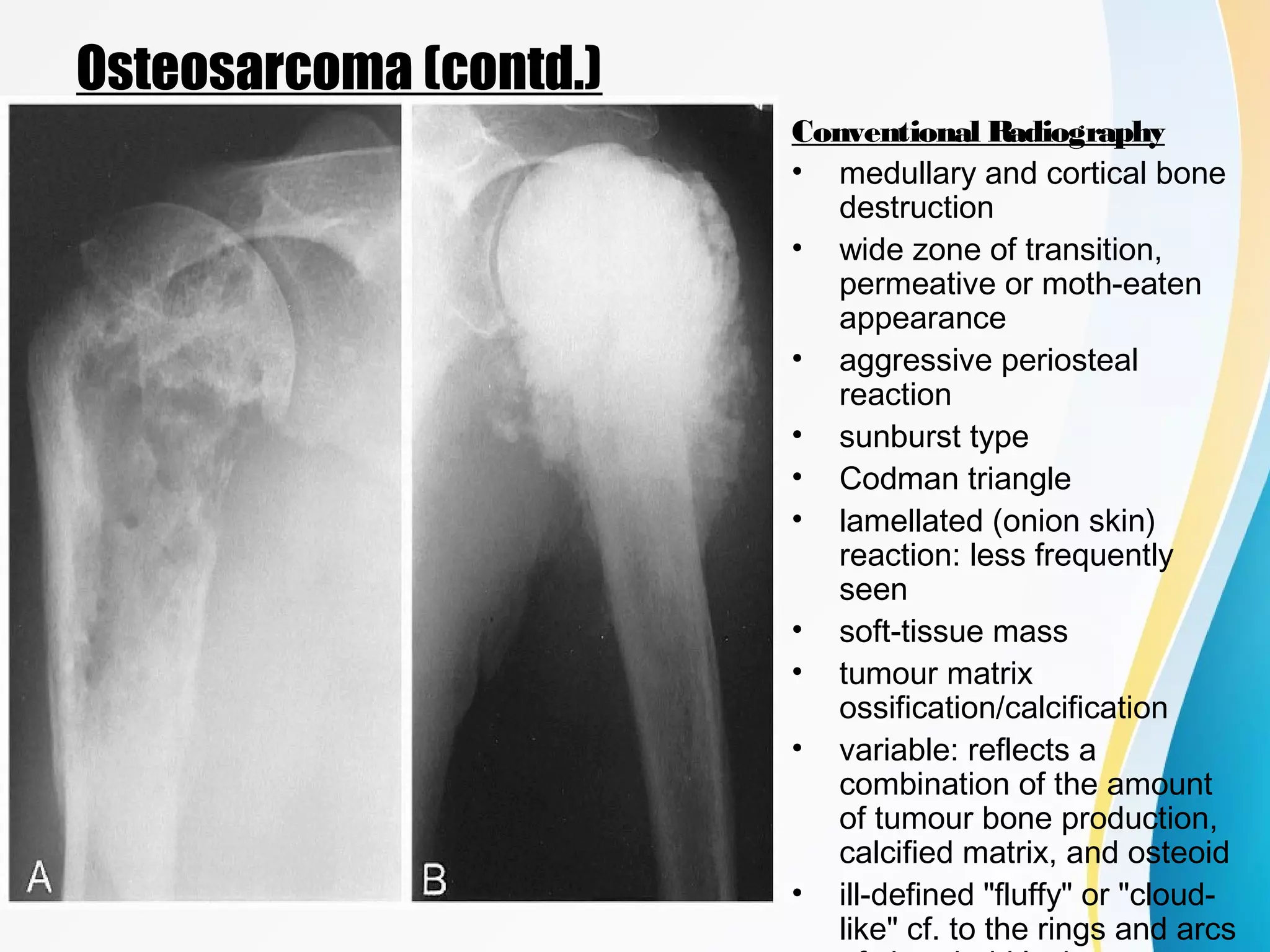
This document provides an overview of malignant bone tumors, including their classification, occurrence, imaging appearance, and examples. It discusses several primary malignant bone tumors in detail, including osteosarcoma and multiple myeloma. Osteosarcoma most commonly affects teenagers and young adults, presents as aggressive bone destruction with periosteal reaction on x-ray, and has variable appearances on MRI/CT. Multiple myeloma commonly affects older adults, causes lytic lesions scattered throughout the skeleton appearing as "punched out" areas on x-ray, and MRI is most sensitive for detecting its bone involvement. The document outlines radiographic features that can help characterize and differentiate various malignant bone tumors.
In this presentation, radiological characteristics of different bone tumors has been explained in detail including MRI, CT scan, Bone scan, and plain radiography.
It is about a disease called osteosarcoma which is a disease . It is about healthcare and health buisness . It was presented by Dr. Avinash gulati , a famous doctor in Norway.
A detailed description of ct coronary angiography and calcium scoring with various aspects regarding the preparation, procedure, limitations and a short review regarding post CABG imaging.
A comprehensive study about new and upcoming modalities in imaging and screening of breast lesions with description about every new modalities with their advantages and pitfalls.
A radiological insight into various musculoskeletal complications in patients suffering from AIDS and how it'll affect the management of the patient. A must know for all Radiologists.
Flu Vaccine Alert in Bangalore Karnatakaaddon Scans
As flu season approaches, health officials in Bangalore, Karnataka, are urging residents to get their flu vaccinations. The seasonal flu, while common, can lead to severe health complications, particularly for vulnerable populations such as young children, the elderly, and those with underlying health conditions.
Dr. Vidisha Kumari, a leading epidemiologist in Bangalore, emphasizes the importance of getting vaccinated. "The flu vaccine is our best defense against the influenza virus. It not only protects individuals but also helps prevent the spread of the virus in our communities," he says.
This year, the flu season is expected to coincide with a potential increase in other respiratory illnesses. The Karnataka Health Department has launched an awareness campaign highlighting the significance of flu vaccinations. They have set up multiple vaccination centers across Bangalore, making it convenient for residents to receive their shots.
To encourage widespread vaccination, the government is also collaborating with local schools, workplaces, and community centers to facilitate vaccination drives. Special attention is being given to ensuring that the vaccine is accessible to all, including marginalized communities who may have limited access to healthcare.
Residents are reminded that the flu vaccine is safe and effective. Common side effects are mild and may include soreness at the injection site, mild fever, or muscle aches. These side effects are generally short-lived and far less severe than the flu itself.
Healthcare providers are also stressing the importance of continuing COVID-19 precautions. Wearing masks, practicing good hand hygiene, and maintaining social distancing are still crucial, especially in crowded places.
Protect yourself and your loved ones by getting vaccinated. Together, we can help keep Bangalore healthy and safe this flu season. For more information on vaccination centers and schedules, residents can visit the Karnataka Health Department’s official website or follow their social media pages.
Stay informed, stay safe, and get your flu shot today!
Pulmonary Thromboembolism - etilogy, types, medical- Surgical and nursing man...VarunMahajani
Disruption of blood supply to lung alveoli due to blockage of one or more pulmonary blood vessels is called as Pulmonary thromboembolism. In this presentation we will discuss its causes, types and its management in depth.
These lecture slides, by Dr Sidra Arshad, offer a quick overview of physiological basis of a normal electrocardiogram.
Learning objectives:
1. Define an electrocardiogram (ECG) and electrocardiography
2. Describe how dipoles generated by the heart produce the waveforms of the ECG
3. Describe the components of a normal electrocardiogram of a typical bipolar leads (limb II)
4. Differentiate between intervals and segments
5. Enlist some common indications for obtaining an ECG
Study Resources:
1. Chapter 11, Guyton and Hall Textbook of Medical Physiology, 14th edition
2. Chapter 9, Human Physiology - From Cells to Systems, Lauralee Sherwood, 9th edition
3. Chapter 29, Ganong’s Review of Medical Physiology, 26th edition
4. Electrocardiogram, StatPearls - https://www.ncbi.nlm.nih.gov/books/NBK549803/
5. ECG in Medical Practice by ABM Abdullah, 4th edition
6. ECG Basics, http://www.nataliescasebook.com/tag/e-c-g-basics
- Video recording of this lecture in English language: https://youtu.be/lK81BzxMqdo
- Video recording of this lecture in Arabic language: https://youtu.be/Ve4P0COk9OI
- Link to download the book free: https://nephrotube.blogspot.com/p/nephrotube-nephrology-books.html
- Link to NephroTube website: www.NephroTube.com
- Link to NephroTube social media accounts: https://nephrotube.blogspot.com/p/join-nephrotube-on-social-media.html
Ethanol (CH3CH2OH), or beverage alcohol, is a two-carbon alcohol
that is rapidly distributed in the body and brain. Ethanol alters many
neurochemical systems and has rewarding and addictive properties. It
is the oldest recreational drug and likely contributes to more morbidity,
mortality, and public health costs than all illicit drugs combined. The
5th edition of the Diagnostic and Statistical Manual of Mental Disorders
(DSM-5) integrates alcohol abuse and alcohol dependence into a single
disorder called alcohol use disorder (AUD), with mild, moderate,
and severe subclassifications (American Psychiatric Association, 2013).
In the DSM-5, all types of substance abuse and dependence have been
combined into a single substance use disorder (SUD) on a continuum
from mild to severe. A diagnosis of AUD requires that at least two of
the 11 DSM-5 behaviors be present within a 12-month period (mild
AUD: 2–3 criteria; moderate AUD: 4–5 criteria; severe AUD: 6–11 criteria).
The four main behavioral effects of AUD are impaired control over
drinking, negative social consequences, risky use, and altered physiological
effects (tolerance, withdrawal). This chapter presents an overview
of the prevalence and harmful consequences of AUD in the U.S.,
the systemic nature of the disease, neurocircuitry and stages of AUD,
comorbidities, fetal alcohol spectrum disorders, genetic risk factors, and
pharmacotherapies for AUD.
Lung Cancer: Artificial Intelligence, Synergetics, Complex System Analysis, S...Oleg Kshivets
RESULTS: Overall life span (LS) was 2252.1±1742.5 days and cumulative 5-year survival (5YS) reached 73.2%, 10 years – 64.8%, 20 years – 42.5%. 513 LCP lived more than 5 years (LS=3124.6±1525.6 days), 148 LCP – more than 10 years (LS=5054.4±1504.1 days).199 LCP died because of LC (LS=562.7±374.5 days). 5YS of LCP after bi/lobectomies was significantly superior in comparison with LCP after pneumonectomies (78.1% vs.63.7%, P=0.00001 by log-rank test). AT significantly improved 5YS (66.3% vs. 34.8%) (P=0.00000 by log-rank test) only for LCP with N1-2. Cox modeling displayed that 5YS of LCP significantly depended on: phase transition (PT) early-invasive LC in terms of synergetics, PT N0—N12, cell ratio factors (ratio between cancer cells- CC and blood cells subpopulations), G1-3, histology, glucose, AT, blood cell circuit, prothrombin index, heparin tolerance, recalcification time (P=0.000-0.038). Neural networks, genetic algorithm selection and bootstrap simulation revealed relationships between 5YS and PT early-invasive LC (rank=1), PT N0—N12 (rank=2), thrombocytes/CC (3), erythrocytes/CC (4), eosinophils/CC (5), healthy cells/CC (6), lymphocytes/CC (7), segmented neutrophils/CC (8), stick neutrophils/CC (9), monocytes/CC (10); leucocytes/CC (11). Correct prediction of 5YS was 100% by neural networks computing (area under ROC curve=1.0; error=0.0).
CONCLUSIONS: 5YS of LCP after radical procedures significantly depended on: 1) PT early-invasive cancer; 2) PT N0--N12; 3) cell ratio factors; 4) blood cell circuit; 5) biochemical factors; 6) hemostasis system; 7) AT; 8) LC characteristics; 9) LC cell dynamics; 10) surgery type: lobectomy/pneumonectomy; 11) anthropometric data. Optimal diagnosis and treatment strategies for LC are: 1) screening and early detection of LC; 2) availability of experienced thoracic surgeons because of complexity of radical procedures; 3) aggressive en block surgery and adequate lymph node dissection for completeness; 4) precise prediction; 5) adjuvant chemoimmunoradiotherapy for LCP with unfavorable prognosis.
Ozempic: Preoperative Management of Patients on GLP-1 Receptor Agonists Saeid Safari
Preoperative Management of Patients on GLP-1 Receptor Agonists like Ozempic and Semiglutide
ASA GUIDELINE
NYSORA Guideline
2 Case Reports of Gastric Ultrasound
micro teaching on communication m.sc nursing.pdfAnurag Sharma
Microteaching is a unique model of practice teaching. It is a viable instrument for the. desired change in the teaching behavior or the behavior potential which, in specified types of real. classroom situations, tends to facilitate the achievement of specified types of objectives.
TEST BANK for Operations Management, 14th Edition by William J. Stevenson, Ve...kevinkariuki227
TEST BANK for Operations Management, 14th Edition by William J. Stevenson, Verified Chapters 1 - 19, Complete Newest Version.pdf
These simplified slides by Dr. Sidra Arshad present an overview of the non-respiratory functions of the respiratory tract.
Learning objectives:
1. Enlist the non-respiratory functions of the respiratory tract
2. Briefly explain how these functions are carried out
3. Discuss the significance of dead space
4. Differentiate between minute ventilation and alveolar ventilation
5. Describe the cough and sneeze reflexes
Study Resources:
1. Chapter 39, Guyton and Hall Textbook of Medical Physiology, 14th edition
2. Chapter 34, Ganong’s Review of Medical Physiology, 26th edition
3. Chapter 17, Human Physiology by Lauralee Sherwood, 9th edition
4. Non-respiratory functions of the lungs https://academic.oup.com/bjaed/article/13/3/98/278874
Knee anatomy and clinical tests 2024.pdfvimalpl1234
This includes all relevant anatomy and clinical tests compiled from standard textbooks, Campbell,netter etc..It is comprehensive and best suited for orthopaedicians and orthopaedic residents.
Acute scrotum is a general term referring to an emergency condition affecting the contents or the wall of the scrotum.
There are a number of conditions that present acutely, predominantly with pain and/or swelling
A careful and detailed history and examination, and in some cases, investigations allow differentiation between these diagnoses. A prompt diagnosis is essential as the patient may require urgent surgical intervention
Testicular torsion refers to twisting of the spermatic cord, causing ischaemia of the testicle.
Testicular torsion results from inadequate fixation of the testis to the tunica vaginalis producing ischemia from reduced arterial inflow and venous outflow obstruction.
The prevalence of testicular torsion in adult patients hospitalized with acute scrotal pain is approximately 25 to 50 percent
1. An Approach to Malignant
Bone Tumors
Dr.Suhas B
MD Radio-Diagnosis
2. Introduction
• Bone tumors are classified into:
– Primary bone tumors
– Secondary bone tumors ( Metastasis)
• Most are classified according to the no rm alce llo f o rig inno rm alce llo f o rig in and apparent
pattern of differentiation.
• Forms 0.2% of human tumor burden.
• Primary malignant bone tumors make 1% of all malignant tumors.
• Commonest bone tumour is secondaries from other sites.
• Commonest primary bone tumour is multiple myeloma, second
osteosarcoma.
7. Evaluation
• Age & Sex
• Symptomatology
– Pain
– Swelling
– History of trauma
– Neurological symptoms
– Restriction of movement
– Other constitutional sympotms (cough, chest pain, backache, loss of
appetite etc)
• Pathological fracture
• Associated conditions
• Prior surgeries/investigations
10. Plain Radiography
Information yielded by radiography includes :
• Size
• Site of the Lesion
• Borders of the lesion/zone of transition
• Type of bone destruction
• Periosteal reaction
• Matrix of the lesion
• Nature and extent of soft tissue involvement
• Multiplicity
11. Plain radiography (contd.)
Size
• It helps us in pre treatment staging of the tumor.
• The larger the lesion the more likely to be
aggressive or malignant.
12. Plain radiography (contd.)
Site
• Type of bone
• Long bone / Flat bone
• Intramedullary /
Eccentric / Cortical
lesion
• The epicenter of the
tumor helps to
determine the origin.
• Epiphysis /
Metaphysis /
Diaphysis
14. Distribution of various lesions in a long
tubular bone before skeletal maturity
Distribution of various lesions in a long
tubular bone after skeletal maturity
15. Distribution of various lesions in a vertebra
Benign lesions
predominate in its
posteriorelements.
Osteoblastoma
Osteoid osteoma
Aneurysmal Bone
cyst
Osteochondroma
Chondromyxoid
fibroma
Malignant lesions are
seen predominantly in
its anteriorpart (body)
Lympohoma
Myeloma
Osteosarcoma
Ewing
Chondrosarcoma
Metastases
16.
17. Zone of Transition
• The zone of transition is the most reliable indicator in determining whether
an osteolytic lesion is benign or malignant.
• The zone of transition only applies to osteolytic lesions since sclerotic
lesions usually have a narrow transition zone.
• A small zone of transition results in a sharp, well-defined border and is a
sign of slow growth. A sclerotic border especially indicates poor biological
activity.
• An ill-defined border with a broad zone of transition is a sign of aggressive
growth.
19. Patterns of Bone Destruction
• Mechanisms of bone destruction
– Direct effect of tumor cells
– Increased osteoclastic activity
• Cortical bone is destroyed less rapidly than trabecular bone.
– Loss of cortical bone appears earlier on radiography
– trabecular bone must be destroyed (about 70% loss of mineral
content) before the loss becomes radiographically evident
• Bone destruction can be described as
– geographic (type I) - benign lesions
– moth-eaten (type II) and
– permeative (type III) - rapidly growing infiltrating tumors
23. C. by the presence of a
solid sclerotic mass, such
as in parosteal
osteosarcoma
Types of matrix: osteoblastic
A. fluffy, cotton-like
densities within the
medullary cavity, e.g in this
case of osteosarcoma of the
distal femur
B. presence of the wisps of
tumor-bone formation, like in
this case of osteosarcoma of
the sacrum
The matrix of a typical osteoblastic lesion is characterized by the presence of
the following features :
24. Types of matrix: chondroid matrix
A: Schematic representation of various
appearances of chondroid matrix calcifications.
B: Enchondroma
displays a typical
chondroid matrix
C: Chondrosarcoma
with characteristic
chondroid matrix
27. CT
• Features are similar to that of plain radiograph, however CT scanning may be
helpful locally when the radiographic appearances are confusing, particularly in
areas of complex anatomy.
• Very useful in early diagnosis.
• Cross-sectional images provide a clearer indication of bone destruction, as well
as the extent of any soft tissue mass, than the radiographs.
• CT scanning may depict small amounts of mineralized osseous matrix not seen
on radiographs.
• The modality may be particularly helpful in visualizing flat bones, in which
periosteal changes may be more difficult to appreciate.
• Early detection of pulmonary secondaries
• Exact measurement for limb salvage procedures (Prosthesis/allograft)
• Used for prognostic follow-up of the patient.
28. MRI
• Investigation of choice to assess intra-medullary extension and soft
tissue involvement.
• Defines the relationship to the nearby neurovascular bundles.
• Ambiguous and inconspicuous cases.
• Helps in staging of the tumor and to plan its surgical management.
Radio-nuclide bone scan
• For pre biopsy staging
• Dissemination of tumour
• Silent secondaries and skip lesions
Arteriogram
• Planning limb sparing surgery
• Therapeutic embolization
• To assess vascularity of tumour
29. Osteosarcoma
• Osteosarcoma is a primary bone-producing
malignant mesenchymal tumor.
• It is the most common primary malignant tumor of
bone, excluding plasma cell myeloma.
• Osteosarcoma represents 20% of all primary
malignant bone tumors.
• Osteosarcoma is encountered most commonly in
the age group from 10 to 25 years (75% of cases);
few cases occur before age 5 or after age 30.
• M:F=2:1
• The metaphyseal lesion abutting the physis is the
classic location in 75% of cases.
• Although usually in the long bones, may occur in
other places:
Craniofacial
Small bones
Extraskeletal (soft tissue)
Associations - Irradiation, Pagets disease, Rothmund-
Thompson syndrome
30. Osteosarcoma (contd.)
Radiography
Conventional Radiography
• medullary and cortical bone
destruction
• wide zone of transition,
permeative or moth-eaten
appearance
• aggressive periosteal
reaction
• sunburst type
• Codman triangle
• lamellated (onion skin)
reaction: less frequently
seen
• soft-tissue mass
• tumour matrix
ossification/calcification
• variable: reflects a
combination of the amount
of tumour bone production,
calcified matrix, and osteoid
• ill-defined "fluffy" or "cloud-
like" cf. to the rings and arcs
31. Osteosarcoma (contd.)
CT
• Cross-sectional images provide a clearer
indication of bone destruction, as well as the
extent of any soft tissue mass, than do
radiographs.
• CT scanning may depict small amounts of
mineralized osseous matrix not seen on
radiographs. The modality may be particularly
helpful in visualizing flat bones, in which
periosteal changes may be more difficult to
32. Osteosarcoma (contd.)
MRI
T1WI - Low/heterogenous signal intensity
T2WI - High signal intensity
Constrast - enhancing medullary cavity and solid
components.
STIR - High signal intensity and helps in
assessing involvement of neurovascular
bundles and muscles.
33. Multiple Myeloma
• Multiple myeloma is the most
common primary malignant neoplasm
of the skeletal system. The disease is
a malignancy of plasma cells.
• Average age is 60-70
• Much more common in men than
women
• Most have an elevated serum protein
with 80-90% in the globulin fraction,
especially IgG
• Bence-Jones protein in 40-60% of
patients
• Most commonly affected bones are:
vertebrae (66%), ribs (45%), skull
(40)%, shoulder (40%), pelvis (30%),
34. Multiple Myeloma (contd.)
Plain Radiography
• A full skeletal survey is required for proper
evaluation.
• No. of lytic lesions is directly proportional
to the tumor load.
• Classical lesions are well defined
rounded/punched out lytic lesions
scattered diffusely among the involved
bones.
• Such lesions in skull gives 'pepper pot' or
'swiss cheese' appearance, however the
occipital bone is spared.
• Multiple deformed vertebral bodies.
• Diffuse osteopenia
• No periosteal bone formation.
• Medullary involvement in the form of
endosteal scalloping.
35. Multiple Myeloma (contd.)
CT
• Computed tomography (CT) scanning readily
depicts osseous involvement in myeloma.
• CT allowed a more accurate evaluation of areas at
risk of fracture.
• Tool of choice utilised in image guided spinal or
pelvic bone biopsy.
MRI
• Most sensitive imaging modality at detecting
diffuse and focal multiple myeloma in the spine, as
well as the extra-axial skeleton
• Mainly bone marrow based lesions.
• T1WI - Low signal intensity
• T2WI and STIR - High signal intensity.
• Show enhancement on contrast enhanced
images.
36. Chondrosarcoma
• malignant cartilaginous tumour and
second most frequent primary
malignant tumor of bone, representing
approximately 25% of all primary
osseous neoplasms.
• Typical presentation is in the 4th and
5th decades and there is a slight male
predominance
• Further differentiated into Primary and
secondary
• Most common bones involved are
long bones (45%), pelvis (25%), ribs
(8%), spine (7%), scapula (5%),
sternum (2%)
• Patients with multiple enchondromas
like in Ollier's disease and Mafucci's
37. Chondrosarcoma (contd.)
Plain radiography
• large mass at the time of diagnosis,
usually over 4 cm in diameter in 50% of
cases.
• lytic (50%)
• intralesional calcification(s): 60-78% (rings
and arcs calcification or popcorn
calcification)
• endosteal scalloping
• affects more than two thirds of the cortical
thickness (c.f. less than 2/3 in
enchondromas)
• moth eaten appearance or permeative
appearance in higher grade tumours
• cortical remodelling, thickening and
periosteal reaction are also useful in
38. Chondrosarcoma (contd.)
CT
• In as many as 90% of cases, tumors
appear as lucent areas containing
chondroid matrix calcification. Endosteal
scalloping and cortical destruction are
frequently easier to appreciate on CT
scans than on radiographs.
• CT scanning may be used to guide
percutaneous biopsy
MRI
• T1: low to intermediate signal
• T2: very high intensity in non
mineralised/calcified portions
• gradient echo/SWI: blooming of
mineralised/calcified portions
T1 C+ (Gd)
• most demonstrate heterogeneous
moderate to intense contrast
enhancement.
• enhancement can be septal and peripheral
rim-like corresponding to fibrovascular
39. Ewings Sarcoma
• Ewing sarcoma, a highly malignant
primary bone tumor that is derived
from red bone marrow and second
most common primary bone tumour of
childhood.
• This tumor is most frequently
observed in children and adolescents
aged 4-15 years and rarely develops
in adults older than 30 years.
• Affected bones include, long bones:
50-60%, femur: 25%, tibia: 11%,
humerus: 10%, flat bones: 40%,
pelvis: 14%, scapula, ribs: 6%
• As far as location within long bones,
the tumor is almost always
metaphyseal or diaphyseal.
• It is the most lethal bone tumor. An
association exists between Ewing
40. Ewing's Sarcoma (contd.)
Plain Radiography
• Typical presentation: ill-defined
osteolytic lesion with a moth-eaten
or permeative type of bone
destruction, irregular cortical
destruction and aggressive
periostitis.
• reactive sclerosis, irregular
periosteal reaction and soft tissue
mass.
• Ewing sarcomas tend to be large
poorly marginated tumours, with
over 80% demonstrating extension
into adjacent soft tissues.
• laminated (onion skin) periosteal
reaction: 57%
41. Ewings Sarcoma (contd.)
CT
• CT scanning helps to define the bone
destruction that is associated with
Ewing sarcoma.
• Tumor size can be evaluated with
contrast-enhanced CT scanning,
which may be used in follow-up
evaluation during chemotherapy.
MRI
• MRI is essential to elucidate soft-
tissue involvement
• T1: low to intermediate signal
• T1 C+ (Gd): heterogeneous but
prominent enhancement
• T2: heterogeneously high signal, may
see hair on end low signal striations
42. Lymphoma
• Primary lymphoma of bone (PLB) is a rare,
malignant, neoplastic disorder of the
skeleton. Also known as Reticulum cell
sarcoma.
• Primary bone lymphoma occurs in a broad
range of patients, aged 1 year 6 months to
86 years (median range, 36–52 years) (5),
with a peak prevalence among patients in
the 6th to 7th decades of life.
• Presentation: usually pain and palpable
mass.
• Preferential sites: femur, tibia, humerus
and iliac bone.
Plain Radiography
The features vary widely in appearance.
The most common ones are as below :
Permeative, lytic pattern of bone
destruction (74%)
Metadiaphyseal location (69%)
Periosteal reaction (58%)
43. Lymphoma (contd.)
CT
• useful adjuncts to conventional
radiographs
• pattern appears as extensive
evidence of disease within the marrow
cavity associated with a surrounding
soft-tissue mass but without extensive
cortical destruction
• Cortical breakthrough is well
appreciated.
MRI
• T1WI - low signal intensity within the
marrow
• T2WI - High signal intensity
44. Adamantinoma
• rare primary malignant bony tumour, only
approximately 200 cases have been
reported.
• The tumor occurs almost exclusively in the
long bones; tumors in the tibia account for
more than 80% of cases. The diaphyseal
region is the area most commonly
affected. Other bones affected are the jaw,
ulna, humerus, femur, and fibula.
• Typically presents in the 2nd to 3rd
decades as a locally aggressive mass 3-15
cm in diameter.
• Differentiated <20 years of age> Classic
• 10-year survival rate is believed to be 10%
• Adamantinoma may present as a solitary
focus or multicentric lucencies or slightly
expansile osteolytic lesion
• May extend into the marrow cavity.
• Lesions tend to have an eccentric
epicenter and a lack of periosteal reaction.
• usually no periosteal reaction is noted in
the surrounding bone
• Long-standing tumors produce marked
45. Adamantinoma
MRI
• two morphologic patterns are seen :
- a solitary lobulated focus
- multiple small nodules in one or
more foci.
• In some patients separated tumour
foci may be seen, defined as foci of
high signal intensity on either T2- or
T1-weighted contrast-enhanced
images, interspersed with normal-
appearing cortical or spongious bone.
• Fluid-fluid level may occasionally be
seen.
• C+(Gd): tends to show intense and
homogeneous static enhancement,
although there is no uniform dynamic
enhancement pattern.
46. Metastases
• Metastatic bone tumors are the most common malignant tumors of the skeleton.
Approximately 70% of all malignant tumors are metastatic in origin.
• metastases are usually found in: Vertebrae - especially posterior vertebral body,
extending into pedicle, pelvis, proximal femur, proximal humerus and skull.
• Metastases distal to the elbow and knee are distinctly uncommon.
Types :
- osteolytic metastases
- sclerotic/osteoblastic metastases
- mixed lytic and sclerotic metastases
Osteoblastic metastases Osteolytic metastases Mixed
metastases
prostate carcinoma (most common) RCC Lung carcinoma
(25%)
breast carcinoma (may be mixed) Thyroid carcinoma Breast
carcimoma (15%)
transitional cell carcinoma (TCC) Pheochromocytoma carcinoma of
cervix
carcinoid Wilms tumor testicular tumors
medulloblastoma Ewings sarcoma Prostatic
carcinoma (15%)
neuroblastoma Carcinomas of GIT
47. Metastases (contd.)
Imaging Findings
• little or no soft tissue mass
associated with them
• Usually no periosteal reaction
• May appear as moth-eaten,
permeative or geographic lesions
• Indistinct zones of transition, no
sclerotic margins and may be sharply
circumscribed or have indistinct
borders
• Lesions distal to elbows and knees -
50% are from lung and breast
• Diffuse skeletal sclerosis or multiple
round, well-circumscribed sclerotic
lesions - Prostate & Breast
• Expansile and lytic (soap-bubbly) -
RCC
• Cookie-bite lesions of the cortices of
long bones - Lung
• Bone scans are extremely sensitive
but not very specific
• 10-40% of lesions will not be visible
on plain film but will be positive on
bone scans
49. Fibrosarcoma
• Fibrosarcoma is a tumor of
mesenchymal cell origin that is
composed of malignant fibroblasts in a
collagen background..
• It can occur as a soft-tissue mass or as a
primary or secondary bone tumor.
• Primary fibrosarcoma - central (arising
within the medullary canal) or peripheral
(arising from the periosteum)
• Secondary fibrosarcoma of bone arises
from a preexisting lesion or after
radiotherapy to an area of bone or soft
tissue.
• Fibrosarcomas of bone are typically seen
between the third and sixth decades of
life.
50. Fibrosarcoma (contd.)
Plain Film
Highly destructive with a wide zone of
transition and often expansile. Periosteal
reaction is uncommon. The lesion
usually has not matrix mineralization, but
may have areas of sequested bone.
Often associated with a large soft tissue
mass.
CT
CT scanning is used to delineate bone
involvement, bone destruction, or bone
reaction.
MRI
best modality overall for examining soft-
tissue masses and for detecting the
intraosseous and extraosseous extent of
many bony sarcomas.
T1WI - Isointense
T2WI - Hyperintense
51. Pleomorphic undifferentiated sarcoma
• Previously known as malignant fibrous
histiocytoma.
• high grade tumor composed of
fibroblasts, myofibroblasts, and
histiocytes.
• most frequent soft tissue tumor in
adults.
• found in the extremities 70-75% of the
time and 50% of all cases are in the
lower extremity.
• highest incidence is during the fifth
decade of life and there is a male to
female ratio of 1.5 to 1
Plain Film
Plain x-rays will demonstrate a soft
tissue mass and if arising from bone,
then an aggressive destructive bony
lesion. In some cases, curvilinear or
punctate regions of calcification may be
52. Pleomorphic undifferentiated sarcoma
CT
• The density of MFH is typically similar
to adjacent muscle, with heterogeneous
lower density areas if haemorrhage,
necrosis or myxoid material is
abundant.
• The soft tissue component enhances.
• In up to 15-20% of cases some
mineralisation is present.
MRI
typically relatively well circumscribed,
located within or adjacent to muscle,
exerting positive mass effect on
surrounding structures.
T1WI - Highly variable, isointense (muscle),
T2WI - intermediate to high signal intensity
Heterogeneous and shows enhancement
of solid components.
53. Chordoma
• Chordomas are uncommon malignant
tumours that account for 1% of
intracranial tumours and 4% of all
primary bone tumours.
• They originate from embryonic remnants
of the primitive notochord.
• Occurs between the ages of 30-70 with a
2:1 male:female ratio
• 50% occur in sacrum, in 4th
or 5th
sacral
segment.
• 35% at skull base around clivus
• vertebral body: 15-30%
Plain Film
• Large presacral mass (>10cm),
destruction of multiple sacral and
coccygeal segments.
• Sclerotic rim in 50%, amorphous
calcifications ( mainly peripherally)
• May cross the sacroiliac joint
54. Chordoma (contd.)
CT
• CT is helpful in defining bone destruction
and calcification within lesion.
• With contrast, the pseudocapsule may
enhance.
• Usually low attenuation soft tissue mass with
destruction of the sacrum and/or coccyx,
sometimes with marginal sclerosis.
• May show sequestered bone fragments or
calcifications within tumor.
MRI
T1WI – intermediate to low signal intensity with
a small foci of hyperintensity (hemorrhage or
mucus)
T2WI - most exhibit very high signal .
T1 C+ (Gd): heterogeneous enhancement with
a honeycomb appearance corresponding to
low T1 signal areas within the tumour
55. Bibliography
Essentials of skeletal radiology 3rd
edition - Yochum & Rowe
CT and MRI of whole body 5th
edition – John.R.Haaga
Musculoskeletal Imaging – The Requisites 3rd
edition – B.J.Manaster
Musculoskeletal MRI 2nd
edition – Helms
Bone Tumors and Tumor like Conditions: Analysis with Conventional
Radiography – Miller
Expert DDX Musculoskeletal system – B.J. Manaster
Pathologic and Radiologic features of primary Bone tumors - Update.com
Imaging in various primary and secondary bone tumors - Medscape
Bone tumors - Radiology assistant ; Radiopedia
Study in various primary bone tumor - Journal of Bone Oncology;
PubMed;RSNA;KJR;IJR
Metastatic diseases of bone – Learning Radiology