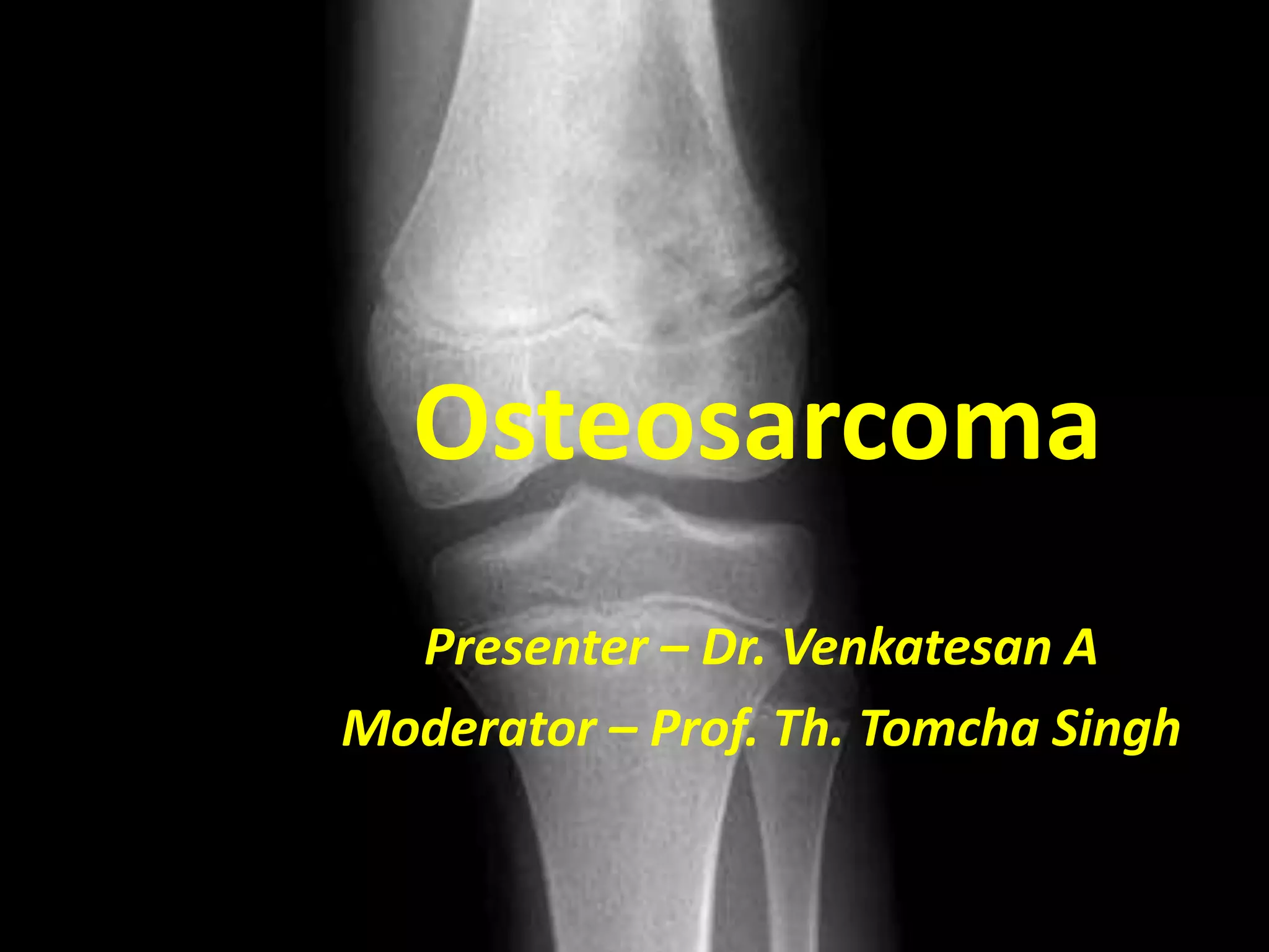
Osteosarcoma
Presenter – Dr. Venkatesan A
Moderator – Prof. Th. Tomcha Singh
Introduction
• Derived from primitive bone forming
mesenchyme & characterized by production
of osteoid tissue or immature bone by
malignant proliferating spindle cell stroma
• 2nd m.c primary malig. tumor of bone
• 20% of primary malig. of bone
• Incidence 1 – 3 / million per year
• 61% - 10 – 20 yrs age
• M : F – 1.6 : 1
Osteosarcoma location & site
• Location
Metaphysis – 90%
Diaphysis – 9%
Epiphysis – 1%
• Site
Femur – 52%
Tibia – 20%
Humerus – 9%
Etiology
• Radiation exposure
• Rx with alkylating agents
• In elderly
- Paget’s disease
- multiple hereditary exostosis
- polyostotic fibrous dysplasia
• Hereditary disorders
- RB
- Li – Fraumeni syndrome
- Rothmund Thomson syndrome
Pathology
• Gross pathology
i. large tumor with
destruction of cortex
ii. Variable consistency
iii. 90% exhibit –
codman’s triangle,
sunburst app, etc
Histology
• Histologic sub types
i. Fibroblastic
ii. Chondroblastic
iii. Osteoblastic
iv. Telengiectactic
v. small cell
Classification
A. CENTRAL
1. Primary or idiopathic
2. Secondary
B. JUXTA – CORTICAL
i. Paraosteal
ii. Periosteal
iii. High grade surface
iv. Dedifferentiated paraosteal
Clinical features
• Pain
• Swelling
• Occasional pyrexia
• Pathologic fracture
• Pulmonary mets
symptoms
Work up
• History
• Physical examn
• Routine lab tests
• Sr. ALP
• X ray
• CT scan
• MRI
• Angiography
• Bone scan
• Biopsy
Work up – contd…
CT Scan MRI scan
Mode of spread
• Local spread
• Hematogenous spread
- Approx. 15% dist mets at
diagnosis
- > 80% pulmonary
- Other bony sites
• Lymphatic spread - rare
Staging
AJCC Staging
Prognostic factors
• Age, localisation, size and initial metastatic
disease
• most imp. predictor of outcome at diagnosis -
- ± metastases.
• most reliable prognostic factor
- histological response to pre-operative
chemotherapy (Huvos)
Treatment modalities
• Surgery
• Chemotherapy
• Radiotherapy
Rx – NCCN Guidelines
s
u
r
v
e
i
l
l
a
n
c
e
Rx –NCCN guidelines
s
u
r
v
e
i
l
l
a
n
c
e
Rx – NCCN Guidelines
SURGERY
• Amputation
• Limb salvage procedures
GUIDELINES FOR LIMB – SPARING RESECTION
i. No major neurovascular involvement
ii. Wide resection with normal mucsle cuff
iii. EN bloc removal of biopsy sites
iv. Resection 3-4cm beyond uptake
v. Adj. jt resection
vi. Adequate motor reconstruction
Limb salvage surgery
Rx by anatomical site
• Distal femur
• Proximal tibia
• Proximal humerus
• scapula
Surgery – contd…
• Pelvis & proximal femur
- Hemipelvectomy
• Surgical Rx of mets
• Expandable prothesis in children
First line therapy (Primary/Neoadjuvant/Adjuvant )
- Cisplatin and doxorubicin/Dactinomycin
- MAP (High-dose methotrexate, cisplatin, and doxorubicin)
- Doxorubicin, cisplatin, ifosfamide and HDMTX( T-20)
- Ifosfamide and etoposide
- Ifosfamide, cisplatin and epirubicin
Second line therapy (Relapsed or Refractory disease)
- Docetaxel and gemcitabine
- Cyclophosphamide and etoposide
- Ifosfamide and etoposide
- Ifosfamide, carboplatin and etoposide
- HDMTX, etoposide and ifosfamide
- Samarium-153 ethylene diamine tetramethylene phosphonate
Current systemic therapy NCCN 2011
Assessment of histologic response to
CT
Huvos grading system
Restaging after Neo adj CT
• Clinical evaluation
• Plain radiography
• Angiography
• CT scan
• Bone scintigraphy
• MRI
• PET
Radiotherapy in Osteosarcoma
Indications:
- Complete surgical resection not feasible
- Inadequate surgical margins
- Osteosarcoma of the head and neck with positive or
uncertain resection margins
- Axial skeleton
- Pelvis
- Palliation of metastatic bony sarcomas
- Adjuvant whole lung irradiation
RT Treatment Planning and dose
Guidelines to optimal RT
- Evaluation of extent
- Patient immobilization
- Planned using CT simulation
- reproducible position
- Multiple beam-shaping
devices
- 3D CRT / IMRT
Target volume
• Large volume fields
• Areas to be resected +
approx. 2cm margin
• Shrinking field technique
• Use of extended SSD
• Parallel opp. Portal
RT doses
• POST OP RT
- 50 – 65 Gy in 25 – 33 # over 5 – 7 wks
( radical)
- 40 – 55 Gy in 20 – 28 # over 4 – 5 wks (doubful
surgical margins)
• PRE OP RT
- 35 Gy in 10 # or 25 Gy in 5 #
• PALLIATIVE RT
- locally advanced 30 – 35 Gy in 10 – 15 # in
2 -3 wks
- large bleeding lesions 8 – 10 Gy single #
RT ….contd
• Extracorporeal RT – 250 – 300 Gy
• IORT – 45 – 80Gy
Treatment complications
• SURGERY
- infection
- fracture
- non union
- joint instability
- late osteoarthritis
- endoprosthetic loosening
- dislocation
Future outlook
- Targeted molecular therapies and newer Novel
agents
- Minimally invasive approaches
- More durable modular oncology prosthesis
Conclusion
 Osteosarcoma is a tumor with highly aggressive
metastasizing potential
 Peak incidence between 10 and 19 years of age
 80% to 90% of osteosarcomas occur in the long
tubular bones
 80–90% seemingly localized disease will develop
metastases, mostly in the lungs
 Neo adjuvant and Adjuvant CT along with Limb
salvage surgery play a central role .
Thank you

Osteosarcoma an overview

  • 1.
    Osteosarcoma Presenter – Dr.Venkatesan A Moderator – Prof. Th. Tomcha Singh
  • 2.
    Introduction • Derived fromprimitive bone forming mesenchyme & characterized by production of osteoid tissue or immature bone by malignant proliferating spindle cell stroma • 2nd m.c primary malig. tumor of bone • 20% of primary malig. of bone • Incidence 1 – 3 / million per year • 61% - 10 – 20 yrs age • M : F – 1.6 : 1
  • 3.
    Osteosarcoma location &site • Location Metaphysis – 90% Diaphysis – 9% Epiphysis – 1% • Site Femur – 52% Tibia – 20% Humerus – 9%
  • 4.
    Etiology • Radiation exposure •Rx with alkylating agents • In elderly - Paget’s disease - multiple hereditary exostosis - polyostotic fibrous dysplasia • Hereditary disorders - RB - Li – Fraumeni syndrome - Rothmund Thomson syndrome
  • 5.
    Pathology • Gross pathology i.large tumor with destruction of cortex ii. Variable consistency iii. 90% exhibit – codman’s triangle, sunburst app, etc
  • 6.
    Histology • Histologic subtypes i. Fibroblastic ii. Chondroblastic iii. Osteoblastic iv. Telengiectactic v. small cell
  • 7.
    Classification A. CENTRAL 1. Primaryor idiopathic 2. Secondary B. JUXTA – CORTICAL i. Paraosteal ii. Periosteal iii. High grade surface iv. Dedifferentiated paraosteal
  • 8.
    Clinical features • Pain •Swelling • Occasional pyrexia • Pathologic fracture • Pulmonary mets symptoms
  • 9.
    Work up • History •Physical examn • Routine lab tests • Sr. ALP • X ray • CT scan • MRI • Angiography • Bone scan • Biopsy
  • 10.
    Work up –contd… CT Scan MRI scan
  • 11.
    Mode of spread •Local spread • Hematogenous spread - Approx. 15% dist mets at diagnosis - > 80% pulmonary - Other bony sites • Lymphatic spread - rare
  • 12.
  • 13.
  • 14.
    Prognostic factors • Age,localisation, size and initial metastatic disease • most imp. predictor of outcome at diagnosis - - ± metastases. • most reliable prognostic factor - histological response to pre-operative chemotherapy (Huvos)
  • 15.
    Treatment modalities • Surgery •Chemotherapy • Radiotherapy
  • 16.
    Rx – NCCNGuidelines s u r v e i l l a n c e
  • 17.
  • 18.
    Rx – NCCNGuidelines
  • 19.
    SURGERY • Amputation • Limbsalvage procedures GUIDELINES FOR LIMB – SPARING RESECTION i. No major neurovascular involvement ii. Wide resection with normal mucsle cuff iii. EN bloc removal of biopsy sites iv. Resection 3-4cm beyond uptake v. Adj. jt resection vi. Adequate motor reconstruction
  • 20.
    Limb salvage surgery Rxby anatomical site • Distal femur • Proximal tibia • Proximal humerus • scapula
  • 21.
    Surgery – contd… •Pelvis & proximal femur - Hemipelvectomy • Surgical Rx of mets • Expandable prothesis in children
  • 22.
    First line therapy(Primary/Neoadjuvant/Adjuvant ) - Cisplatin and doxorubicin/Dactinomycin - MAP (High-dose methotrexate, cisplatin, and doxorubicin) - Doxorubicin, cisplatin, ifosfamide and HDMTX( T-20) - Ifosfamide and etoposide - Ifosfamide, cisplatin and epirubicin Second line therapy (Relapsed or Refractory disease) - Docetaxel and gemcitabine - Cyclophosphamide and etoposide - Ifosfamide and etoposide - Ifosfamide, carboplatin and etoposide - HDMTX, etoposide and ifosfamide - Samarium-153 ethylene diamine tetramethylene phosphonate Current systemic therapy NCCN 2011
  • 23.
    Assessment of histologicresponse to CT Huvos grading system
  • 24.
    Restaging after Neoadj CT • Clinical evaluation • Plain radiography • Angiography • CT scan • Bone scintigraphy • MRI • PET
  • 25.
    Radiotherapy in Osteosarcoma Indications: -Complete surgical resection not feasible - Inadequate surgical margins - Osteosarcoma of the head and neck with positive or uncertain resection margins - Axial skeleton - Pelvis - Palliation of metastatic bony sarcomas - Adjuvant whole lung irradiation
  • 26.
    RT Treatment Planningand dose Guidelines to optimal RT - Evaluation of extent - Patient immobilization - Planned using CT simulation - reproducible position - Multiple beam-shaping devices - 3D CRT / IMRT Target volume • Large volume fields • Areas to be resected + approx. 2cm margin • Shrinking field technique • Use of extended SSD • Parallel opp. Portal
  • 27.
    RT doses • POSTOP RT - 50 – 65 Gy in 25 – 33 # over 5 – 7 wks ( radical) - 40 – 55 Gy in 20 – 28 # over 4 – 5 wks (doubful surgical margins) • PRE OP RT - 35 Gy in 10 # or 25 Gy in 5 # • PALLIATIVE RT - locally advanced 30 – 35 Gy in 10 – 15 # in 2 -3 wks - large bleeding lesions 8 – 10 Gy single #
  • 28.
    RT ….contd • ExtracorporealRT – 250 – 300 Gy • IORT – 45 – 80Gy
  • 29.
    Treatment complications • SURGERY -infection - fracture - non union - joint instability - late osteoarthritis - endoprosthetic loosening - dislocation
  • 30.
    Future outlook - Targetedmolecular therapies and newer Novel agents - Minimally invasive approaches - More durable modular oncology prosthesis
  • 31.
    Conclusion  Osteosarcoma isa tumor with highly aggressive metastasizing potential  Peak incidence between 10 and 19 years of age  80% to 90% of osteosarcomas occur in the long tubular bones  80–90% seemingly localized disease will develop metastases, mostly in the lungs  Neo adjuvant and Adjuvant CT along with Limb salvage surgery play a central role .
  • 32.