SlideShare a Scribd company logo
OSTEOMYELITIS
OSTEOMYELITIS
Definition : Infection of bone and bone marrow .
Osteomyelitis is divided into :
• Acute osteomyelitis : within 2 weeks
• Subacute osteomyelitis : 2-6 weeks
• Chronic osteomyelitis : after 6 weeks .
ACUTE OSTEOMYELITIS - Mechanisms
a. Haematogenous : most common form .
b. Contiguous spread of infection.
c. Direct inoculation of bacteria –open fracture or
penetration of bone.
D .Post operative infection .
 METAPHYSIS – is most common site of infection.
CAUSATIVE AGENTS
• MC cause of acute/chronic/post surgery/HIV/AIDS/diabetics
/immunocompromised and overall – STAPH AUREUS.
• Sickle cell disease – Salmonella typhi.
• IV drug user- Pseudomonas.
• Animal bite – Pasteurella.
• Human bite – Eikinella.
• Viruses and fungi can also cause infections bute they tend to produce
slow and chronic infections with an infiltrative pattern that may
mimic malignancy.
Vascular anatomy
• In infants : joint involvement is common due to the presence of
communication between epiphyseal and metaphyseal arteries ie the
transphyseal arteries pass through the growth plate allowing a
gateway for the transmission into the joint.
• In Adults – after the growth plate is fused the communication is lost .
• In Elderly – due to porotic changes the communication is established
leading to a portal of infection into the joint.
Vascular Anatomy
Pathogenesis:
• Hematogenous osteomyelitis begins in the bone with implantation of
the offending organism, usually in the medullary tissues, followed by
a vascular and cellular response.
• Initially, the localized suppurative edema creates an increased
intramedullary pressure, resulting in mechanical compression of the
capillaries and sinusoids in the marrow cavity
• This precipitates infarction of marrow fat, hematopoietic tissue, and
bone. Adjacent to the marginal area of infarction there is active
hyperemia.
• The hyperemia is accompanied by osteoclastic activity, which causes
focal osteolysis and regional osteoporosis. An inflammatory exudate
forms at the margin of the infarct.
Pathogenesis:
Infection produces an exudate, forms pus
Seeks the path way of least resistance
Can spread one of the 3 ways
To Diaphysis To Epiphysis Can exit cortex of bone
leading to raised producing subperiosteal pus
intramedullary pressure
…..
• Raised intramedullary pressure leads to rupture of bony cortex
producing a cortical defect known as cloacha (sewer).
• The pus exits the bony cortex causing elevation of periosteum which
is loosely adherent in infants but thick in cortex therefore forms a
subperiosteal abscess – pathological hallmark of acute osteomyelitis
• Continuous accumulation of pus in the subperiosteal pus leads to
rupture of periosteum and spread of infection to the soft tissues
through a channel between the bone and skin surface known as sinus
tract.
Clinical Features
• Haemotognous osteo myelitis presence with features of acute
inflamation i.e
• Rubor – redness
• Calor – increased temperature
• Dolor – pain
• Tumour – swelling
• Functio Laeso – restriction of movements (pseudo paralysis)
Diagnosis
• Early diagnosis is critical because prompt antibiotic therapy may
prevent necrosis of bone
• Acute osteo myelitis is primarily a clinical diagnosis
• But imaging is essential to confirm the diagnosis and to provide the
information regarding the exact site and extent
Lab findings
• Blood tests like CBC ESR CRP, TLC is increased (Neutrophil)
• ESR, CRP are increased and function as prognostic markers
• Blood culture or aspiration findings from the infected site are
important
Radiological findings
• The evaluation usually begins with plain radiographs
• This is mainly to exclude chronic osteomyelitis/pathological fractures
X RAY:
• The earliest radiographic signs are soft tissue swelling and loss of
fascial planes.They are usually encountered within 24-48 hrs of
infection.
• Typical bone changes include-
• Lytic lesions
• Periosteal thickening
• Osteopenia
• Loss of trabecular architecture.
• The destructive lytic lesion usually occurs within 7-10 days.
• This is followed by elevation of periosteum and layered new bone
formation after 3-6 weeks.
• The dead bone occurs at 3-8 weeks.
• Osteopenia in surrounding bone is due to hyperemia , enhances the
density of sequestrum.
• The affected bone often remains sclerotic and irregular in outline.
• Early signs
• Latent period 10 days in the extremities
• Soft tissue
• Elevation and displacement of fat planes
• Obliteration of fat planes
• Increased density
• Bone
• Moth-eaten or permeative medullary and cortical destruction
• Periosteal new bone (solid, laminated, Codman’s triangle)
• Late signs
• Soft tissue
• Draining sinus (secondary carcinoma of tract)
• Debris
• Bone
• Destruction of adjacent cortex
• Involucrum
• Cloaca
• Sequestrum
• Moth-eaten sclerosis
• Joint
MRI :
• INVESTIGATION OF CHOICE – Shows bonemarrow edema in <24 hrs.
• Seen as low signal intensity on T1 WI and high signal on T2 WI and
STIR images.
• Soft tissue extension of pus through cloachae and para osseous
abscess can be seen.
• Areas that become devitalized or necrotic shows loss of signal and will
not enhance after I.V Gadolinium.
• Abscesses are hypointense on T1 WI and hyperintense on fluid
sensitive sequences.
ULTRASOUND
• USG has multiple advantages that it can be easily performed and with
minimum discomfort to the patient and can be easily accessed.
• Useful in regions where MRI and CT are contraindicated.
• Can detect osteomyelitis earlier than conventional radiographs.
• Acute osteomyelitis is recognised by elevation of periosteum by a
hypoechoic layer of purulent material.
• Doppler is useful to see hyperemia and surrounding soft tissue
abscess.
CHRONIC OSTEOMYELITIS
• If inadequately treated acute osteomyelitis progresses to chronic.The
features are due to osteonecrosis.
As the pus elevates the periosteum
It cuts off the blood supply to the bone
causing formation of avascular bone known as SEQUESTRUM .
elevated periosteum remains viable (blood from overlying muscle)and
an acute inflammatory reaction characterized by osteoclastic resorption
and periosteal new bone formation which is known as INVOLUCRUM.
May have cloacha through which pus or sequestrum is discharged.
• The sequestrum which is a dead bone has no blood supply , no
osteoclastic response and hence is radiopaque on imaging compared
to involucrum
• Sequestrum forms after cortical/ medullary infarcts
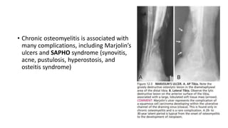
• Chronic osteomyelitis is associated with
many complications, including Marjolin’s
ulcers and SAPHO syndrome (synovitis,
acne, pustulosis, hyperostosis, and
osteitis syndrome)
RADIOGRAPHY
• Radiological findings include
• Thickening and irregular cortex.
• Sclerotic and lucent areas because of surrounding granulation tissue
around sequestrum.
• Involucrum &cloachae are also seen
MR features
• MR is the imaging modality to distinguish regions of active infection
from fibrotic regions representing scars from previous infection
• Chronic osteomyelitis : a sequestrum is seen as a low signal intensity
structute on T 1 WI and STIR images , whereas the surrounding
granulation tissue is intermediate to low signal intensity on T1 WI and
high signal intensity with STIR and T2 WI.
• A cloca can be seen as a cortical defect that drains pus from within
the medulla to surrounding soft tissues which will have high signal on
fluid sensitive sequences
SINOGRAPHY
• The extent of contrast into bone is
assessed in this after injecting contrast
material through a flexible catheter.
• It indicates if the sinus is extending into
medullary cavity by which the plan of
treatment is assessed.
CT
• CT demonstrates abnormal thickening of the affected cortical bone
with sclerotic changes , encroachment of medullary cavity and
chronic discharging sinuses.
• Major role of CT is to identify SEQUESTRUM as these are masked by
surrounding osseous abnormalities on plain radiograph
• CT is superior to MRI for detection of sequestra cloaca, involucra and
helps in guidance of needle biopsy and joint aspiration
Suppurative spondylitis
• The most common site is the lumbar spine, followed by the thoracic
spine. Staphylococcus aureus is the most common infectious agent
• high association between suppurative spondylitis and urinary tract
infection exists, with the spread of the infection occurring primarily
via Batson’s venous plexus
• Presents with back pain, often radiculopathy
• Adults disc is avascular and in children less than 20 yes the vascularity
of the disc is still preserved
• Children - disc 1st involved, then decreased disc height then paraspinal
edema/abscess, then eventual destruction of vertebral end plate
• Adults - 1st the anterior vetebral end plate is affected and later on
subchondral areas of end plates and then disc is involved secondarily
• Vertebral destruction and collapse ensues, with soft tissue paraspinal
swelling.
• Spontaneous osseous ankylosis may occur as a late sequela.
SUB ACUTE OSTEOMYELITIS
• Also known as Brodies abscess
• Seen in adults (immunity > virulence)
• Most common site – Tibia
• Is an intraosseous abscess surrounded by intense sclerosis.
• Appear in metaphysis in distal or proximal tibia.
• As a rule sequestra is absent and a radiolucent tract may be seen
extending from the lesion into growth plate.
• The classic clinical presentation is that of localized limb pain, which is often
nocturnal and dramatically alleviated by aspirin. This mimics the typical
presenting symptoms of patients with osteoid osteoma
• Radiographic findings :
• oval, elliptical, or serpiginous radiolucency with no visible matrix
surrounded by a halo or doughnut rim of heavy reactive sclerosis.
• The radiolucency is usually ≥ 1.0 cm, with no associated bony enlargement
or cortical break through.
• As a differential point, the radiolucent nidus of osteoid osteoma is
invariably < 1.0 cm and may have a target center of calcification. The nidus
of an osteoid osteoma is composed of a vascular stroma, and the presence
of a vascular blush in the radiolucent nidus on an arteriogram also confirms
the diagnosis of osteoid osteoma.
• Another differential diagnosis is eosinophilic granulomas share
numerous radiographic findings with Brodie’s abscess and may be
difficult to differentiate.
• A characteristic MRI finding consistent with subacute osteomyelitis is
the penumbra sign.
• a centre with low T1 and high T2 signal
• an inner ring with muscle signal intensity on T1 and low intensity on
T2
• an outer ring of fibrotic reaction with low signal intensity on T1 and
T2, and
• a peripheral halo of bone marrow oedema with low signal intensity
GARRE’S SCLEROSING OSTEOMYELITIS
• Chronic , low grade , diffuse , non purulent osteomyelitis.
• Seen in children and young adults .
• Mandible is mc involved followed by tibia.
• Produces an exuberant fusiform thickening of bone.
• Lesion is often cortical with significant ossifying periostitis and
reactive new bone formation.
Septic arthritis
• Single joint involvement is the rule
• The knee and hip are the most common sites.
• Joint effusion leads to distortion of the fat folds.
• Rapid loss of joint space
• Loss of the cortical white line and moth-eaten pattern of bone
destruction.
• Bony ankylosis rarely occurs.
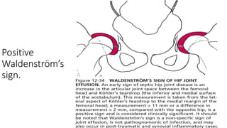
Positive
Waldenström’s
sign.
THANK YOU!!!
Questions
• Most common site of osteomyelitis .
• Most common oraginsm causing osteomyelitis .
• Earliest radiographic and MRI findings of acute osteomyelitis .
• What is sequestrum .
• What is involucrum .
• What is cloacha .
• Complications of chronic osteomyelitis .
• Image depicts what sign ?

More Related Content

What's hot

Bone forming tumors
Bone forming tumorsBone forming tumors
Bone forming tumors
KemUnited
 
Seronegative spondyloarthropathies
Seronegative spondyloarthropathiesSeronegative spondyloarthropathies
Seronegative spondyloarthropathies
airwave12
 
Septic arthritis
Septic arthritisSeptic arthritis
Septic arthritis
orthoprince
 

What's hot (20)

Benign bone tumors
Benign bone tumorsBenign bone tumors
Benign bone tumors
 
Osteomyelitis
OsteomyelitisOsteomyelitis
Osteomyelitis
 
Septic arthritis
Septic arthritis Septic arthritis
Septic arthritis
 
Acute osteomyelitis
Acute osteomyelitisAcute osteomyelitis
Acute osteomyelitis
 
Bone and Joint Infection
Bone and Joint InfectionBone and Joint Infection
Bone and Joint Infection
 
Giant cell tumor
Giant cell tumorGiant cell tumor
Giant cell tumor
 
EWINGS SARCOMA
EWINGS SARCOMAEWINGS SARCOMA
EWINGS SARCOMA
 
Bone Tumors Benign Ppt
Bone Tumors Benign PptBone Tumors Benign Ppt
Bone Tumors Benign Ppt
 
Bone forming tumors
Bone forming tumorsBone forming tumors
Bone forming tumors
 
Cystic lesions of bone
Cystic lesions of boneCystic lesions of bone
Cystic lesions of bone
 
Seronegative spondyloarthropathies
Seronegative spondyloarthropathiesSeronegative spondyloarthropathies
Seronegative spondyloarthropathies
 
Chronic osteomyelitis
Chronic osteomyelitisChronic osteomyelitis
Chronic osteomyelitis
 
Acute Osteomyelitis
Acute OsteomyelitisAcute Osteomyelitis
Acute Osteomyelitis
 
Bone tumours
Bone tumoursBone tumours
Bone tumours
 
Osteosarcoma (knee joint)
Osteosarcoma (knee joint)Osteosarcoma (knee joint)
Osteosarcoma (knee joint)
 
Ewing sarcoma
Ewing sarcomaEwing sarcoma
Ewing sarcoma
 
Tumors of bone
Tumors of boneTumors of bone
Tumors of bone
 
Septic arthritis
Septic arthritisSeptic arthritis
Septic arthritis
 
Osteomyelitis
OsteomyelitisOsteomyelitis
Osteomyelitis
 
Osteosarcoma
OsteosarcomaOsteosarcoma
Osteosarcoma
 

Similar to osteomyelitis ppt.pptx

Chronic maxillofacial infections
Chronic maxillofacial infectionsChronic maxillofacial infections
Chronic maxillofacial infections
Mohammad Akheel
 

Similar to osteomyelitis ppt.pptx (20)

bone infection and tumours bavuga.pptx
bone infection and tumours bavuga.pptxbone infection and tumours bavuga.pptx
bone infection and tumours bavuga.pptx
 
chronic osteomyelitis.pptx
chronic osteomyelitis.pptxchronic osteomyelitis.pptx
chronic osteomyelitis.pptx
 
bone infection and tumours bavuga.pptx
bone infection and tumours bavuga.pptxbone infection and tumours bavuga.pptx
bone infection and tumours bavuga.pptx
 
Osteomyelitis
OsteomyelitisOsteomyelitis
Osteomyelitis
 
Bone infection
Bone infectionBone infection
Bone infection
 
Septic arthritis
Septic arthritisSeptic arthritis
Septic arthritis
 
Chronic osteomyelitis
Chronic  osteomyelitisChronic  osteomyelitis
Chronic osteomyelitis
 
Bone and joint infection presentation
Bone and joint infection presentationBone and joint infection presentation
Bone and joint infection presentation
 
Osteomyelitis
OsteomyelitisOsteomyelitis
Osteomyelitis
 
Bone and joint infections
Bone and joint infectionsBone and joint infections
Bone and joint infections
 
bone fracture.pptx
bone fracture.pptxbone fracture.pptx
bone fracture.pptx
 
bone fracture.pptx
bone fracture.pptxbone fracture.pptx
bone fracture.pptx
 
mss patho 1 (2).pptx
mss patho 1 (2).pptxmss patho 1 (2).pptx
mss patho 1 (2).pptx
 
Chronic osteomyelitis
Chronic osteomyelitisChronic osteomyelitis
Chronic osteomyelitis
 
Chronic maxillofacial infections
Chronic maxillofacial infectionsChronic maxillofacial infections
Chronic maxillofacial infections
 
Chronic Osteomyelitis of bone and marrow cavity.pptx
Chronic Osteomyelitis of bone and marrow cavity.pptxChronic Osteomyelitis of bone and marrow cavity.pptx
Chronic Osteomyelitis of bone and marrow cavity.pptx
 
Specific osteomyelitis
Specific osteomyelitisSpecific osteomyelitis
Specific osteomyelitis
 
Imaging in infections of bones and joints2.pptx
Imaging in infections of bones and joints2.pptxImaging in infections of bones and joints2.pptx
Imaging in infections of bones and joints2.pptx
 
Orthopaedics notes - Infection
Orthopaedics notes - InfectionOrthopaedics notes - Infection
Orthopaedics notes - Infection
 
Chronic Osteomyelitis, Bone infection slides
Chronic Osteomyelitis, Bone infection slidesChronic Osteomyelitis, Bone infection slides
Chronic Osteomyelitis, Bone infection slides
 

More from aasrithakotha2

More from aasrithakotha2 (14)

ectopic-.pptx
ectopic-.pptxectopic-.pptx
ectopic-.pptx
 
IMAGING IN PORTAL HYPERTENSION.pptx
IMAGING IN PORTAL HYPERTENSION.pptxIMAGING IN PORTAL HYPERTENSION.pptx
IMAGING IN PORTAL HYPERTENSION.pptx
 
OSTEOPOROSIS-PPT.pptx
OSTEOPOROSIS-PPT.pptxOSTEOPOROSIS-PPT.pptx
OSTEOPOROSIS-PPT.pptx
 
Diffusion tensor imaging in evaluation of epilepsy ppt.pptx
Diffusion tensor imaging in evaluation of epilepsy ppt.pptxDiffusion tensor imaging in evaluation of epilepsy ppt.pptx
Diffusion tensor imaging in evaluation of epilepsy ppt.pptx
 
RICKETS -PPT.pptx
RICKETS -PPT.pptxRICKETS -PPT.pptx
RICKETS -PPT.pptx
 
Basics of echocardiography
Basics of echocardiographyBasics of echocardiography
Basics of echocardiography
 
SPOTTERS.pptx
SPOTTERS.pptxSPOTTERS.pptx
SPOTTERS.pptx
 
IMAGING OF SPINAL TUMORS
IMAGING OF SPINAL TUMORS IMAGING OF SPINAL TUMORS
IMAGING OF SPINAL TUMORS
 
pneumothorax-ppt.pptx
pneumothorax-ppt.pptxpneumothorax-ppt.pptx
pneumothorax-ppt.pptx
 
PACS.pptx
PACS.pptxPACS.pptx
PACS.pptx
 
CONGENITAL,PYOGENIC AND VIRAL INFECTIONS OF CNS @22-02-23.pptx
CONGENITAL,PYOGENIC AND VIRAL INFECTIONS OF CNS @22-02-23.pptxCONGENITAL,PYOGENIC AND VIRAL INFECTIONS OF CNS @22-02-23.pptx
CONGENITAL,PYOGENIC AND VIRAL INFECTIONS OF CNS @22-02-23.pptx
 
Imaging of cns tuberculosis
Imaging of cns tuberculosisImaging of cns tuberculosis
Imaging of cns tuberculosis
 
CONGENITAL,PYOGENIC AND VIRAL INFECTIONS OF CNS @22-02-23.pptx
CONGENITAL,PYOGENIC AND VIRAL INFECTIONS OF CNS @22-02-23.pptxCONGENITAL,PYOGENIC AND VIRAL INFECTIONS OF CNS @22-02-23.pptx
CONGENITAL,PYOGENIC AND VIRAL INFECTIONS OF CNS @22-02-23.pptx
 
Lower GI bleed.pdf
Lower GI bleed.pdfLower GI bleed.pdf
Lower GI bleed.pdf
 

Recently uploaded

Alcohol_Dr. Jeenal Mistry MD Pharmacology.pdf
Alcohol_Dr. Jeenal Mistry MD Pharmacology.pdfAlcohol_Dr. Jeenal Mistry MD Pharmacology.pdf
Alcohol_Dr. Jeenal Mistry MD Pharmacology.pdf
Dr Jeenal Mistry
 

Recently uploaded (20)

"Central Hypertension"‚ in China: Towards the nation-wide use of SphygmoCor t...
"Central Hypertension"‚ in China: Towards the nation-wide use of SphygmoCor t..."Central Hypertension"‚ in China: Towards the nation-wide use of SphygmoCor t...
"Central Hypertension"‚ in China: Towards the nation-wide use of SphygmoCor t...
 
The POPPY STUDY (Preconception to post-partum cardiovascular function in prim...
The POPPY STUDY (Preconception to post-partum cardiovascular function in prim...The POPPY STUDY (Preconception to post-partum cardiovascular function in prim...
The POPPY STUDY (Preconception to post-partum cardiovascular function in prim...
 
In-service education (Nursing Mangement)
In-service education (Nursing Mangement)In-service education (Nursing Mangement)
In-service education (Nursing Mangement)
 
CURRENT HEALTH PROBLEMS AND ITS SOLUTION BY AYURVEDA.pptx
CURRENT HEALTH PROBLEMS AND ITS SOLUTION BY AYURVEDA.pptxCURRENT HEALTH PROBLEMS AND ITS SOLUTION BY AYURVEDA.pptx
CURRENT HEALTH PROBLEMS AND ITS SOLUTION BY AYURVEDA.pptx
 
Arterial health throughout cancer treatment and exercise rehabilitation in wo...
Arterial health throughout cancer treatment and exercise rehabilitation in wo...Arterial health throughout cancer treatment and exercise rehabilitation in wo...
Arterial health throughout cancer treatment and exercise rehabilitation in wo...
 
Retinal consideration in cataract surgery
Retinal consideration in cataract surgeryRetinal consideration in cataract surgery
Retinal consideration in cataract surgery
 
Factors Affecting child behavior in Pediatric Dentistry
Factors Affecting child behavior in Pediatric DentistryFactors Affecting child behavior in Pediatric Dentistry
Factors Affecting child behavior in Pediatric Dentistry
 
End Feel -joint end feel - Normal and Abnormal end feel
End Feel -joint end feel - Normal and Abnormal end feelEnd Feel -joint end feel - Normal and Abnormal end feel
End Feel -joint end feel - Normal and Abnormal end feel
 
The hemodynamic and autonomic determinants of elevated blood pressure in obes...
The hemodynamic and autonomic determinants of elevated blood pressure in obes...The hemodynamic and autonomic determinants of elevated blood pressure in obes...
The hemodynamic and autonomic determinants of elevated blood pressure in obes...
 
DECIPHERING COMMON ECG FINDINGS IN ED.pptx
DECIPHERING COMMON ECG FINDINGS IN ED.pptxDECIPHERING COMMON ECG FINDINGS IN ED.pptx
DECIPHERING COMMON ECG FINDINGS IN ED.pptx
 
TEST BANK For Wong’s Essentials of Pediatric Nursing, 11th Edition by Marilyn...
TEST BANK For Wong’s Essentials of Pediatric Nursing, 11th Edition by Marilyn...TEST BANK For Wong’s Essentials of Pediatric Nursing, 11th Edition by Marilyn...
TEST BANK For Wong’s Essentials of Pediatric Nursing, 11th Edition by Marilyn...
 
Vaccines: A Powerful and Cost-Effective Tool Protecting Americans Against Dis...
Vaccines: A Powerful and Cost-Effective Tool Protecting Americans Against Dis...Vaccines: A Powerful and Cost-Effective Tool Protecting Americans Against Dis...
Vaccines: A Powerful and Cost-Effective Tool Protecting Americans Against Dis...
 
Alcohol_Dr. Jeenal Mistry MD Pharmacology.pdf
Alcohol_Dr. Jeenal Mistry MD Pharmacology.pdfAlcohol_Dr. Jeenal Mistry MD Pharmacology.pdf
Alcohol_Dr. Jeenal Mistry MD Pharmacology.pdf
 
Aptopadesha Pramana / Pariksha: The Verbal Testimony
Aptopadesha Pramana / Pariksha: The Verbal TestimonyAptopadesha Pramana / Pariksha: The Verbal Testimony
Aptopadesha Pramana / Pariksha: The Verbal Testimony
 
180-hour Power Capsules For Men In Ghana
180-hour Power Capsules For Men In Ghana180-hour Power Capsules For Men In Ghana
180-hour Power Capsules For Men In Ghana
 
Antiplatelets in IHD, Dose Duration, DAPT vs SAPT
Antiplatelets in IHD, Dose Duration, DAPT vs SAPTAntiplatelets in IHD, Dose Duration, DAPT vs SAPT
Antiplatelets in IHD, Dose Duration, DAPT vs SAPT
 
Final CAPNOCYTOPHAGA INFECTION by Gauri Gawande.pptx
Final CAPNOCYTOPHAGA INFECTION by Gauri Gawande.pptxFinal CAPNOCYTOPHAGA INFECTION by Gauri Gawande.pptx
Final CAPNOCYTOPHAGA INFECTION by Gauri Gawande.pptx
 
Relationship between vascular system disfunction, neurofluid flow and Alzheim...
Relationship between vascular system disfunction, neurofluid flow and Alzheim...Relationship between vascular system disfunction, neurofluid flow and Alzheim...
Relationship between vascular system disfunction, neurofluid flow and Alzheim...
 
Scientificity and feasibility study of non-invasive central arterial pressure...
Scientificity and feasibility study of non-invasive central arterial pressure...Scientificity and feasibility study of non-invasive central arterial pressure...
Scientificity and feasibility study of non-invasive central arterial pressure...
 
Couples presenting to the infertility clinic- Do they really have infertility...
Couples presenting to the infertility clinic- Do they really have infertility...Couples presenting to the infertility clinic- Do they really have infertility...
Couples presenting to the infertility clinic- Do they really have infertility...
 

osteomyelitis ppt.pptx

  • 2. OSTEOMYELITIS Definition : Infection of bone and bone marrow . Osteomyelitis is divided into : • Acute osteomyelitis : within 2 weeks • Subacute osteomyelitis : 2-6 weeks • Chronic osteomyelitis : after 6 weeks .
  • 3. ACUTE OSTEOMYELITIS - Mechanisms a. Haematogenous : most common form . b. Contiguous spread of infection. c. Direct inoculation of bacteria –open fracture or penetration of bone. D .Post operative infection .  METAPHYSIS – is most common site of infection.
  • 4.
  • 5. CAUSATIVE AGENTS • MC cause of acute/chronic/post surgery/HIV/AIDS/diabetics /immunocompromised and overall – STAPH AUREUS. • Sickle cell disease – Salmonella typhi. • IV drug user- Pseudomonas. • Animal bite – Pasteurella. • Human bite – Eikinella. • Viruses and fungi can also cause infections bute they tend to produce slow and chronic infections with an infiltrative pattern that may mimic malignancy.
  • 6. Vascular anatomy • In infants : joint involvement is common due to the presence of communication between epiphyseal and metaphyseal arteries ie the transphyseal arteries pass through the growth plate allowing a gateway for the transmission into the joint. • In Adults – after the growth plate is fused the communication is lost . • In Elderly – due to porotic changes the communication is established leading to a portal of infection into the joint.
  • 8.
  • 9. Pathogenesis: • Hematogenous osteomyelitis begins in the bone with implantation of the offending organism, usually in the medullary tissues, followed by a vascular and cellular response. • Initially, the localized suppurative edema creates an increased intramedullary pressure, resulting in mechanical compression of the capillaries and sinusoids in the marrow cavity
  • 10. • This precipitates infarction of marrow fat, hematopoietic tissue, and bone. Adjacent to the marginal area of infarction there is active hyperemia. • The hyperemia is accompanied by osteoclastic activity, which causes focal osteolysis and regional osteoporosis. An inflammatory exudate forms at the margin of the infarct.
  • 11. Pathogenesis: Infection produces an exudate, forms pus Seeks the path way of least resistance Can spread one of the 3 ways To Diaphysis To Epiphysis Can exit cortex of bone leading to raised producing subperiosteal pus intramedullary pressure
  • 12. ….. • Raised intramedullary pressure leads to rupture of bony cortex producing a cortical defect known as cloacha (sewer). • The pus exits the bony cortex causing elevation of periosteum which is loosely adherent in infants but thick in cortex therefore forms a subperiosteal abscess – pathological hallmark of acute osteomyelitis • Continuous accumulation of pus in the subperiosteal pus leads to rupture of periosteum and spread of infection to the soft tissues through a channel between the bone and skin surface known as sinus tract.
  • 13.
  • 14. Clinical Features • Haemotognous osteo myelitis presence with features of acute inflamation i.e • Rubor – redness • Calor – increased temperature • Dolor – pain • Tumour – swelling • Functio Laeso – restriction of movements (pseudo paralysis)
  • 15. Diagnosis • Early diagnosis is critical because prompt antibiotic therapy may prevent necrosis of bone • Acute osteo myelitis is primarily a clinical diagnosis • But imaging is essential to confirm the diagnosis and to provide the information regarding the exact site and extent
  • 16. Lab findings • Blood tests like CBC ESR CRP, TLC is increased (Neutrophil) • ESR, CRP are increased and function as prognostic markers • Blood culture or aspiration findings from the infected site are important
  • 17. Radiological findings • The evaluation usually begins with plain radiographs • This is mainly to exclude chronic osteomyelitis/pathological fractures X RAY: • The earliest radiographic signs are soft tissue swelling and loss of fascial planes.They are usually encountered within 24-48 hrs of infection.
  • 18. • Typical bone changes include- • Lytic lesions • Periosteal thickening • Osteopenia • Loss of trabecular architecture.
  • 19. • The destructive lytic lesion usually occurs within 7-10 days. • This is followed by elevation of periosteum and layered new bone formation after 3-6 weeks. • The dead bone occurs at 3-8 weeks. • Osteopenia in surrounding bone is due to hyperemia , enhances the density of sequestrum. • The affected bone often remains sclerotic and irregular in outline.
  • 20. • Early signs • Latent period 10 days in the extremities • Soft tissue • Elevation and displacement of fat planes • Obliteration of fat planes • Increased density • Bone • Moth-eaten or permeative medullary and cortical destruction • Periosteal new bone (solid, laminated, Codman’s triangle)
  • 21. • Late signs • Soft tissue • Draining sinus (secondary carcinoma of tract) • Debris • Bone • Destruction of adjacent cortex • Involucrum • Cloaca • Sequestrum • Moth-eaten sclerosis • Joint
  • 22.
  • 23.
  • 24.
  • 25.
  • 26.
  • 27. MRI : • INVESTIGATION OF CHOICE – Shows bonemarrow edema in <24 hrs. • Seen as low signal intensity on T1 WI and high signal on T2 WI and STIR images.
  • 28. • Soft tissue extension of pus through cloachae and para osseous abscess can be seen. • Areas that become devitalized or necrotic shows loss of signal and will not enhance after I.V Gadolinium. • Abscesses are hypointense on T1 WI and hyperintense on fluid sensitive sequences.
  • 29.
  • 30. ULTRASOUND • USG has multiple advantages that it can be easily performed and with minimum discomfort to the patient and can be easily accessed. • Useful in regions where MRI and CT are contraindicated. • Can detect osteomyelitis earlier than conventional radiographs. • Acute osteomyelitis is recognised by elevation of periosteum by a hypoechoic layer of purulent material. • Doppler is useful to see hyperemia and surrounding soft tissue abscess.
  • 31.
  • 32. CHRONIC OSTEOMYELITIS • If inadequately treated acute osteomyelitis progresses to chronic.The features are due to osteonecrosis. As the pus elevates the periosteum It cuts off the blood supply to the bone causing formation of avascular bone known as SEQUESTRUM . elevated periosteum remains viable (blood from overlying muscle)and
  • 33. an acute inflammatory reaction characterized by osteoclastic resorption and periosteal new bone formation which is known as INVOLUCRUM. May have cloacha through which pus or sequestrum is discharged. • The sequestrum which is a dead bone has no blood supply , no osteoclastic response and hence is radiopaque on imaging compared to involucrum • Sequestrum forms after cortical/ medullary infarcts
  • 34. • Chronic osteomyelitis is associated with many complications, including Marjolin’s ulcers and SAPHO syndrome (synovitis, acne, pustulosis, hyperostosis, and osteitis syndrome)
  • 35. RADIOGRAPHY • Radiological findings include • Thickening and irregular cortex. • Sclerotic and lucent areas because of surrounding granulation tissue around sequestrum. • Involucrum &cloachae are also seen
  • 36.
  • 37.
  • 38. MR features • MR is the imaging modality to distinguish regions of active infection from fibrotic regions representing scars from previous infection • Chronic osteomyelitis : a sequestrum is seen as a low signal intensity structute on T 1 WI and STIR images , whereas the surrounding granulation tissue is intermediate to low signal intensity on T1 WI and high signal intensity with STIR and T2 WI. • A cloca can be seen as a cortical defect that drains pus from within the medulla to surrounding soft tissues which will have high signal on fluid sensitive sequences
  • 39.
  • 40. SINOGRAPHY • The extent of contrast into bone is assessed in this after injecting contrast material through a flexible catheter. • It indicates if the sinus is extending into medullary cavity by which the plan of treatment is assessed.
  • 41. CT • CT demonstrates abnormal thickening of the affected cortical bone with sclerotic changes , encroachment of medullary cavity and chronic discharging sinuses. • Major role of CT is to identify SEQUESTRUM as these are masked by surrounding osseous abnormalities on plain radiograph • CT is superior to MRI for detection of sequestra cloaca, involucra and helps in guidance of needle biopsy and joint aspiration
  • 42. Suppurative spondylitis • The most common site is the lumbar spine, followed by the thoracic spine. Staphylococcus aureus is the most common infectious agent • high association between suppurative spondylitis and urinary tract infection exists, with the spread of the infection occurring primarily via Batson’s venous plexus • Presents with back pain, often radiculopathy
  • 43. • Adults disc is avascular and in children less than 20 yes the vascularity of the disc is still preserved • Children - disc 1st involved, then decreased disc height then paraspinal edema/abscess, then eventual destruction of vertebral end plate • Adults - 1st the anterior vetebral end plate is affected and later on subchondral areas of end plates and then disc is involved secondarily • Vertebral destruction and collapse ensues, with soft tissue paraspinal swelling. • Spontaneous osseous ankylosis may occur as a late sequela.
  • 44.
  • 45.
  • 46.
  • 47. SUB ACUTE OSTEOMYELITIS • Also known as Brodies abscess • Seen in adults (immunity > virulence) • Most common site – Tibia • Is an intraosseous abscess surrounded by intense sclerosis. • Appear in metaphysis in distal or proximal tibia. • As a rule sequestra is absent and a radiolucent tract may be seen extending from the lesion into growth plate.
  • 48. • The classic clinical presentation is that of localized limb pain, which is often nocturnal and dramatically alleviated by aspirin. This mimics the typical presenting symptoms of patients with osteoid osteoma • Radiographic findings : • oval, elliptical, or serpiginous radiolucency with no visible matrix surrounded by a halo or doughnut rim of heavy reactive sclerosis. • The radiolucency is usually ≥ 1.0 cm, with no associated bony enlargement or cortical break through. • As a differential point, the radiolucent nidus of osteoid osteoma is invariably < 1.0 cm and may have a target center of calcification. The nidus of an osteoid osteoma is composed of a vascular stroma, and the presence of a vascular blush in the radiolucent nidus on an arteriogram also confirms the diagnosis of osteoid osteoma.
  • 49. • Another differential diagnosis is eosinophilic granulomas share numerous radiographic findings with Brodie’s abscess and may be difficult to differentiate. • A characteristic MRI finding consistent with subacute osteomyelitis is the penumbra sign. • a centre with low T1 and high T2 signal • an inner ring with muscle signal intensity on T1 and low intensity on T2 • an outer ring of fibrotic reaction with low signal intensity on T1 and T2, and • a peripheral halo of bone marrow oedema with low signal intensity
  • 50.
  • 51.
  • 52.
  • 53.
  • 54. GARRE’S SCLEROSING OSTEOMYELITIS • Chronic , low grade , diffuse , non purulent osteomyelitis. • Seen in children and young adults . • Mandible is mc involved followed by tibia. • Produces an exuberant fusiform thickening of bone. • Lesion is often cortical with significant ossifying periostitis and reactive new bone formation.
  • 55.
  • 56. Septic arthritis • Single joint involvement is the rule • The knee and hip are the most common sites. • Joint effusion leads to distortion of the fat folds. • Rapid loss of joint space • Loss of the cortical white line and moth-eaten pattern of bone destruction. • Bony ankylosis rarely occurs.
  • 59. Questions • Most common site of osteomyelitis . • Most common oraginsm causing osteomyelitis . • Earliest radiographic and MRI findings of acute osteomyelitis . • What is sequestrum . • What is involucrum . • What is cloacha . • Complications of chronic osteomyelitis . • Image depicts what sign ?