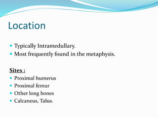
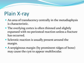
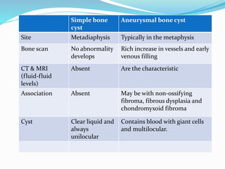
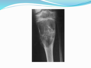
SIMPLE BONE CYST
Dr. Manmohan Bir Shrestha
MD Resident, Phase-A
Department of Radiology and Imaging
BSMMU.
Simple Bone Cyst
 Also known as Unicameral Bone Cyst.
 Are entirely benign lesion of unknown aetiology.
 Are always unilocular.
Age
 Childhood and early adolescense
 Before the epiphyseal fusion occur.
 In adults, some lesions occur after skeletal maturation
in such bones as the Calcaneus, Talus
Location
 Typically Intramedullary.
 Most frequently found in the metaphysis.
Sites :
 Proximal humerus
 Proximal femur
 Other long bones
 Calcaneus, Talus.
Sex Prevalence
 Male>Female (2:1)
Clinical Features
 Asymptomatic and found incidentally.
 Only a few produce minor discomfort. May be pain,
swelling and stiffness of adjacent joint.
 More than half present due to a pathological fracture.
Pathology
 Cyst contains clear liquid unless there has been
contamination by bleeding following a fracture .
 Cyst is lined by a thin layer of connective tissue.
Investigations
 Plain X-ray film
 CT Scan
 MRI
 Bone Scan.
Plain X-ray
 An area of translucency centrally in the metadiaphysis
is characteristic.
 The overlying cortex is often thinned and slightly
expansed with no periosteal reaction unless a fracture
has occurred.
 Sclerotic reaction is usually present around the
margin.
 A serpiginous margin (by prominent ridges of bone)
may cause the cyst to appear multilocular.
Fallen fragment sign
 If there is fracture through this lesion, a dependent
bony fragment may be seen, and this is known as the
fallen fragment sign.
MRI
 T1 -low signal intensity.
 T2 -high signal intensity.
Bone scan
 No abnormality develops in the blood-pool phase.
Treatment
1)If small and low risk of fracture
 Some cases resolve spontaneously with age.
 Intralesional Steroid
An injection of Methylprednisolone Acetate into the
lesion in several intervals for a time span of 6-12 months.
Complications are infection, fracture or recurrence.
Cont.
2)If risk of fracture or large lesion
 Curettage
 Bone grafting
It is proceeded after curettage. The empty cavity
is transplanted with donor bone tissue, bone chips or
artificial material.
Differential diagnosis
1. Fibrous dysplasia
2. Aneurysmal bone cyst
3. Giant cell tumor
4. Eosinophilic granuloma
5. Non ossifying fibroma
6. Chondroma.
Simple bone
cyst
Aneurysmal bone cyst
Site Metadiaphysis Typically in the metaphysis
Bone scan No abnormality
develops
Rich increase in vessels and early
venous filling
CT & MRI
(fluid-fluid
levels)
Absent Are the characteristic
Association Absent May be with non-ossifying
fibroma, fibrous dysplasia and
chondromyxoid fibroma
Cyst Clear liquid and
always
unilocular
Contains blood with giant cells
and multilocular.
Simple bone
cyst
Giant cell tumor
Age Before
epiphyseal
fusion .
Childhood and
early adolescent
Majority between 20-40 yrs of
age. Only 3% in immature
skeleton.
Anatomical
distribution
Proximal
humerus
proximal femur
Other long
bones rarely
calcaneum
Majority occurs around knee and
wrist
Extension Do not extends
to the articular
surface and is
central
Extension is subarticular and
eccentric in nature.
Simple bone cyst Fibrous dysplasia
Sites Proximal humerus
Proximal femur
Other long bones
Calcaneus
Commonly pelvis, femur, ribs. Often
skull
Plain film An area of
translucency.
Centrally in the
metadiaphysis.
Lesions may be lucent, dense or a
mixture with small flecks of density
due to ossification
Pathology Cyst filled with
clear fluid
Medullary bone is replaced by well
defined area of fibrous tissue and cysts
containing blood or serous fluid
Endocrine
complications
Absent May be associated with-
Skin pigmentation,precocious puberty,
acromegaly, hyperthyroidism, cushings
syndrome.
Non-ossifying fibroma-
eccentric and cortical based.
Simple bone cyst Eosinophilic
granuloma
Site Proximal humerus
Proximal femur
Other long bones
Calcaneus
Any bone may be affected
commonly –skull
- pelvis
-femur.
Clinically Asymptomatic often
presents with
pathological fracture
Pain, swelling and mild
fever.
Histology Cyst filled with clear
liquid
Contains eosinophilic
infiltration.
Simple bone cyst Chondroma
Age Childhood and
early adolescence.
Adult
Plain film Area of
translucency
Centrally in
metadiaphysis
Flecks of calcification
occur. As they become
mature, may assume a
pathognomic popcorn or
annular configuration.
Simple bone cyst

Simple bone cyst

  • 2.
    SIMPLE BONE CYST Dr.Manmohan Bir Shrestha MD Resident, Phase-A Department of Radiology and Imaging BSMMU.
  • 3.
    Simple Bone Cyst Also known as Unicameral Bone Cyst.  Are entirely benign lesion of unknown aetiology.  Are always unilocular.
  • 4.
    Age  Childhood andearly adolescense  Before the epiphyseal fusion occur.  In adults, some lesions occur after skeletal maturation in such bones as the Calcaneus, Talus
  • 5.
    Location  Typically Intramedullary. Most frequently found in the metaphysis. Sites :  Proximal humerus  Proximal femur  Other long bones  Calcaneus, Talus.
  • 6.
  • 7.
    Clinical Features  Asymptomaticand found incidentally.  Only a few produce minor discomfort. May be pain, swelling and stiffness of adjacent joint.  More than half present due to a pathological fracture.
  • 8.
    Pathology  Cyst containsclear liquid unless there has been contamination by bleeding following a fracture .  Cyst is lined by a thin layer of connective tissue.
  • 9.
    Investigations  Plain X-rayfilm  CT Scan  MRI  Bone Scan.
  • 10.
    Plain X-ray  Anarea of translucency centrally in the metadiaphysis is characteristic.  The overlying cortex is often thinned and slightly expansed with no periosteal reaction unless a fracture has occurred.  Sclerotic reaction is usually present around the margin.  A serpiginous margin (by prominent ridges of bone) may cause the cyst to appear multilocular.
  • 14.
    Fallen fragment sign If there is fracture through this lesion, a dependent bony fragment may be seen, and this is known as the fallen fragment sign.
  • 18.
    MRI  T1 -lowsignal intensity.  T2 -high signal intensity.
  • 19.
    Bone scan  Noabnormality develops in the blood-pool phase.
  • 20.
    Treatment 1)If small andlow risk of fracture  Some cases resolve spontaneously with age.  Intralesional Steroid An injection of Methylprednisolone Acetate into the lesion in several intervals for a time span of 6-12 months. Complications are infection, fracture or recurrence.
  • 21.
    Cont. 2)If risk offracture or large lesion  Curettage  Bone grafting It is proceeded after curettage. The empty cavity is transplanted with donor bone tissue, bone chips or artificial material.
  • 22.
    Differential diagnosis 1. Fibrousdysplasia 2. Aneurysmal bone cyst 3. Giant cell tumor 4. Eosinophilic granuloma 5. Non ossifying fibroma 6. Chondroma.
  • 23.
    Simple bone cyst Aneurysmal bonecyst Site Metadiaphysis Typically in the metaphysis Bone scan No abnormality develops Rich increase in vessels and early venous filling CT & MRI (fluid-fluid levels) Absent Are the characteristic Association Absent May be with non-ossifying fibroma, fibrous dysplasia and chondromyxoid fibroma Cyst Clear liquid and always unilocular Contains blood with giant cells and multilocular.
  • 25.
    Simple bone cyst Giant celltumor Age Before epiphyseal fusion . Childhood and early adolescent Majority between 20-40 yrs of age. Only 3% in immature skeleton. Anatomical distribution Proximal humerus proximal femur Other long bones rarely calcaneum Majority occurs around knee and wrist Extension Do not extends to the articular surface and is central Extension is subarticular and eccentric in nature.
  • 27.
    Simple bone cystFibrous dysplasia Sites Proximal humerus Proximal femur Other long bones Calcaneus Commonly pelvis, femur, ribs. Often skull Plain film An area of translucency. Centrally in the metadiaphysis. Lesions may be lucent, dense or a mixture with small flecks of density due to ossification Pathology Cyst filled with clear fluid Medullary bone is replaced by well defined area of fibrous tissue and cysts containing blood or serous fluid Endocrine complications Absent May be associated with- Skin pigmentation,precocious puberty, acromegaly, hyperthyroidism, cushings syndrome.
  • 28.
  • 29.
    Simple bone cystEosinophilic granuloma Site Proximal humerus Proximal femur Other long bones Calcaneus Any bone may be affected commonly –skull - pelvis -femur. Clinically Asymptomatic often presents with pathological fracture Pain, swelling and mild fever. Histology Cyst filled with clear liquid Contains eosinophilic infiltration.
  • 30.
    Simple bone cystChondroma Age Childhood and early adolescence. Adult Plain film Area of translucency Centrally in metadiaphysis Flecks of calcification occur. As they become mature, may assume a pathognomic popcorn or annular configuration.