SlideShare a Scribd company logo
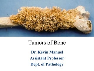
Tumors of Bone
Dr. Kevin Manuel
Assistant Professor
Dept. of Pathology
Introduction
• Bone tumors are diverse in their gross and morphologic
features
• Innocuous to the rapidly fatal.
• Critical to diagnose these tumors correctly, stage them
accurately and treat them appropriately
• Affected patients should survive and maintain optimal
function of the affected body parts.
• Most bone tumors are classified according to the normal cell
or tissue type they arise from.
• Most frequent benign tumors - Osteochondroma
and fibrous cortical defect.
• Most common malignant tumor – Osteosarcoma
followed by chondrosarcoma and Ewing sarcoma
(excluding malignant neoplasms of marrow origin
such as myeloma, lymphoma and leukemia)
• Precise incidence of specific bone tumors is not
known
• Relatively infrequent
• Great diversity
• Occur at all ages / any part of body
• Certain tumors target particular age group and sites
• Diagnosis requires integration of
• Clinical history
• Radiographic appearance
• Histopathology
Age and location
• Most develop during the first several decades
of life and have a propensity to originate in the
long bones of the extremities.
• Location of a tumor provides important
diagnostic information.
Bone Tumours
Sites of Occurrence
Clinical presentation of Bone tumors
• Pain
• Slow-growing mass
• Sudden pathologic fracture
• Radiologic imaging studies - Important role in
diagnosing these lesions. In addition to
– Provides exact location
– Tumor extent
– Aggressiveness of the tumor
Classification of Bone tumors
Bone Tumours
Sites of Occurrence
Giant cell tumour
Chondroblastoma
Ewing’s
Osteosarcoma
BONE-FORMING TUMORS
• Production of bone by the neoplastic cells.
• The tumor bone is usually deposited as woven
trabeculae (except in osteomas) and is variably
mineralized.
• Benign – Osteoma, Osteoid Osteoma and
Osteoblastoma
• Malignant - Osteosarcoma
Osteoma
1. Benign, Often craniofacial in
location
2. Hamartomatous / reactive &
not true tumor.
3. Histologically dense
lamellar bone (closely
resemble normal bone).
4. Gardner Syndrome:
Autosomal Dominant
condition associated with
multiple, Osteoma,
Osteochondroma, GIT
polyps, skin tumors. Colon
Cancer may occur
Osteoid Osteoma
1. Osteoid osteoma are less than 2 cm in greatest dimension
and usually occur in patients in their teens and twenties. 90%
of patients are teens or in their 20’s.
2. Osteoid osteomas can arise in any bone but 50% of cases
involve the femur or tibia, affecting mainly the cortical bone
(diaphysis or metaphysis)
3. Osteoid osteomas are characteristically painful at night. The
pain is caused by excess prostaglandin E2 (due to
proliferating osteoblasts) and is relieved by aspirin.
Osteoid Osteoma
X-Ray: This is the central nidus of an
osteoid osteoma.
Radiographically, there is a small
round central lucent area in the
femoral cortex surrounded by
sclerotic bone.
Micro: the central nidus of an osteoid
osteoma is composed of irregular
reactive new woven bone
dispersed in a highly vascular
stroma
X-Ray & Microscopy
Central nidus – Actual tumor
Microscopy of Osteoid osteoma
Osteoid osteoma composed of haphazardly interconnecting trabeculae of woven bone
that are rimmed by prominent osteoblasts. The intertrabecular spaces are filled by
vascularized loose connective tissue
Osteoblastoma
• Osteoblastoma is larger than 2 cm and
involves the spine more frequently
• The pain is dull, achy, and unresponsive to
salicylates
• Tumor usually does not induce a marked bony
reaction.
Osteosarcoma
• Osteosarcoma is a malignant mesenchymal tumor in
which the cancerous cells produce bone matrix.
• Most common primary malignant tumor of bone
• Occurs in all age groups but has a bimodal age
distribution
– 1st peak - 75% occur in persons younger than 20 years of
age
– 2nd peak occurs in the elderly – Predisposing conditions -
Pagets disease, bone infarcts and prior irradiation
• Men are more commonly affected than women
Major sites of origin of osteosarcoma
Usually arise in the metaphyseal region of the long bones of the extremities, and almost 50%
occur about the knee
Pathogenesis
• 70% of osteosarcomas have acquired genetic
abnormalities such as ploidy changes and
chromosomal aberrations
– RB, the retinoblastoma gene, a critical cell cycle
regulator
– p53, a gene whose product regulates DNA repair and
certain aspects of cellular metabolism. Li-Fraumeni
syndrome
• Abnormalities in INK4a, which encodes p16 (a cell
cycle regulator) and p14 (which aids and abets p53
function), also are seen in osteosarcoma
Morphology
• Site of origin (intramedullary, intracortical, or
surface)
• Degree of differentiation
• Multicentricity (synchronous, metachronous)
• Primary (underlying bone is unremarkable) or
secondary to preexisting disorders such as benign
tumors, Paget disease, bone infarcts, previous
irradiation
• Histologic features (osteoblastic, chondroblastic,
fibroblastic, telangiectatic, small cell, and giant
cell).
Classification of Osteosarcomas
Intramedullary osteosarcomas
• Conventional osteosarcoma
• Telangiectatic osteosarcoma
• Small cell osteosarcoma
• Osteosarcoma in Paget disease
• Post-irradiation osteosarcoma
Surface osteosarcomas
• Parosteal osteosarcoma
• Periosteal osteosarcoma
Osteosarcoma
• Typically present as painful and
progressively enlarging
masses. Sometimes a
sudden fracture of the bone is
the first symptom.
1. Classic X ray findings:
a) Codman’s triangle
(periosteal elevation)
b) Sunburst pattern
c) Bone destruction
Clinical & X-ray findings
Codman Triangle
Sunray
appearance
Sunray
appearance
Gross
• Big bulky tumors that are gritty, gray-white, and
often contain areas of hemorrhage and cystic
degeneration.
• Destroy the surrounding cortices and produce soft-
tissue masses. They spread extensively in the
medullary canal, infiltrating and replacing the
marrow surrounding the preexisting bone trabeculae.
Osteosarcoma of the upper end of the tibia. The tan-white tumor fills most of the medullary
cavity of the metaphysis and proximal diaphysis. It has infiltrated through the cortex, lifted the
periosteum, and formed soft-tissue masses on both sides of the bone.
Microscopy
• Tumor cells vary in size and shape and frequently
have large hyperchromatic nuclei.
• Bizarre tumor giant cells are common along with
mitoses.
• The formation of bone by the tumor cells is
characteristic.
• Neoplastic bone usually has a coarse, lace-like
architecture
• When malignant cartilage is abundant, the tumor is
called chondroblastic osteosarcoma.
• Vascular invasion and necrotic areas are present.
Coarse, lacelike pattern of neoplastic bone produced by anaplastic malignant tumor cells.
Note the mitotic figures.
Metastasis, Treatment and Prognosis
• Highly aggressive neoplasms
• Hematogenous mode of spread
• 90% have metastases to the lungs, bones, brain and
elsewhere.
• Treated with a multimodality approach that includes
chemotherapy
• 5-year survival rate – 20%
CARTILAGE-FORMING TUMORS
• Characterized by the formation of hyaline or myxoid
cartilage
• Benign – Osteochondroma, Chondroma,
Chondroblastoma, Chondromyxoid fibroma
• Malignant - Chondrosarcoma
Osteochondroma
• Also known as an exostosis
• Benign cartilage-capped tumor that is attached to the
underlying skeleton by a bony stalk.
• Most common benign bone tumor; about 85% are
solitary
• Multiple hereditary exostosis syndrome, which is an
autosomal dominant hereditary disease. Hereditary
exostoses are caused by germline loss-of-function
mutations in either the EXT1 or EXT2 genes
• Solitary osteochondromas are usually first diagnosed
in late adolescence and early adulthood, but multiple
osteochondromas become apparent during
childhood.
• Men are affected three times more often than
women
• Develop only in bones of endochondral origin and
arise from the metaphysis near the growth plate of
long tubular bones, especially about the knee
• Incidental finding or presents as slow growing
masses
Osteochondroma
1. Hereditary (multiple) or sporadic (single)
2. Benign bone growths capped with
cartilage
3. affects children/ adolescent males; may
be asymptomatic or cause pain,
producing deformity
4. hereditary type can undergo malignant
transformation (Chondrosarcoma )
Exostosis
Morphology
• Sessile or mushroom shaped
• Range in size from 1 to 20 cm.
• The cap is composed of benign hyaline cartilage
varying in thickness and is covered peripherally by
perichondrium. The cartilage has the appearance of
disorganized growth plate and undergoes enchondral
ossification, with the newly made bone forming the
inner portion of the head and stalk.
A, X-ray of an osteochondroma arising off the posterior surface of the tibia. B, Axial
CT scan shows continuity of the cortex of the bone and the center of the
osteochondroma. The fibula is adjacent to the mass. C, Gross specimen of sessile
osteochondroma composed of a cap of hyaline cartilage undergoing enchondral
ossification. D, The cartilage cap has the histologic appearance of disorganized growth
plate-like cartilage.
Chondromas
• Benign tumors of hyaline cartilage that usually
occur in bones of enchondral origin.
• Enchondromas - Arise within the medullary
cavity.
• Subperiosteal or juxtacortical chondromas -
Surface of bone.
• Age – 20s to 40s
Enchondroma
• Benign
• Single or multiple sites
• Often involves small bones of hands and
feet.
• Well demarcated, mature cartilage.
• Hereditary – multiple enchondromatosis.
Usually over one side of the body.
(Ollier’s disease).
• Maffucci's syndrome - multiple bone
chondromas and hemangiomas of soft
tissue
• Increased risk for chondrosarcoma
Chondroma
Morphology
• Enchondromas are usually smaller than 3 cm
and grossly are gray-blue and translucent
• Composed of well-circumscribed nodules of
cyto logically benign hyaline cartilage
Enchondroma with a nodule of hyaline cartilage encased by a thin layer of reactive
bone
Enchondroma of the phalanx with a pathologic fracture. The radiolucent nodules of
hyaline cartilage scallop the endosteal surface.
Chondrosarcoma
• Production of neoplastic cartilage
• Subclassified according to site as central
(intramedullary) and peripheral (juxtacortical and
surface).
• Histologically, they include conventional (hyaline
and/or myxoid), clear cell, dedifferentiated, and
mesenchymal variants.
• Age > 40 years
• Men
• 15% of conventional chondrosarcomas arise from a
preexisting enchondroma or osteochondroma.
• Commonly arise in the central portions of the
skeleton, including the pelvis, shoulder, and ribs
• Present as painful, progressively enlarging masses.
• Slow-growing, low-grade tumor causes reactive
thickening of the cortex, whereas a more aggressive
high-grade neoplasm destroys the cortex and forms a
soft-tissue mass
Morphology - Gross
• Large bulky tumors are made up of nodules of
gray-white, somewhat translucent glistening
tissue
Microscopy
• Tumors vary in degree of cellularity, cytologic
atypia, and mitotic activity. Presence of anaplastic
chondrocytes
– Grade 1, 2 and 3
Metastasis and Prognosis
• Direct correlation between the grade and the
biologic behavior of the tumor
• 5-year survival rates were 90%, 81%, and 43%
for grades 1 through 3, respectively
• Spread preferentially to the lungs and skeleton
• Treatment of conventional chondrosarcoma is
wide surgical excision
Miscellaneous tumors of bone
• GIANT-CELL TUMOR
• EWING SARCOMA/PRIMITIVE
EUROECTODERMAL TUMOR
• ANEURYSMAL BONE CYST
Giant cell Tumour of Bone
• Known as osteoclastoma
• Common tumour – 20% of all benign bone
tumors
• Age - 20 -40 years
• Slight female preponderence
• Histogenesis – not known
• Epiphysis of long bones affected
• Radiolucent lesion involving end of long bones
• Almost always solitary
• Grossly dark brown - due to abundant
vascularity
• Areas of necrosis and cystic change present
Giant cell tumor
Magnetic resonance image of a giant-cell tumor that replaces most of the
femoral condyle and extends to the subchondral bone plate.
Morphology
• HPE - 2 major population of cells
• Multinucleated giant cells - reactive component
• Neoplastic component – round to spindle shaped
mononuclear cells
• Large number of osteoclast likes giant cells with
mononuclear cells.
Giant cell tumor
• Clinical features
• Local pain – mistaken for arthritis
• Wide variety of bone disorder may contain
multinucleated giant cells
– Brown tumor
– Aneurysmal bone cyst
• Unpredictable behaviour
• Recurrence common after curettage
Ewing’s Sarcoma
• Most common form of bone tumour in children / adolescent
• Peak incidence 2nd decade
• Highly aggressive tumour
• Must be differentiated from other small blue cell tumours.
• Translocation involving the EWS gene on chromosome 22
and a gene encoding an ETS family transcription factor; the
most commonly involved ETS gene is FLI1
• Present as painful enlarging masses
• Tender, warm, and swollen.
• Some affected individuals have systemic findings,
including fever, elevated sedimentation rate, anemia,
and leukocytosis, which mimic infection.
• Plain radiograms show a destructive lytic tumor that
has permeative margins and extension into the
surrounding soft tissues.
• The characteristic periosteal reaction produces
layers of reactive bone deposited in an onion-skin
fashion.
Morphology
• Arise in medullary cavity
• Soft, expansive mass
• Site – femur, tibia, pelvis – diaphysis
commonly affected
• Extends beyond medullary cavity
Ewings tumor
• HPE
• Sheets of small round cells
• Small, fairly uniform nuclei
• Scant cytoplasm
• Cytoplasm contain glycogen (PAS stain)
• Produce reactive bone / not osteoid
• Presence of Homer-Wright rosettes
(tumor cells arranged in a circle about a
central fibrillary space) is indicative of
neural differentiation
Ewings tumour
Ewings tumour (PAS stain)
• Immunohistochemical study needed
to distinguish from
• Neuroblastoma
• Rhabdomyosarcoma
• Lymphoma
• EWS express neural marker
• Clinical features
• Pain and local inflammation
• Fever is common
• Biopsy needed for diagnosis
• Recent advances in treatment
improved outlook of patients
• 5 year survival rate is 75%
ANEURYSMAL BONE CYST
• Benign tumor of bone characterized by
multiloculated blood-filled cystic spaces that may
present as a rapidly growing expansile tumor
• First 2 decades of life and has no sex predilection
• Metaphyses of long bones and the posterior
elements of vertebral bodies
• Most common signs and symptoms are pain and
swelling
Coronal computed axial tomography scan showing eccentric aneurysmal
bone cyst of tibia
Aneurysmal bone cyst with blood-filled cystic space surrounded by wall containing
proliferating fibroblasts, reactive woven bone, and osteoclast-type giant cells.
Metastatic tumors to bone
• Pathways of spread
– Direct extension
– Lymphatic or hematogenous dissemination
– intraspinal seeding (via the Batson plexus of veins)
• Adults more than 75% of skeletal metastases
originate from cancers of the prostate, breast,
kidney, and lung.
• In children, metastases to bone originate from
neuroblastoma, Wilms tumor, osteosarcoma, Ewing
sarcoma, and rhabdomyosarcoma.
Thank You…

More Related Content

What's hot

Malignant bone tumors
Malignant bone tumorsMalignant bone tumors
Malignant bone tumors
SaachiGupta4
 
Malignant bone tumors
Malignant bone tumorsMalignant bone tumors
Malignant bone tumors
Eneutron
 
Osteosarcoma
OsteosarcomaOsteosarcoma
Osteosarcoma
nicholasfrane
 
Synovial biopsy
Synovial biopsySynovial biopsy
Synovial biopsy
Appy Akshay Agarwal
 
Bone tumors part one
Bone tumors part oneBone tumors part one
Bone tumors part one
Ramin Sadeghi
 
Bone Tumors
Bone TumorsBone Tumors
Bone Tumors
Muhammad Eimaduddin
 
Bone tumour , enchondroma , osteochondroma
Bone tumour , enchondroma , osteochondromaBone tumour , enchondroma , osteochondroma
Bone tumour , enchondroma , osteochondroma
Sagar Savsani
 
Cystic lesions of bone
Cystic lesions of boneCystic lesions of bone
Cystic lesions of bone
kesarkar88
 
Bone tumours
Bone tumoursBone tumours
Bone tumours
Usman Shams
 
Ewing's sarcoma
Ewing's sarcomaEwing's sarcoma
Ewing's sarcoma
Sreekanth Nallam
 
Metabolic bone diseases, pathology dept
Metabolic bone diseases, pathology deptMetabolic bone diseases, pathology dept
Metabolic bone diseases, pathology dept
regional institute of medical sciences
 
Malignant bone tumours
Malignant bone tumoursMalignant bone tumours
Malignant bone tumours
drharshwardhandawar
 
Osteolytic lesions of Bone
Osteolytic lesions of BoneOsteolytic lesions of Bone
Osteolytic lesions of Bone
Vineel Bezawada
 
Bone tumours
Bone tumoursBone tumours
Bone tumours
Surya Prakash
 
Soft tissue tumor.ppt
Soft tissue tumor.pptSoft tissue tumor.ppt
Soft tissue tumor.ppt
WillyChangchien2
 
Benign bone tumours
Benign bone tumoursBenign bone tumours
Benign bone tumours
Arif S
 
Multiple myeloma final 2018 updated
Multiple myeloma final 2018 updatedMultiple myeloma final 2018 updated
Multiple myeloma final 2018 updated
AmrinderSingh248
 
Bone tumours
Bone tumoursBone tumours
Bone tumours
Abhilash Gavarraju
 
Bone tumors
Bone tumorsBone tumors
Bone tumors
Eileesha Belarso
 

What's hot (20)

Malignant bone tumors
Malignant bone tumorsMalignant bone tumors
Malignant bone tumors
 
Malignant bone tumors
Malignant bone tumorsMalignant bone tumors
Malignant bone tumors
 
Osteosarcoma
OsteosarcomaOsteosarcoma
Osteosarcoma
 
Synovial biopsy
Synovial biopsySynovial biopsy
Synovial biopsy
 
Bone tumors part one
Bone tumors part oneBone tumors part one
Bone tumors part one
 
Bone Tumors
Bone TumorsBone Tumors
Bone Tumors
 
Bone tumour , enchondroma , osteochondroma
Bone tumour , enchondroma , osteochondromaBone tumour , enchondroma , osteochondroma
Bone tumour , enchondroma , osteochondroma
 
Cystic lesions of bone
Cystic lesions of boneCystic lesions of bone
Cystic lesions of bone
 
Osteosarcoma
OsteosarcomaOsteosarcoma
Osteosarcoma
 
Bone tumours
Bone tumoursBone tumours
Bone tumours
 
Ewing's sarcoma
Ewing's sarcomaEwing's sarcoma
Ewing's sarcoma
 
Metabolic bone diseases, pathology dept
Metabolic bone diseases, pathology deptMetabolic bone diseases, pathology dept
Metabolic bone diseases, pathology dept
 
Malignant bone tumours
Malignant bone tumoursMalignant bone tumours
Malignant bone tumours
 
Osteolytic lesions of Bone
Osteolytic lesions of BoneOsteolytic lesions of Bone
Osteolytic lesions of Bone
 
Bone tumours
Bone tumoursBone tumours
Bone tumours
 
Soft tissue tumor.ppt
Soft tissue tumor.pptSoft tissue tumor.ppt
Soft tissue tumor.ppt
 
Benign bone tumours
Benign bone tumoursBenign bone tumours
Benign bone tumours
 
Multiple myeloma final 2018 updated
Multiple myeloma final 2018 updatedMultiple myeloma final 2018 updated
Multiple myeloma final 2018 updated
 
Bone tumours
Bone tumoursBone tumours
Bone tumours
 
Bone tumors
Bone tumorsBone tumors
Bone tumors
 

Similar to Tumors of bone

Pathology of bone tumors
Pathology of bone tumorsPathology of bone tumors
Pathology of bone tumors
Subhash Das
 
Bone tumors ug
Bone tumors ugBone tumors ug
Bone tumors ug
vichand8
 
bone tumor
bone tumorbone tumor
bone tumor
Dr Manoj Prajapati
 
bone%20tumor%20ppt.pptx
bone%20tumor%20ppt.pptxbone%20tumor%20ppt.pptx
bone%20tumor%20ppt.pptx
Dr Manoj Prajapati
 
bone tumors.pptx
bone tumors.pptxbone tumors.pptx
bone tumors.pptx
DharmendraSingh78468
 
Bone.pptx
Bone.pptxBone.pptx
Bone.pptx
santoshgoit2
 
BT.pptx
BT.pptxBT.pptx
BT.pptx
santoshgoit2
 
Bone forming tumors
Bone forming tumorsBone forming tumors
Bone forming tumorsKemUnited
 
bonetumors...............-180123083659.pptx
bonetumors...............-180123083659.pptxbonetumors...............-180123083659.pptx
bonetumors...............-180123083659.pptx
sacootcbe
 
Oncology oite-review-2012
Oncology oite-review-2012Oncology oite-review-2012
Oncology oite-review-2012mghbonephone
 
bonetumors-180123083659.pdf
bonetumors-180123083659.pdfbonetumors-180123083659.pdf
bonetumors-180123083659.pdf
VijeshBichu
 
Bone tumors
Bone tumorsBone tumors
Bone tumors
ANJANI WALIA
 
Bone tumors and tumor-like lesions.ppt
Bone tumors and tumor-like lesions.pptBone tumors and tumor-like lesions.ppt
Bone tumors and tumor-like lesions.ppt
drqazi7777
 
Benign bone lesion 1
Benign bone lesion 1Benign bone lesion 1
Benign bone lesion 1
sabir khadka
 
Tumor and tumor like conditions 2.
Tumor and tumor like conditions 2.Tumor and tumor like conditions 2.
Tumor and tumor like conditions 2.
Ali Jiwani
 
Bone tumors.pptx
Bone tumors.pptxBone tumors.pptx
Bone tumors.pptx
ydnxq7zfcz
 
Bone tumour seminar ,ewing sarcoma, chordoma,
Bone tumour seminar ,ewing sarcoma, chordoma,Bone tumour seminar ,ewing sarcoma, chordoma,
Bone tumour seminar ,ewing sarcoma, chordoma,Narmada Tiwari
 
Bone tumors
Bone tumors   Bone tumors
Bone tumors
K BHATTACHARJEE
 
Bone Tumor
Bone TumorBone Tumor
Bone Tumor (Benign and malignant)
Bone Tumor (Benign and malignant)Bone Tumor (Benign and malignant)
Bone Tumor (Benign and malignant)
Nassr ALBarhi
 

Similar to Tumors of bone (20)

Pathology of bone tumors
Pathology of bone tumorsPathology of bone tumors
Pathology of bone tumors
 
Bone tumors ug
Bone tumors ugBone tumors ug
Bone tumors ug
 
bone tumor
bone tumorbone tumor
bone tumor
 
bone%20tumor%20ppt.pptx
bone%20tumor%20ppt.pptxbone%20tumor%20ppt.pptx
bone%20tumor%20ppt.pptx
 
bone tumors.pptx
bone tumors.pptxbone tumors.pptx
bone tumors.pptx
 
Bone.pptx
Bone.pptxBone.pptx
Bone.pptx
 
BT.pptx
BT.pptxBT.pptx
BT.pptx
 
Bone forming tumors
Bone forming tumorsBone forming tumors
Bone forming tumors
 
bonetumors...............-180123083659.pptx
bonetumors...............-180123083659.pptxbonetumors...............-180123083659.pptx
bonetumors...............-180123083659.pptx
 
Oncology oite-review-2012
Oncology oite-review-2012Oncology oite-review-2012
Oncology oite-review-2012
 
bonetumors-180123083659.pdf
bonetumors-180123083659.pdfbonetumors-180123083659.pdf
bonetumors-180123083659.pdf
 
Bone tumors
Bone tumorsBone tumors
Bone tumors
 
Bone tumors and tumor-like lesions.ppt
Bone tumors and tumor-like lesions.pptBone tumors and tumor-like lesions.ppt
Bone tumors and tumor-like lesions.ppt
 
Benign bone lesion 1
Benign bone lesion 1Benign bone lesion 1
Benign bone lesion 1
 
Tumor and tumor like conditions 2.
Tumor and tumor like conditions 2.Tumor and tumor like conditions 2.
Tumor and tumor like conditions 2.
 
Bone tumors.pptx
Bone tumors.pptxBone tumors.pptx
Bone tumors.pptx
 
Bone tumour seminar ,ewing sarcoma, chordoma,
Bone tumour seminar ,ewing sarcoma, chordoma,Bone tumour seminar ,ewing sarcoma, chordoma,
Bone tumour seminar ,ewing sarcoma, chordoma,
 
Bone tumors
Bone tumors   Bone tumors
Bone tumors
 
Bone Tumor
Bone TumorBone Tumor
Bone Tumor
 
Bone Tumor (Benign and malignant)
Bone Tumor (Benign and malignant)Bone Tumor (Benign and malignant)
Bone Tumor (Benign and malignant)
 

Recently uploaded

Physiology of Chemical Sensation of smell.pdf
Physiology of Chemical Sensation of smell.pdfPhysiology of Chemical Sensation of smell.pdf
Physiology of Chemical Sensation of smell.pdf
MedicoseAcademics
 
New Directions in Targeted Therapeutic Approaches for Older Adults With Mantl...
New Directions in Targeted Therapeutic Approaches for Older Adults With Mantl...New Directions in Targeted Therapeutic Approaches for Older Adults With Mantl...
New Directions in Targeted Therapeutic Approaches for Older Adults With Mantl...
i3 Health
 
Ozempic: Preoperative Management of Patients on GLP-1 Receptor Agonists
Ozempic: Preoperative Management of Patients on GLP-1 Receptor Agonists  Ozempic: Preoperative Management of Patients on GLP-1 Receptor Agonists
Ozempic: Preoperative Management of Patients on GLP-1 Receptor Agonists
Saeid Safari
 
POST OPERATIVE OLIGURIA and its management
POST OPERATIVE OLIGURIA and its managementPOST OPERATIVE OLIGURIA and its management
POST OPERATIVE OLIGURIA and its management
touseefaziz1
 
Hemodialysis: Chapter 3, Dialysis Water Unit - Dr.Gawad
Hemodialysis: Chapter 3, Dialysis Water Unit - Dr.GawadHemodialysis: Chapter 3, Dialysis Water Unit - Dr.Gawad
Hemodialysis: Chapter 3, Dialysis Water Unit - Dr.Gawad
NephroTube - Dr.Gawad
 
ANATOMY AND PHYSIOLOGY OF URINARY SYSTEM.pptx
ANATOMY AND PHYSIOLOGY OF URINARY SYSTEM.pptxANATOMY AND PHYSIOLOGY OF URINARY SYSTEM.pptx
ANATOMY AND PHYSIOLOGY OF URINARY SYSTEM.pptx
Swetaba Besh
 
Couples presenting to the infertility clinic- Do they really have infertility...
Couples presenting to the infertility clinic- Do they really have infertility...Couples presenting to the infertility clinic- Do they really have infertility...
Couples presenting to the infertility clinic- Do they really have infertility...
Sujoy Dasgupta
 
Knee anatomy and clinical tests 2024.pdf
Knee anatomy and clinical tests 2024.pdfKnee anatomy and clinical tests 2024.pdf
Knee anatomy and clinical tests 2024.pdf
vimalpl1234
 
Report Back from SGO 2024: What’s the Latest in Cervical Cancer?
Report Back from SGO 2024: What’s the Latest in Cervical Cancer?Report Back from SGO 2024: What’s the Latest in Cervical Cancer?
Report Back from SGO 2024: What’s the Latest in Cervical Cancer?
bkling
 
NVBDCP.pptx Nation vector borne disease control program
NVBDCP.pptx Nation vector borne disease control programNVBDCP.pptx Nation vector borne disease control program
NVBDCP.pptx Nation vector borne disease control program
Sapna Thakur
 
BENIGN PROSTATIC HYPERPLASIA.BPH. BPHpdf
BENIGN PROSTATIC HYPERPLASIA.BPH. BPHpdfBENIGN PROSTATIC HYPERPLASIA.BPH. BPHpdf
BENIGN PROSTATIC HYPERPLASIA.BPH. BPHpdf
DR SETH JOTHAM
 
Prix Galien International 2024 Forum Program
Prix Galien International 2024 Forum ProgramPrix Galien International 2024 Forum Program
Prix Galien International 2024 Forum Program
Levi Shapiro
 
Are There Any Natural Remedies To Treat Syphilis.pdf
Are There Any Natural Remedies To Treat Syphilis.pdfAre There Any Natural Remedies To Treat Syphilis.pdf
Are There Any Natural Remedies To Treat Syphilis.pdf
Little Cross Family Clinic
 
Evaluation of antidepressant activity of clitoris ternatea in animals
Evaluation of antidepressant activity of clitoris ternatea in animalsEvaluation of antidepressant activity of clitoris ternatea in animals
Evaluation of antidepressant activity of clitoris ternatea in animals
Shweta
 
Charaka Samhita Sutra sthana Chapter 15 Upakalpaniyaadhyaya
Charaka Samhita Sutra sthana Chapter 15 UpakalpaniyaadhyayaCharaka Samhita Sutra sthana Chapter 15 Upakalpaniyaadhyaya
Charaka Samhita Sutra sthana Chapter 15 Upakalpaniyaadhyaya
Dr KHALID B.M
 
Alcohol_Dr. Jeenal Mistry MD Pharmacology.pdf
Alcohol_Dr. Jeenal Mistry MD Pharmacology.pdfAlcohol_Dr. Jeenal Mistry MD Pharmacology.pdf
Alcohol_Dr. Jeenal Mistry MD Pharmacology.pdf
Dr Jeenal Mistry
 
Non-respiratory Functions of the Lungs.pdf
Non-respiratory Functions of the Lungs.pdfNon-respiratory Functions of the Lungs.pdf
Non-respiratory Functions of the Lungs.pdf
MedicoseAcademics
 
Superficial & Deep Fascia of the NECK.pptx
Superficial & Deep Fascia of the NECK.pptxSuperficial & Deep Fascia of the NECK.pptx
Superficial & Deep Fascia of the NECK.pptx
Dr. Rabia Inam Gandapore
 
Novas diretrizes da OMS para os cuidados perinatais de mais qualidade
Novas diretrizes da OMS para os cuidados perinatais de mais qualidadeNovas diretrizes da OMS para os cuidados perinatais de mais qualidade
Novas diretrizes da OMS para os cuidados perinatais de mais qualidade
Prof. Marcus Renato de Carvalho
 
ARTIFICIAL INTELLIGENCE IN HEALTHCARE.pdf
ARTIFICIAL INTELLIGENCE IN  HEALTHCARE.pdfARTIFICIAL INTELLIGENCE IN  HEALTHCARE.pdf
ARTIFICIAL INTELLIGENCE IN HEALTHCARE.pdf
Anujkumaranit
 

Recently uploaded (20)

Physiology of Chemical Sensation of smell.pdf
Physiology of Chemical Sensation of smell.pdfPhysiology of Chemical Sensation of smell.pdf
Physiology of Chemical Sensation of smell.pdf
 
New Directions in Targeted Therapeutic Approaches for Older Adults With Mantl...
New Directions in Targeted Therapeutic Approaches for Older Adults With Mantl...New Directions in Targeted Therapeutic Approaches for Older Adults With Mantl...
New Directions in Targeted Therapeutic Approaches for Older Adults With Mantl...
 
Ozempic: Preoperative Management of Patients on GLP-1 Receptor Agonists
Ozempic: Preoperative Management of Patients on GLP-1 Receptor Agonists  Ozempic: Preoperative Management of Patients on GLP-1 Receptor Agonists
Ozempic: Preoperative Management of Patients on GLP-1 Receptor Agonists
 
POST OPERATIVE OLIGURIA and its management
POST OPERATIVE OLIGURIA and its managementPOST OPERATIVE OLIGURIA and its management
POST OPERATIVE OLIGURIA and its management
 
Hemodialysis: Chapter 3, Dialysis Water Unit - Dr.Gawad
Hemodialysis: Chapter 3, Dialysis Water Unit - Dr.GawadHemodialysis: Chapter 3, Dialysis Water Unit - Dr.Gawad
Hemodialysis: Chapter 3, Dialysis Water Unit - Dr.Gawad
 
ANATOMY AND PHYSIOLOGY OF URINARY SYSTEM.pptx
ANATOMY AND PHYSIOLOGY OF URINARY SYSTEM.pptxANATOMY AND PHYSIOLOGY OF URINARY SYSTEM.pptx
ANATOMY AND PHYSIOLOGY OF URINARY SYSTEM.pptx
 
Couples presenting to the infertility clinic- Do they really have infertility...
Couples presenting to the infertility clinic- Do they really have infertility...Couples presenting to the infertility clinic- Do they really have infertility...
Couples presenting to the infertility clinic- Do they really have infertility...
 
Knee anatomy and clinical tests 2024.pdf
Knee anatomy and clinical tests 2024.pdfKnee anatomy and clinical tests 2024.pdf
Knee anatomy and clinical tests 2024.pdf
 
Report Back from SGO 2024: What’s the Latest in Cervical Cancer?
Report Back from SGO 2024: What’s the Latest in Cervical Cancer?Report Back from SGO 2024: What’s the Latest in Cervical Cancer?
Report Back from SGO 2024: What’s the Latest in Cervical Cancer?
 
NVBDCP.pptx Nation vector borne disease control program
NVBDCP.pptx Nation vector borne disease control programNVBDCP.pptx Nation vector borne disease control program
NVBDCP.pptx Nation vector borne disease control program
 
BENIGN PROSTATIC HYPERPLASIA.BPH. BPHpdf
BENIGN PROSTATIC HYPERPLASIA.BPH. BPHpdfBENIGN PROSTATIC HYPERPLASIA.BPH. BPHpdf
BENIGN PROSTATIC HYPERPLASIA.BPH. BPHpdf
 
Prix Galien International 2024 Forum Program
Prix Galien International 2024 Forum ProgramPrix Galien International 2024 Forum Program
Prix Galien International 2024 Forum Program
 
Are There Any Natural Remedies To Treat Syphilis.pdf
Are There Any Natural Remedies To Treat Syphilis.pdfAre There Any Natural Remedies To Treat Syphilis.pdf
Are There Any Natural Remedies To Treat Syphilis.pdf
 
Evaluation of antidepressant activity of clitoris ternatea in animals
Evaluation of antidepressant activity of clitoris ternatea in animalsEvaluation of antidepressant activity of clitoris ternatea in animals
Evaluation of antidepressant activity of clitoris ternatea in animals
 
Charaka Samhita Sutra sthana Chapter 15 Upakalpaniyaadhyaya
Charaka Samhita Sutra sthana Chapter 15 UpakalpaniyaadhyayaCharaka Samhita Sutra sthana Chapter 15 Upakalpaniyaadhyaya
Charaka Samhita Sutra sthana Chapter 15 Upakalpaniyaadhyaya
 
Alcohol_Dr. Jeenal Mistry MD Pharmacology.pdf
Alcohol_Dr. Jeenal Mistry MD Pharmacology.pdfAlcohol_Dr. Jeenal Mistry MD Pharmacology.pdf
Alcohol_Dr. Jeenal Mistry MD Pharmacology.pdf
 
Non-respiratory Functions of the Lungs.pdf
Non-respiratory Functions of the Lungs.pdfNon-respiratory Functions of the Lungs.pdf
Non-respiratory Functions of the Lungs.pdf
 
Superficial & Deep Fascia of the NECK.pptx
Superficial & Deep Fascia of the NECK.pptxSuperficial & Deep Fascia of the NECK.pptx
Superficial & Deep Fascia of the NECK.pptx
 
Novas diretrizes da OMS para os cuidados perinatais de mais qualidade
Novas diretrizes da OMS para os cuidados perinatais de mais qualidadeNovas diretrizes da OMS para os cuidados perinatais de mais qualidade
Novas diretrizes da OMS para os cuidados perinatais de mais qualidade
 
ARTIFICIAL INTELLIGENCE IN HEALTHCARE.pdf
ARTIFICIAL INTELLIGENCE IN  HEALTHCARE.pdfARTIFICIAL INTELLIGENCE IN  HEALTHCARE.pdf
ARTIFICIAL INTELLIGENCE IN HEALTHCARE.pdf
 

Tumors of bone

  • 1. Tumors of Bone Dr. Kevin Manuel Assistant Professor Dept. of Pathology
  • 2.
  • 3. Introduction • Bone tumors are diverse in their gross and morphologic features • Innocuous to the rapidly fatal. • Critical to diagnose these tumors correctly, stage them accurately and treat them appropriately • Affected patients should survive and maintain optimal function of the affected body parts. • Most bone tumors are classified according to the normal cell or tissue type they arise from.
  • 4. • Most frequent benign tumors - Osteochondroma and fibrous cortical defect. • Most common malignant tumor – Osteosarcoma followed by chondrosarcoma and Ewing sarcoma (excluding malignant neoplasms of marrow origin such as myeloma, lymphoma and leukemia)
  • 5. • Precise incidence of specific bone tumors is not known • Relatively infrequent • Great diversity • Occur at all ages / any part of body • Certain tumors target particular age group and sites • Diagnosis requires integration of • Clinical history • Radiographic appearance • Histopathology
  • 6. Age and location • Most develop during the first several decades of life and have a propensity to originate in the long bones of the extremities. • Location of a tumor provides important diagnostic information.
  • 8. Clinical presentation of Bone tumors • Pain • Slow-growing mass • Sudden pathologic fracture • Radiologic imaging studies - Important role in diagnosing these lesions. In addition to – Provides exact location – Tumor extent – Aggressiveness of the tumor
  • 10. Bone Tumours Sites of Occurrence Giant cell tumour Chondroblastoma Ewing’s Osteosarcoma
  • 11.
  • 12. BONE-FORMING TUMORS • Production of bone by the neoplastic cells. • The tumor bone is usually deposited as woven trabeculae (except in osteomas) and is variably mineralized. • Benign – Osteoma, Osteoid Osteoma and Osteoblastoma • Malignant - Osteosarcoma
  • 13. Osteoma 1. Benign, Often craniofacial in location 2. Hamartomatous / reactive & not true tumor. 3. Histologically dense lamellar bone (closely resemble normal bone). 4. Gardner Syndrome: Autosomal Dominant condition associated with multiple, Osteoma, Osteochondroma, GIT polyps, skin tumors. Colon Cancer may occur
  • 14. Osteoid Osteoma 1. Osteoid osteoma are less than 2 cm in greatest dimension and usually occur in patients in their teens and twenties. 90% of patients are teens or in their 20’s. 2. Osteoid osteomas can arise in any bone but 50% of cases involve the femur or tibia, affecting mainly the cortical bone (diaphysis or metaphysis) 3. Osteoid osteomas are characteristically painful at night. The pain is caused by excess prostaglandin E2 (due to proliferating osteoblasts) and is relieved by aspirin.
  • 15. Osteoid Osteoma X-Ray: This is the central nidus of an osteoid osteoma. Radiographically, there is a small round central lucent area in the femoral cortex surrounded by sclerotic bone. Micro: the central nidus of an osteoid osteoma is composed of irregular reactive new woven bone dispersed in a highly vascular stroma X-Ray & Microscopy
  • 16. Central nidus – Actual tumor
  • 17. Microscopy of Osteoid osteoma Osteoid osteoma composed of haphazardly interconnecting trabeculae of woven bone that are rimmed by prominent osteoblasts. The intertrabecular spaces are filled by vascularized loose connective tissue
  • 18. Osteoblastoma • Osteoblastoma is larger than 2 cm and involves the spine more frequently • The pain is dull, achy, and unresponsive to salicylates • Tumor usually does not induce a marked bony reaction.
  • 19. Osteosarcoma • Osteosarcoma is a malignant mesenchymal tumor in which the cancerous cells produce bone matrix. • Most common primary malignant tumor of bone • Occurs in all age groups but has a bimodal age distribution – 1st peak - 75% occur in persons younger than 20 years of age – 2nd peak occurs in the elderly – Predisposing conditions - Pagets disease, bone infarcts and prior irradiation • Men are more commonly affected than women
  • 20. Major sites of origin of osteosarcoma Usually arise in the metaphyseal region of the long bones of the extremities, and almost 50% occur about the knee
  • 21. Pathogenesis • 70% of osteosarcomas have acquired genetic abnormalities such as ploidy changes and chromosomal aberrations – RB, the retinoblastoma gene, a critical cell cycle regulator – p53, a gene whose product regulates DNA repair and certain aspects of cellular metabolism. Li-Fraumeni syndrome • Abnormalities in INK4a, which encodes p16 (a cell cycle regulator) and p14 (which aids and abets p53 function), also are seen in osteosarcoma
  • 22. Morphology • Site of origin (intramedullary, intracortical, or surface) • Degree of differentiation • Multicentricity (synchronous, metachronous) • Primary (underlying bone is unremarkable) or secondary to preexisting disorders such as benign tumors, Paget disease, bone infarcts, previous irradiation • Histologic features (osteoblastic, chondroblastic, fibroblastic, telangiectatic, small cell, and giant cell).
  • 23. Classification of Osteosarcomas Intramedullary osteosarcomas • Conventional osteosarcoma • Telangiectatic osteosarcoma • Small cell osteosarcoma • Osteosarcoma in Paget disease • Post-irradiation osteosarcoma Surface osteosarcomas • Parosteal osteosarcoma • Periosteal osteosarcoma
  • 24. Osteosarcoma • Typically present as painful and progressively enlarging masses. Sometimes a sudden fracture of the bone is the first symptom. 1. Classic X ray findings: a) Codman’s triangle (periosteal elevation) b) Sunburst pattern c) Bone destruction Clinical & X-ray findings Codman Triangle Sunray appearance Sunray appearance
  • 25. Gross • Big bulky tumors that are gritty, gray-white, and often contain areas of hemorrhage and cystic degeneration. • Destroy the surrounding cortices and produce soft- tissue masses. They spread extensively in the medullary canal, infiltrating and replacing the marrow surrounding the preexisting bone trabeculae.
  • 26. Osteosarcoma of the upper end of the tibia. The tan-white tumor fills most of the medullary cavity of the metaphysis and proximal diaphysis. It has infiltrated through the cortex, lifted the periosteum, and formed soft-tissue masses on both sides of the bone.
  • 27.
  • 28. Microscopy • Tumor cells vary in size and shape and frequently have large hyperchromatic nuclei. • Bizarre tumor giant cells are common along with mitoses. • The formation of bone by the tumor cells is characteristic. • Neoplastic bone usually has a coarse, lace-like architecture • When malignant cartilage is abundant, the tumor is called chondroblastic osteosarcoma. • Vascular invasion and necrotic areas are present.
  • 29. Coarse, lacelike pattern of neoplastic bone produced by anaplastic malignant tumor cells. Note the mitotic figures.
  • 30.
  • 31. Metastasis, Treatment and Prognosis • Highly aggressive neoplasms • Hematogenous mode of spread • 90% have metastases to the lungs, bones, brain and elsewhere. • Treated with a multimodality approach that includes chemotherapy • 5-year survival rate – 20%
  • 32. CARTILAGE-FORMING TUMORS • Characterized by the formation of hyaline or myxoid cartilage • Benign – Osteochondroma, Chondroma, Chondroblastoma, Chondromyxoid fibroma • Malignant - Chondrosarcoma
  • 33. Osteochondroma • Also known as an exostosis • Benign cartilage-capped tumor that is attached to the underlying skeleton by a bony stalk. • Most common benign bone tumor; about 85% are solitary • Multiple hereditary exostosis syndrome, which is an autosomal dominant hereditary disease. Hereditary exostoses are caused by germline loss-of-function mutations in either the EXT1 or EXT2 genes
  • 34. • Solitary osteochondromas are usually first diagnosed in late adolescence and early adulthood, but multiple osteochondromas become apparent during childhood. • Men are affected three times more often than women • Develop only in bones of endochondral origin and arise from the metaphysis near the growth plate of long tubular bones, especially about the knee • Incidental finding or presents as slow growing masses
  • 35. Osteochondroma 1. Hereditary (multiple) or sporadic (single) 2. Benign bone growths capped with cartilage 3. affects children/ adolescent males; may be asymptomatic or cause pain, producing deformity 4. hereditary type can undergo malignant transformation (Chondrosarcoma ) Exostosis
  • 36. Morphology • Sessile or mushroom shaped • Range in size from 1 to 20 cm. • The cap is composed of benign hyaline cartilage varying in thickness and is covered peripherally by perichondrium. The cartilage has the appearance of disorganized growth plate and undergoes enchondral ossification, with the newly made bone forming the inner portion of the head and stalk.
  • 37.
  • 38. A, X-ray of an osteochondroma arising off the posterior surface of the tibia. B, Axial CT scan shows continuity of the cortex of the bone and the center of the osteochondroma. The fibula is adjacent to the mass. C, Gross specimen of sessile osteochondroma composed of a cap of hyaline cartilage undergoing enchondral ossification. D, The cartilage cap has the histologic appearance of disorganized growth plate-like cartilage.
  • 39. Chondromas • Benign tumors of hyaline cartilage that usually occur in bones of enchondral origin. • Enchondromas - Arise within the medullary cavity. • Subperiosteal or juxtacortical chondromas - Surface of bone. • Age – 20s to 40s
  • 40. Enchondroma • Benign • Single or multiple sites • Often involves small bones of hands and feet. • Well demarcated, mature cartilage. • Hereditary – multiple enchondromatosis. Usually over one side of the body. (Ollier’s disease). • Maffucci's syndrome - multiple bone chondromas and hemangiomas of soft tissue • Increased risk for chondrosarcoma Chondroma
  • 41. Morphology • Enchondromas are usually smaller than 3 cm and grossly are gray-blue and translucent • Composed of well-circumscribed nodules of cyto logically benign hyaline cartilage
  • 42. Enchondroma with a nodule of hyaline cartilage encased by a thin layer of reactive bone
  • 43. Enchondroma of the phalanx with a pathologic fracture. The radiolucent nodules of hyaline cartilage scallop the endosteal surface.
  • 44. Chondrosarcoma • Production of neoplastic cartilage • Subclassified according to site as central (intramedullary) and peripheral (juxtacortical and surface). • Histologically, they include conventional (hyaline and/or myxoid), clear cell, dedifferentiated, and mesenchymal variants. • Age > 40 years • Men • 15% of conventional chondrosarcomas arise from a preexisting enchondroma or osteochondroma.
  • 45. • Commonly arise in the central portions of the skeleton, including the pelvis, shoulder, and ribs • Present as painful, progressively enlarging masses. • Slow-growing, low-grade tumor causes reactive thickening of the cortex, whereas a more aggressive high-grade neoplasm destroys the cortex and forms a soft-tissue mass
  • 46. Morphology - Gross • Large bulky tumors are made up of nodules of gray-white, somewhat translucent glistening tissue
  • 47.
  • 48. Microscopy • Tumors vary in degree of cellularity, cytologic atypia, and mitotic activity. Presence of anaplastic chondrocytes – Grade 1, 2 and 3
  • 49. Metastasis and Prognosis • Direct correlation between the grade and the biologic behavior of the tumor • 5-year survival rates were 90%, 81%, and 43% for grades 1 through 3, respectively • Spread preferentially to the lungs and skeleton • Treatment of conventional chondrosarcoma is wide surgical excision
  • 50. Miscellaneous tumors of bone • GIANT-CELL TUMOR • EWING SARCOMA/PRIMITIVE EUROECTODERMAL TUMOR • ANEURYSMAL BONE CYST
  • 51. Giant cell Tumour of Bone • Known as osteoclastoma • Common tumour – 20% of all benign bone tumors • Age - 20 -40 years • Slight female preponderence • Histogenesis – not known
  • 52. • Epiphysis of long bones affected • Radiolucent lesion involving end of long bones • Almost always solitary • Grossly dark brown - due to abundant vascularity • Areas of necrosis and cystic change present
  • 54. Magnetic resonance image of a giant-cell tumor that replaces most of the femoral condyle and extends to the subchondral bone plate.
  • 55. Morphology • HPE - 2 major population of cells • Multinucleated giant cells - reactive component • Neoplastic component – round to spindle shaped mononuclear cells • Large number of osteoclast likes giant cells with mononuclear cells.
  • 57.
  • 58. • Clinical features • Local pain – mistaken for arthritis • Wide variety of bone disorder may contain multinucleated giant cells – Brown tumor – Aneurysmal bone cyst • Unpredictable behaviour • Recurrence common after curettage
  • 59. Ewing’s Sarcoma • Most common form of bone tumour in children / adolescent • Peak incidence 2nd decade • Highly aggressive tumour • Must be differentiated from other small blue cell tumours. • Translocation involving the EWS gene on chromosome 22 and a gene encoding an ETS family transcription factor; the most commonly involved ETS gene is FLI1
  • 60. • Present as painful enlarging masses • Tender, warm, and swollen. • Some affected individuals have systemic findings, including fever, elevated sedimentation rate, anemia, and leukocytosis, which mimic infection. • Plain radiograms show a destructive lytic tumor that has permeative margins and extension into the surrounding soft tissues. • The characteristic periosteal reaction produces layers of reactive bone deposited in an onion-skin fashion.
  • 61. Morphology • Arise in medullary cavity • Soft, expansive mass • Site – femur, tibia, pelvis – diaphysis commonly affected • Extends beyond medullary cavity
  • 63.
  • 64. • HPE • Sheets of small round cells • Small, fairly uniform nuclei • Scant cytoplasm • Cytoplasm contain glycogen (PAS stain) • Produce reactive bone / not osteoid • Presence of Homer-Wright rosettes (tumor cells arranged in a circle about a central fibrillary space) is indicative of neural differentiation
  • 67. • Immunohistochemical study needed to distinguish from • Neuroblastoma • Rhabdomyosarcoma • Lymphoma • EWS express neural marker
  • 68. • Clinical features • Pain and local inflammation • Fever is common • Biopsy needed for diagnosis • Recent advances in treatment improved outlook of patients • 5 year survival rate is 75%
  • 69. ANEURYSMAL BONE CYST • Benign tumor of bone characterized by multiloculated blood-filled cystic spaces that may present as a rapidly growing expansile tumor • First 2 decades of life and has no sex predilection • Metaphyses of long bones and the posterior elements of vertebral bodies • Most common signs and symptoms are pain and swelling
  • 70. Coronal computed axial tomography scan showing eccentric aneurysmal bone cyst of tibia
  • 71.
  • 72. Aneurysmal bone cyst with blood-filled cystic space surrounded by wall containing proliferating fibroblasts, reactive woven bone, and osteoclast-type giant cells.
  • 73. Metastatic tumors to bone • Pathways of spread – Direct extension – Lymphatic or hematogenous dissemination – intraspinal seeding (via the Batson plexus of veins) • Adults more than 75% of skeletal metastases originate from cancers of the prostate, breast, kidney, and lung. • In children, metastases to bone originate from neuroblastoma, Wilms tumor, osteosarcoma, Ewing sarcoma, and rhabdomyosarcoma.