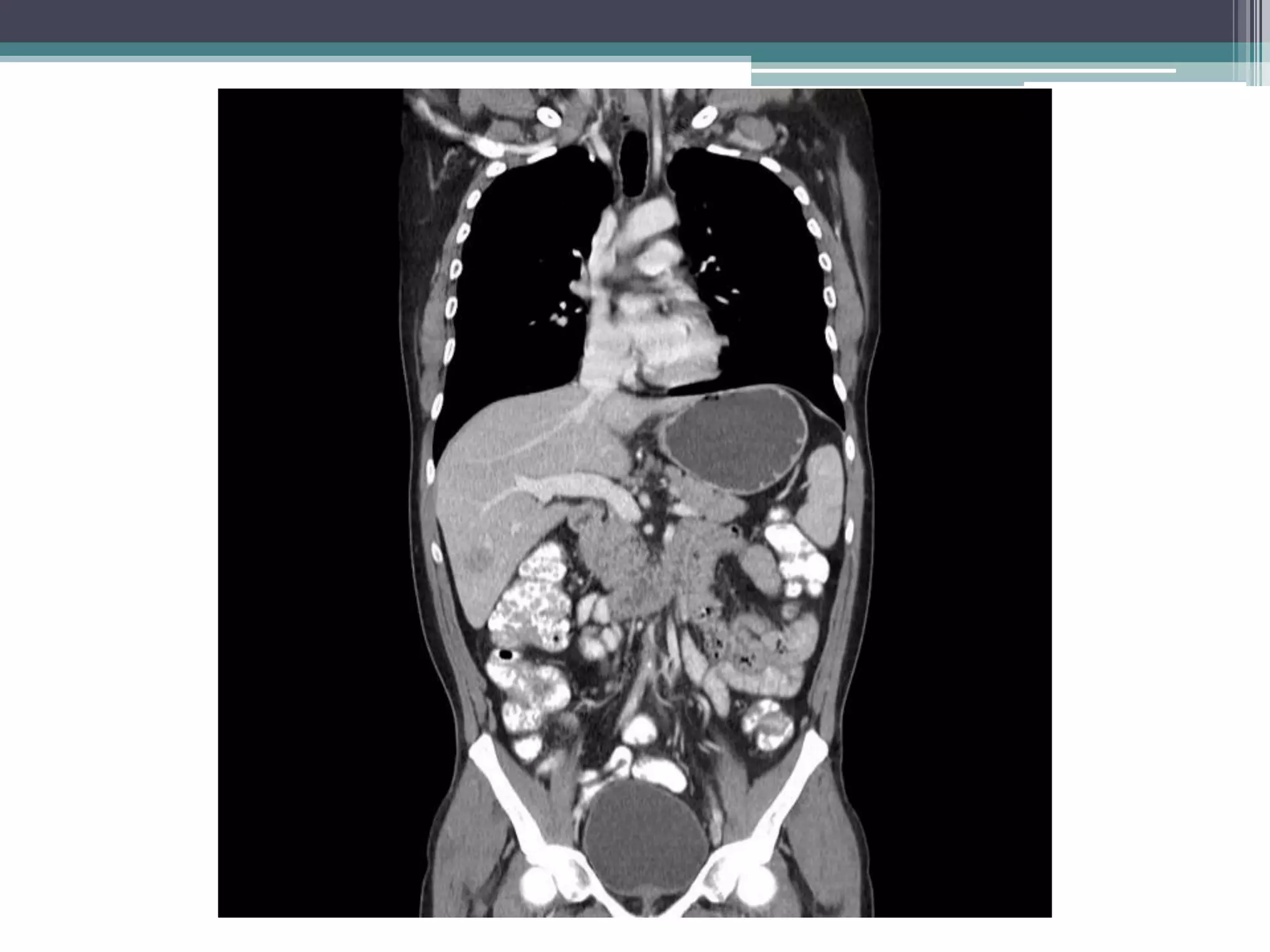
Lower gastrointestinal bleeding refers to bleeding from the small and large intestines. Common causes include hemorrhoids, diverticulosis, angiodysplasia, inflammatory bowel disease, colon polyps and cancer. Diagnostic evaluation involves colonoscopy, CT/CT angiography, capsule endoscopy, nuclear scintigraphy and angiography. Colonoscopy allows for both diagnosis and treatment but other imaging modalities are useful when colonoscopy is non-diagnostic or not possible. Angiography can localize the bleeding site and provide therapeutic embolization in unstable patients. Surgical or endoscopic intervention may be needed depending on the cause of bleeding.
This document provides information on gastrointestinal bleeding, including:
- Causes of upper and lower GI bleeding such as peptic ulcers, varices, angiodysplasia
- Clinical presentations including hematemesis, melena, hematochezia
- Evaluations including endoscopy, angiography, CT angiography
- Anatomy of arteries supplying the GI tract including celiac, SMA, IMA
- Imaging findings of active bleeding and various pathologies
- Management of different causes such as endoscopic therapy for ulcers or TIPS procedure for variceal bleeding
Rectal bleeding has many potential causes, both minor and major. Minor bleeding may be due to hemorrhoids or fissures, while more severe bleeding requires emergency treatment. In cases of massive bleeding, initial steps include admission to the hospital, insertion of IV lines, monitoring of vitals, and blood transfusions as needed to stabilize the patient. Further tests such as colonoscopy or angiography aim to locate the source of bleeding so it can be addressed through methods like cauterization or surgery. Surgical intervention may be needed if other measures do not stop severe or persistent bleeding.
This document discusses lower gastrointestinal (GI) bleeding, including:
- Causes such as angiodysplasia, carcinoma, diverticulosis, and anorectal diseases.
- Clinical evaluation involving history of presenting symptoms, physical exam, investigations to localize the source of bleeding such as colonoscopy, and management including resuscitation, treating the underlying cause, and potential surgical intervention for massive or recurrent bleeding.
- Diagnostic tools like colonoscopy, mesenteric angiography, and radionuclide scanning along with their advantages and disadvantages.
This document discusses lower gastrointestinal bleeding, including its definition, causes, symptoms, diagnosis, and management. The most common causes of lower GI bleeding are diverticular disease and angiodysplasia. Diagnostic tests include colonoscopy, angiography, and radionuclide scanning. Colonoscopy allows for diagnosis and treatment but requires bowel prep, while angiography and scanning are less invasive but also less accurate. Management depends on the severity and cause of bleeding, and may include blood transfusions, endoscopic therapies, angiography, or surgery in severe cases.
Imaging and intervention in hemetemesisSindhu Gowdar
This document discusses various imaging modalities for evaluating gastrointestinal bleeding, including hematemesis. It provides details on angiography, computed tomography angiography, and endoscopy. The key points are:
- Endoscopy is the primary initial investigation but additional techniques like CT angiography and catheter angiography may be needed when endoscopy is negative or fails to identify the bleeding source.
- CT angiography has advantages over catheter angiography as it is more widely available, non-invasive, and allows detection of bleeding sources throughout the GI tract.
- Both endoscopy and CT angiography play important roles in evaluating GI bleeding, with endoscopy also allowing for therapeutic interventions when a source is identified.
1. Acute gastrointestinal bleeding is a potentially life-threatening emergency that is commonly caused by variceal bleeding from liver disease or non-variceal bleeding from peptic ulcers.
2. Initial management involves stabilization, identifying the source of bleeding through endoscopy within 24 hours, and treating the underlying cause.
3. Ongoing resuscitation may require blood transfusions, antibiotics, gastric acid suppression, and vasoconstrictors while the source of bleeding is addressed endoscopically or surgically.
Acute mesenteric arterial disease can result from occlusion of the mesenteric arteries or veins, reducing or stopping blood flow to the intestines. It has high morbidity and mortality rates of 60-70% despite aggressive treatment. Diagnosis involves clinical evaluation, lab tests, CT angiography and mesenteric angiography. Treatment depends on the severity and includes resuscitation, antibiotics, surgery to remove dead bowel and restore blood flow, and endovascular procedures in some cases. Prompt diagnosis and treatment is needed to prevent intestinal infarction and improve outcomes.
This document discusses the anatomy, physiology, and clinical management of splenic injuries. It describes the spleen's location, ligament attachments, blood supply, and functions. Evaluation for splenic injury involves history, physical exam, FAST ultrasound, and CT scan. Management depends on hemodynamic stability and includes non-operative care, embolization, or splenectomy. Splenectomy can be performed open or laparoscopically, involving ligation of vessels and removal of the spleen. Post-operative vaccination is needed due to loss of immune function.
Acute abdoment contains all traumatic and non traumatic routine workup done at radiology center along with all the causes regarding abdominal pain refrence takent from manorama berry book of radiology
This document provides an overview of mesenteric vascular occlusion including:
- It affects the blood supply to the gastrointestinal tract and has high mortality.
- CT scanning is the preferred diagnostic method and can detect bowel wall thickening or lack of enhancement.
- Treatment involves resuscitation, antibiotics, surgery to remove clots or place stents, and resecting non-viable bowel.
- Goals are to diagnose the cause, restore blood flow, and assess bowel viability.
The document discusses portal hypertension in children. It covers the anatomy of the portal system, causes/classifications of portal hypertension, clinical manifestations, diagnosis, and treatment. Regarding diagnosis, it describes using endoscopy to identify varices, ultrasound to detect portal vein thrombosis, and CT/MRI/venography to further evaluate vascular anatomy. Treatment of acute variceal bleeding involves stabilizing the patient and reducing portal pressure to stop bleeding.
1. Colon ischemia is the most common type of mesenteric ischemia and is often caused by non-occlusive hypoperfusion.
2. It is frequently underdiagnosed at initial presentation due to non-specific symptoms but requires prompt diagnosis and treatment to improve outcomes.
3. Establishing the diagnosis typically involves CT imaging followed by early colonoscopy within 48 hours to directly visualize mucosal changes and confirm the diagnosis.
Simple and Algorthymic approach ,covering all aspects of gastrointestinal hemorrhage.
A concise discussion of the diagnostic approach to obscure
bleeding.
Fundamental principles of initial evaluation and management followed with a welldefined and logical approach to the patient with GI hemorrhage
is outlined.
Acute mesenteric ischemia; anatomy, pathophysiology and managementPezhman Kharazm
Acute mesenteric ischemia is one of the most problematic causes of acute abdominal pain. In this presentation, etiologies of acute mesenteric ischemia, their diagnostic evaluation and treatment are discussed.
This document provides an overview of mesenteric ischemia. It begins by describing the arterial blood supply to the gut and then introduces and classifies mesenteric ischemia. The main causes are arterial embolism, thrombosis, venous thrombosis, and non-occlusive ischemia. Clinical features are nonspecific abdominal pain but diagnosis involves imaging like CT angiography. Management involves gastrointestinal decompression, fluid resuscitation, antibiotics, and revascularization through endovascular or open techniques. Prognosis is poor with acute mesenteric ischemia having a mortality over 60%.
This document provides an overview of mesenteric ischemia, including its various types, risk factors, clinical features, diagnosis, and management. It begins with definitions of relevant terms like ischemia, infarction, embolism, and thrombosis. It then describes the different types of mesenteric ischemia - acute, chronic, and non-occlusive. For each type, it outlines typical causes, risk factors, clinical presentations, diagnostic approaches, and treatment options, which may involve endovascular or open surgical revascularization procedures. It concludes by noting the generally poor prognosis of acute mesenteric ischemia but improved outcomes with timely diagnosis and treatment.
Portal hypertension occurs when blood pressure in the portal vein system, which carries blood to the liver, exceeds 10 mm Hg. It is usually caused by cirrhosis or scarring of the liver. Complications include variceal bleeding, ascites, and end-stage liver disease. Diagnosis involves assessing the underlying liver disease through clinical examination, labs, and imaging. Doppler ultrasound and angiography can evaluate the portal venous system. Upper endoscopy is important to identify esophageal or gastric varices which are prone to bleeding in portal hypertension. Treatment depends on the severity but may include pharmacotherapy, endoscopic variceal ligation, TIPS procedure, or liver transplantation.
This document discusses imaging in abdominal trauma. It begins by outlining the mechanisms and types of abdominal injuries from blunt and penetrating trauma. It then describes the FAST (Focused Assessment with Sonography for Trauma) exam and its role in the initial assessment of hemodynamically unstable patients. For stable patients, CT is typically used to further evaluate injuries suggested on clinical exam or FAST. The document outlines key CT findings for various intra-abdominal injuries and hemorrhage.
This document provides an overview of the spleen, splenic injuries, and approaches to splenic surgery. It describes the spleen's anatomy, vascular supply, functions, and types of injuries. For splenic injuries, it discusses evaluation with FAST and CT scans, injury grading scales, and management approaches like angiography, embolization, splenorrhaphy versus splenectomy. It then covers surgical techniques for open and laparoscopic splenectomy, including positioning, mobilization, hilar dissection and hemostasis. Postoperative risks are also summarized.
This document discusses lower gastrointestinal bleeding (LGIB), including its sources, diagnostic tests, and treatment goals. The most common causes of LGIB are infectious, colitis, and anorectal diseases in those under 50, and diverticulosis, angiectasias, and malignancy in those over 50. Diagnostic tests include exclusion of upper sources, anoscopy/sigmoidoscopy, colonoscopy, and nuclear bleeding scans. The first treatment goal is resuscitation, while the second is identifying the bleeding source, typically through endoscopy or angiography. Colonoscopy may control bleeding, and colectomy is considered if bleeding persists.
This document provides an overview of lower gastrointestinal bleeding, including definitions, epidemiology, causes, diagnosis, and management. Some key points:
- Lower GI bleed most commonly originates from the colon, with diverticular disease and angiodysplasias being the most frequent underlying causes.
- Diagnosis involves digital rectal exam, endoscopic procedures like sigmoidoscopy and colonoscopy, and imaging tests like radionuclide scanning.
- Specific colonic conditions that may cause bleeding include diverticular disease, angiodysplasias, colitis, infections, radiation proctitis, and anorectal diseases.
- Small bowel sources of bleeding include angiodysplasias,
Ultrasound is useful for evaluating the pancreas and detecting complications of acute and chronic pancreatitis. In acute pancreatitis, ultrasound can identify changes in the pancreas such as areas of hypoechogenicity and peripancreatic inflammation. Complications like pseudocysts and vascular thromboses are also detectable. Chronic pancreatitis is characterized on ultrasound by ductal dilatation, calcifications, and changes in pancreatic echotexture. Differentiating chronic pancreatitis from pancreatic cancer can be challenging. CT or MRI may be needed when ultrasound findings are inconclusive or to further evaluate necrosis in acute pancreatitis.
Massive lower gastrointestinal bleeding is a life-threatening condition defined by transfusing at least 4 units of blood in 24 hours, hemodynamic instability, or a hematocrit of less than 6g/dl. The main causes are diverticulosis (60%), unknown (13%), hemorrhoids (11%), and neoplasia (9%). Management involves resuscitation, risk assessment, blood transfusions, endoscopy for diagnosis and treatment, and angiography for patients with ongoing bleeding or when endoscopy fails to identify the source. Colonoscopy has high sensitivity but requires bowel preparation, while angiography can localize active bleeding but the patient must be stable. Endoscopic treatments include clips, thermal coagulation, and injections.
Word ectopic comes from the Greek word meaning "out of place". Ectopic pregnancy occurs when a fertilized egg implants outside of the uterus, most commonly in the fallopian tubes. Ultrasound is the gold standard for diagnosing ectopic pregnancies and identifying whether the pregnancy is intrauterine or extrauterine, ruptured or unruptured. Early diagnosis of ectopic pregnancy is lifesaving as it allows for elective surgery or non-surgical treatment options to be used. Common risk factors for ectopic pregnancy include previous pelvic inflammatory disease, infertility, and prior ectopic pregnancies.
This document discusses portal hypertension and its imaging approach. It defines portal hypertension as increased portal venous pressure due to increased resistance or blood flow. Imaging findings can help identify the underlying etiology, which includes cirrhosis, noncirrhotic portal hypertension, extrahepatic portal vein obstruction, and Budd-Chiari syndrome. Ultrasound, CT, MRI, and Doppler ultrasound are used to evaluate the liver, portal and hepatic vasculature, and for signs of portal hypertension like splenomegaly and collateral vessels. Liver stiffness measurements and hemodynamic studies can also aid diagnosis. The document reviews characteristic imaging findings for each cause of portal hypertension.
Osteoporosis was presented by Dr. Anuradha. The moderator, Dr. Bagyalakshmi, an Assistant Professor, introduced Dr. Anuradha to discuss osteoporosis. The presentation covered osteoporosis and concluded with the moderator thanking Dr. Anuradha.
Here are the answers to your questions:
1. Most common site of osteomyelitis - Metaphysis of long bones, especially distal femur and proximal tibia.
2. Most common organism causing osteomyelitis - Staphylococcus aureus.
3. Earliest radiographic and MRI findings of acute osteomyelitis - Soft tissue swelling and loss of fascial planes seen within 24-48 hours on radiographs. Bone marrow edema seen as low signal on T1 and high signal on T2/STIR sequences in MRI.
4. What is sequestrum - Avascular/necrotic bone fragment formed due to osteonecrosis in chronic osteomyelitis
Diffusion tensor imaging in evaluation of epilepsy ppt.pptxaasrithakotha2
Diffusion tensor imaging (DTI) can be used to evaluate epilepsy by measuring the restricted diffusion of water in brain tissues. DTI generates metrics like mean diffusivity (MD) and fractional anisotropy (FA) that can identify microstructural abnormalities in white matter. Studies have found increased MD and lower FA in mesial temporal lobe epilepsy patients, indicating widespread abnormalities beyond what is seen on conventional MRI. DTI is useful for localizing lesions in MRI-negative epilepsy and assessing connectivity changes. The document outlines the principles and applications of DTI for assessing epilepsy.
Rickets is a disease characterized by defective mineralization of growing bones, usually due to vitamin D deficiency or abnormal vitamin D metabolism. There are two main types: calcipenic rickets associated with elevated PTH and low calcium/phosphate, and hereditary vitamin D resistant rickets caused by mutations. Radiological features include osteopenia, irregular epiphyseal borders, bowed long bones, and a rachitic rosary of enlarged costochondral junctions. Differential diagnoses include scurvy and certain genetic bone diseases.
Echocardiography uses ultrasound technology to produce images of the heart. It was pioneered in the 1950s by Drs. Hertz and Edler in Sweden, who began using an ultrasonoscope to examine the heart. Modern echocardiography machines generate ultrasound images using a transducer that transmits sound waves into the body and receives echoes to produce cardiac images. Standard echocardiograms visualize the heart in 2D, M-Mode, and with Doppler modalities from different transducer positions. Echocardiography is used to assess cardiac structure and function, identify abnormalities, and determine the severity of conditions such as valvular disease or pulmonary hypertension.
This document discusses spotters, which are short notes written by Dr. Sanjiva N H on various medical topics. The 25 topics covered include immunocompromised patients, with the document thanking the reader at the end for their time.
This document provides an overview of approaches to spinal tumors. It begins by discussing how location, clinical presentation, age and gender are important for differential diagnosis. Spinal tumors are classified as intramedullary, intradural extramedullary, or extradural. The approach involves first examining the spinal cord and CSF spaces for expansion or compression. Common intramedullary tumors discussed include ependymomas, astrocytomas, gangliogliomas, and hemangioblastomas. Intradural extramedullary tumors include meningiomas, nerve sheath tumors, and myxopapillary ependymomas. Imaging features that help characterize different tumors are also summarized.
Pneumothorax is the presence of air in the pleural space. It can be caused by spontaneous rupture of lung tissue, trauma, medical procedures, or mechanical ventilation. Chest x-rays are used to diagnose pneumothorax by identifying the visceral pleural line and absent vascular markings beyond it. Different positions and locations of pneumothorax have distinguishing radiographic features, such as an anteromedial pneumothorax appearing as a sharp delineation of vascular structures in that region. Pneumothorax can also outline collapsed lung lobes, appearing as lucencies around structures like the aorta or inferior vena cava for left and right upper lobe collapses respectively.
A picture archiving and communication system (PACS) allows for digital storage, transmission, and display of medical images. It replaces the need for film by acquiring, storing, and transmitting images digitally. A PACS has components like input devices, image storage, a transmission network, and display stations. It provides advantages like simultaneous multilocation viewing, organized data storage, and the ability to manipulate images. PACS installation is expensive but provides benefits like improved efficiency and the ability to access prior images easily.
CONGENITAL,PYOGENIC AND VIRAL INFECTIONS OF CNS @22-02-23.pptxaasrithakotha2
This document discusses various congenital, pyogenic, and viral infections of the central nervous system (CNS). It describes the pathology and imaging findings of several TORCH infections, including toxoplasmosis, rubella, cytomegalovirus (CMV), and herpes. Imaging may reveal parenchymal calcifications, periventricular calcifications (seen in CMV), cysts, and polymicrogyria. Congenital infections like Zika, CMV, and herpes simplex virus can cause microcephaly and calcifications. Herpes simplex virus specifically causes meningoencephalitis in neonates visible on MRI as hyperintensities and hemorrhagic foci. Congenital HIV
This document discusses mycobacterial and fungal infections of the brain. It begins by describing the different types of mycobacteria, including M. tuberculosis which causes tuberculosis (TB), and atypical mycobacteria. TB most commonly presents as TB meningitis (TBM), appearing on imaging as basilar exudates and meningeal enhancement. Tuberculomas also occur and appear as ring-enhancing lesions, while TB abscesses are rare. Nontuberculous mycobacteria can cause cervical lymphadenitis or CNS disease. Fungal infections like cryptococcal meningitis are also discussed, as well as the imaging appearance of different fungal infections.
CONGENITAL,PYOGENIC AND VIRAL INFECTIONS OF CNS @22-02-23.pptxaasrithakotha2
This document discusses various congenital, pyogenic, and viral infections of the central nervous system (CNS). It describes the pathology and imaging findings of several TORCH infections (toxoplasmosis, rubella, cytomegalovirus (CMV), and herpes), including parenchymal calcifications. For CMV specifically, periventricular calcifications are common. Herpes simplex virus (HSV) can cause meningoencephalitis with necrosis and hemorrhage in neonates. Congenital HIV infection typically leads to atrophy of the frontal lobes and basal ganglia calcifications. Other infections discussed include Zika virus, which can result in microcephaly and calcifications.
Local Advanced Lung Cancer: Artificial Intelligence, Synergetics, Complex Sys...Oleg Kshivets
Overall life span (LS) was 1671.7±1721.6 days and cumulative 5YS reached 62.4%, 10 years – 50.4%, 20 years – 44.6%. 94 LCP lived more than 5 years without cancer (LS=2958.6±1723.6 days), 22 – more than 10 years (LS=5571±1841.8 days). 67 LCP died because of LC (LS=471.9±344 days). AT significantly improved 5YS (68% vs. 53.7%) (P=0.028 by log-rank test). Cox modeling displayed that 5YS of LCP significantly depended on: N0-N12, T3-4, blood cell circuit, cell ratio factors (ratio between cancer cells-CC and blood cells subpopulations), LC cell dynamics, recalcification time, heparin tolerance, prothrombin index, protein, AT, procedure type (P=0.000-0.031). Neural networks, genetic algorithm selection and bootstrap simulation revealed relationships between 5YS and N0-12 (rank=1), thrombocytes/CC (rank=2), segmented neutrophils/CC (3), eosinophils/CC (4), erythrocytes/CC (5), healthy cells/CC (6), lymphocytes/CC (7), stick neutrophils/CC (8), leucocytes/CC (9), monocytes/CC (10). Correct prediction of 5YS was 100% by neural networks computing (error=0.000; area under ROC curve=1.0).
Cell Therapy Expansion and Challenges in Autoimmune DiseaseHealth Advances
There is increasing confidence that cell therapies will soon play a role in the treatment of autoimmune disorders, but the extent of this impact remains to be seen. Early readouts on autologous CAR-Ts in lupus are encouraging, but manufacturing and cost limitations are likely to restrict access to highly refractory patients. Allogeneic CAR-Ts have the potential to broaden access to earlier lines of treatment due to their inherent cost benefits, however they will need to demonstrate comparable or improved efficacy to established modalities.
In addition to infrastructure and capacity constraints, CAR-Ts face a very different risk-benefit dynamic in autoimmune compared to oncology, highlighting the need for tolerable therapies with low adverse event risk. CAR-NK and Treg-based therapies are also being developed in certain autoimmune disorders and may demonstrate favorable safety profiles. Several novel non-cell therapies such as bispecific antibodies, nanobodies, and RNAi drugs, may also offer future alternative competitive solutions with variable value propositions.
Widespread adoption of cell therapies will not only require strong efficacy and safety data, but also adapted pricing and access strategies. At oncology-based price points, CAR-Ts are unlikely to achieve broad market access in autoimmune disorders, with eligible patient populations that are potentially orders of magnitude greater than the number of currently addressable cancer patients. Developers have made strides towards reducing cell therapy COGS while improving manufacturing efficiency, but payors will inevitably restrict access until more sustainable pricing is achieved.
Despite these headwinds, industry leaders and investors remain confident that cell therapies are poised to address significant unmet need in patients suffering from autoimmune disorders. However, the extent of this impact on the treatment landscape remains to be seen, as the industry rapidly approaches an inflection point.
Histololgy of Female Reproductive System.pptxAyeshaZaid1
Dive into an in-depth exploration of the histological structure of female reproductive system with this comprehensive lecture. Presented by Dr. Ayesha Irfan, Assistant Professor of Anatomy, this presentation covers the Gross anatomy and functional histology of the female reproductive organs. Ideal for students, educators, and anyone interested in medical science, this lecture provides clear explanations, detailed diagrams, and valuable insights into female reproductive system. Enhance your knowledge and understanding of this essential aspect of human biology.
Does Over-Masturbation Contribute to Chronic Prostatitis.pptxwalterHu5
In some case, your chronic prostatitis may be related to over-masturbation. Generally, natural medicine Diuretic and Anti-inflammatory Pill can help mee get a cure.
share - Lions, tigers, AI and health misinformation, oh my!.pptxTina Purnat
• Pitfalls and pivots needed to use AI effectively in public health
• Evidence-based strategies to address health misinformation effectively
• Building trust with communities online and offline
• Equipping health professionals to address questions, concerns and health misinformation
• Assessing risk and mitigating harm from adverse health narratives in communities, health workforce and health system
These lecture slides, by Dr Sidra Arshad, offer a quick overview of the physiological basis of a normal electrocardiogram.
Learning objectives:
1. Define an electrocardiogram (ECG) and electrocardiography
2. Describe how dipoles generated by the heart produce the waveforms of the ECG
3. Describe the components of a normal electrocardiogram of a typical bipolar lead (limb II)
4. Differentiate between intervals and segments
5. Enlist some common indications for obtaining an ECG
6. Describe the flow of current around the heart during the cardiac cycle
7. Discuss the placement and polarity of the leads of electrocardiograph
8. Describe the normal electrocardiograms recorded from the limb leads and explain the physiological basis of the different records that are obtained
9. Define mean electrical vector (axis) of the heart and give the normal range
10. Define the mean QRS vector
11. Describe the axes of leads (hexagonal reference system)
12. Comprehend the vectorial analysis of the normal ECG
13. Determine the mean electrical axis of the ventricular QRS and appreciate the mean axis deviation
14. Explain the concepts of current of injury, J point, and their significance
Study Resources:
1. Chapter 11, Guyton and Hall Textbook of Medical Physiology, 14th edition
2. Chapter 9, Human Physiology - From Cells to Systems, Lauralee Sherwood, 9th edition
3. Chapter 29, Ganong’s Review of Medical Physiology, 26th edition
4. Electrocardiogram, StatPearls - https://www.ncbi.nlm.nih.gov/books/NBK549803/
5. ECG in Medical Practice by ABM Abdullah, 4th edition
6. Chapter 3, Cardiology Explained, https://www.ncbi.nlm.nih.gov/books/NBK2214/
7. ECG Basics, http://www.nataliescasebook.com/tag/e-c-g-basics
Integrating Ayurveda into Parkinson’s Management: A Holistic ApproachAyurveda ForAll
Explore the benefits of combining Ayurveda with conventional Parkinson's treatments. Learn how a holistic approach can manage symptoms, enhance well-being, and balance body energies. Discover the steps to safely integrate Ayurvedic practices into your Parkinson’s care plan, including expert guidance on diet, herbal remedies, and lifestyle modifications.
Recomendações da OMS sobre cuidados maternos e neonatais para uma experiência pós-natal positiva.
Em consonância com os ODS – Objetivos do Desenvolvimento Sustentável e a Estratégia Global para a Saúde das Mulheres, Crianças e Adolescentes, e aplicando uma abordagem baseada nos direitos humanos, os esforços de cuidados pós-natais devem expandir-se para além da cobertura e da simples sobrevivência, de modo a incluir cuidados de qualidade.
Estas diretrizes visam melhorar a qualidade dos cuidados pós-natais essenciais e de rotina prestados às mulheres e aos recém-nascidos, com o objetivo final de melhorar a saúde e o bem-estar materno e neonatal.
Uma “experiência pós-natal positiva” é um resultado importante para todas as mulheres que dão à luz e para os seus recém-nascidos, estabelecendo as bases para a melhoria da saúde e do bem-estar a curto e longo prazo. Uma experiência pós-natal positiva é definida como aquela em que as mulheres, pessoas que gestam, os recém-nascidos, os casais, os pais, os cuidadores e as famílias recebem informação consistente, garantia e apoio de profissionais de saúde motivados; e onde um sistema de saúde flexível e com recursos reconheça as necessidades das mulheres e dos bebês e respeite o seu contexto cultural.
Estas diretrizes consolidadas apresentam algumas recomendações novas e já bem fundamentadas sobre cuidados pós-natais de rotina para mulheres e neonatos que recebem cuidados no pós-parto em unidades de saúde ou na comunidade, independentemente dos recursos disponíveis.
É fornecido um conjunto abrangente de recomendações para cuidados durante o período puerperal, com ênfase nos cuidados essenciais que todas as mulheres e recém-nascidos devem receber, e com a devida atenção à qualidade dos cuidados; isto é, a entrega e a experiência do cuidado recebido. Estas diretrizes atualizam e ampliam as recomendações da OMS de 2014 sobre cuidados pós-natais da mãe e do recém-nascido e complementam as atuais diretrizes da OMS sobre a gestão de complicações pós-natais.
O estabelecimento da amamentação e o manejo das principais intercorrências é contemplada.
Recomendamos muito.
Vamos discutir essas recomendações no nosso curso de pós-graduação em Aleitamento no Instituto Ciclos.
Esta publicação só está disponível em inglês até o momento.
Prof. Marcus Renato de Carvalho
www.agostodourado.com
Basavarajeeyam is a Sreshta Sangraha grantha (Compiled book ), written by Neelkanta kotturu Basavaraja Virachita. It contains 25 Prakaranas, First 24 Chapters related to Rogas& 25th to Rasadravyas.
Muktapishti is a traditional Ayurvedic preparation made from Shoditha Mukta (Purified Pearl), is believed to help regulate thyroid function and reduce symptoms of hyperthyroidism due to its cooling and balancing properties. Clinical evidence on its efficacy remains limited, necessitating further research to validate its therapeutic benefits.
2. • Gastrointestinal bleeding is typically categorized
as either upper or lower gastrointestinal
bleeding depending on the anatomic location of
the bleeding site.
3.
4.
5.
6.
7. • The celiac axis and the superiormesenteric artery (SMA) are the
first two branches of the abdominal aorta and provide a rich and
well-collateralizednetwork of branch vessels that supply blood to
the upper gastrointestinal tract.
• Extensive collateralizationbetween the celiac artery and the SMA
protects the upper gastrointestinal tract from ischemic insult and
permits surgical and embolizationprocedures to be carried out with
a relatively low risk of ischemic injury.
8. • Upper GI- Esophagus, stomach and duodenum
• Lower GI – Small and large bowels.
9. • Haematemesis : Vomiting of blood whether fresh and red or
digested and black.
• Melaena : Passage of loose,black tarry stools with a characteristic
foul smell.
• Coffee ground vomiting : Blood clot in the vomitus.
• Hematochezia : Passageof bright red blood per rectum (if the
haemorrhage is severe).
10. Blood on its own or streakingthe stool:
• Rectum : polyps or carcinoma,prolapsed
• Anus : Haemorrhoids, Fissure-in-ano,Anal carcinoma.
Stool mixed with blood:
• GIT above sigmoid colon.
• Sigmoid carcinoma or diverticulardisease.
Blood separatefromthe stool:
• Follows defaecation: Anal condition eg: Haemorrhoids.
• Blood is passed by itself : Rapidly bleeding carcinoma,inflammatory
bowel disease, diverticulitis, or passed down from high up in the gut.
Blood is on the surfaceof the stool: suggest a lesion such as polyp or
carcinomafurther proximallyeither in the rectum or descendingcolon
Blood on the toilet paper: Fissure-in-ano, Heamorrhoids.
Loose, black, tarry, foul smelling stool: from the proximal of DJ flexure
13. • Risk factors include :
• medications (e.g. NSAID, warfarin)
• recent colonoscopy with polypectomy (post polypectomy bleeding)
• prior abdominal/pelvic radiation (radiation proctitis/colitis)
• prior surgery
• history of alcoholism or chronic liver disease
• history of abdominal aortic aneurysm with or without surgical repair
(causing an aortoenteric fistula)
14. Imaging
• Colonoscopyis the first-line investigationfor both diagnosticand
therapeutic management.CT angiography(CTA), nuclear medicine
studies,and angiographycan all be used to assess LGIB but have
limited sensitivity when bleeding is intermittent or slow.
15. CT
• On non-contrast CT, hemorrhagemay be visualized as
circumferential thickeningof the bowel wall
• CTA provides a relatively non-invasiveand effective way of
localizing the source of bleeding,especially in patients with
continuous bleeding
16. • In some cases, while there is no evidence of active
extravasation, a sentinel clot (seen as an unchanging
hyperdensity) can be used to localize the site of recent
bleeding.
• Even in situations in which no acute bleeding is identified,
CTA can diagnose abnormalities that may be responsible
for the bleeding, such as ischemia, inflammatory bowel
disease, neoplasms and arteriovenous malformations
(AVMs) .
• CTA therefore provides an excellent road map guiding the
next step in triaging patients to either endoscopic, surgical
or angiographic management.
17.
18. • Multiphase CTE
• MultiphaseCTE is most useful in evaluating hemodynamically
stable patients with intermittentand occult GI Bleeds, which
commonly have an underlyingsmall bowel source.
• The main difference between a multiphaseCTE and routine CTA
is that in a CTE study,the small bowel lumen is distendedwith a
bolus (about 1.5–2.0 litres) of a neutral oral contrast agent. This
luminal distentionallows optimal visualizationof enhancement
of the small bowel mucosa and wall following intravenous
contrast administration,thereby increasing the sensitivityof
detection of bleeding and non -bleedingabnormalities
20. NUCLEAR IMAGING
• Erythrocytes are labeled with technetium-99m, then serial
scintigraphy is performed (a.k.a. 99mTc-labeled RBC
scintigraphy or tagged red blood cell scan) to detect focal
collections of radiolabelled material.
• It can be performed relatively quickly and may help
localize the general area of active bleeding to guide
subsequent endoscopy, angiography or surgery .
• A false-positive result can be produced by a rapid transit of
luminal blood so that labeled blood is detected in
the colon even though it originated from a more proximal
site in the gastrointestinal tract .
21. • Extravasated radiolabeled RBCs within the lumen of
the bowel are identified as an area of activity that
increases in intensity with time, and/or as a focus of
activity that moves in a pattern corresponding to the
lumen of the large or small bowel.
• Small bowel bleeding usually can be distinguished
from large bowel bleeding by its rapid serpiginous
movement
22. Delayed bleeding from a midjejunal site.
Findings at initial imaging performed 0-
60 minutes after injection were
normal. Anterior 5-minute static image
obtained 22 hours after injection
demonstrates activity throughout the
large bowel and in a few small bowel
loops in the pelvis (arrow).
23. Capsule endoscopy
• Also known as video capsule endoscopy (VCE) or wireless
capsule endoscopy,is a non-invasivemeans of investigating
the small bowel, principallyfor identifyingthe underlying cause
of occult gastrointestinal tract bleeding,such as due
to arteriovenous malformations,small bowel tumors, and
ulcers.
• It is also used for the detection of the earliest manifestations
of Crohn disease, such as mucosal ulceration,which are poorly
detected by other imaging modalities such as small bowel MRI or
ultrasound.
24.
25. ANGIOGRAPHY
• Angiographyis an invasive examination that can be used for
accurate localizationas well as treatment of both upper and
lower gastrointestinal bleeding.
• Preffered in acutely unstablepatient,after a negative or failed
endoscopicevaluation,or as a first-lineexamination for lower
gastrointestinalhemorrhage.
• In patients with lower gastrointestinalbleeding who are
haemodynamically-stableand do not have ongoing fresh rectal
bleeding,an RBC-labeled Tc99m scan is recommended as a first
line of investigation.
26. • Angiographycan provide the opportunityfor therapeutic
interventionat the time of diagnosis .
• However, the bleeding rate must be ≥0.5 mL/min to detect
extravasation into the gut, which is significantlyhigher than in
nuclear medicine.
• Additionally,certain patient factors (e.g. contrast allergy,
acute/chronickidney disease) are potential contraindicationsto
angiography
• Extravasation of contrast material into the bowel lumen is
pathognomonicfor active gastrointestinal hemorrhage.
• Indirect signs includedetection of pseudoaneurysm,
arteriovenous fistula, hyperemia,neovascularity,and
extravasation of contrast material into a confined space
27. Active GI hemorrhage in a 67-year-old
woman with melena (a) Selective
angiogram of the gastroduodenal artery
shows contrast material extravasation
into the duodenal lumen(b, c)
Gastroduodenal artery (b) and celiac
trunk (c) angiograms, obtained after coil
embolization show stoppage of the GI
bleeding.
28. Active GI hemorrhage in an 82-year-old
man with hematochezia.
(a) Colonoscopic image of the right colon
shows a large quantity of blood, which
obscures the colonoscopic findings. (b)
Selective angiogram of the ileocolic
artery shows contrast material
extravasation into the colonic lumen . (c)
Ileocolic artery angiogram obtained after
coil embolization (arrow) shows
stoppage of the GI bleeding
29. A 70-year-old male presenting with bleeding per rectum and drop in
hematocrit. Colonoscopy was negative; underwent tagged RBC scan that
demonstrated site of active bleeding in the hepatic flexure of the colon
(A, arrow) which was subsequently embolized via conventional
angiography (B, C: arrowheads).
30. Finding Diagnosis
Extravasation Active bleed site
Filling of spaces outside the bowel
lumen
Aneurysm or pseudoaneurysm
Early arterial filling, accumulation in
vascular tufts, slow draining vessels
Angiodysplasia
Neovascularity Neoplasm
Hyperaemia Colitis
31. Advantages Disadvantages
Does not require bowel preparation
Can be done in emergency situation
Therapeutic uses
Low sensitivity rate
Requires active bleeding
Complication rate
32. Diverticula
• Outpouching of bowel wall
• Diverticulosis,Diverticulitis
• The site of protrusionis that of the
entry points of the bowel vascular
supply throughthe mesentery.
• In some cases, the incoming vessel
runs over the diverticulumdome. This
close relationshipis responsiblefor the
complicationof hemorrhage that results from diverticula.
33.
34.
35.
36. Hemorrhoids
• Swollen veins in the lower
rectum
• Internal hemorrhoids often
result in painless,bright red
rectal bleedingwhen defecating
External hemorrhoids often result in pain and swelling in the area of
the anus
If bleeding occurs it is usually darker.
A skin tag may remain after the healing of an external hemorrhoid.
37. Perianal fistula
• presence of a fistulous tract across/between/adjacent to the anal
sphincters and is usually an inflammatorycondition
• Intersphincteric
• Transsphincteric
• Suprasphincteric
• Extrasphincteric
38. Angiodysplasia
• One of the most common causes of occult GI
bleed
• refers to dilated, thin-walled blood vessels
(capillaries, venules, veins) found in the
mucosa and submucosa of the
gastrointestinal tract
39. CT :
• appear as focal areas (<5 mm) of contrast enhancementin the
bowel wall (most prominent in the enteric phase )
• early filling of an antimesentericvein
Angiography:
• ectatic vessels but no discretelesion
• early venous enhancementindicatingarteriovenous shunting
43. Aorto-Enteric fistula
• Aortoenteric fistulas are pathologiccommunicationsbetween
the aorta (or aortoiliac tree) and the gastrointestinaltract and
represent an uncommon cause of catastrophicgastrointestinal
hemorrhage
44. CT
Primary fistula Secondary fistula
Direct signs include:
• ectopic gas adjacent to or within the
aorta
• presence of vascular contrast within
the gastrointestinal tract
Indirect signs include:
• bowel/esophageal wall thickening
overlying an aneurysm
• disruption of the aortic fat cover
• retroperitoneal/mediastinal
hematoma or hematoma within the
bowel wall or lumen
• increased perigraft soft tissue
• pseudoaneurysm formation
• disruption of aneurysmal wrap
• increased soft tissue between the graft
and aneurysmal wrap
45.
46.
47. Colorectal carcinoma
• most common cancer of the gastrointestinal tract and
the second most frequently diagnosed malignancy in
adults
• Colorectal cancers can be found anywhere from the
cecum to the rectum, in the following distribution :
• rectosigmoid: 55%
• cecum and ascending colon: ~20%
• ileocecal valve: 2%
• transverse colon: ~10%
• descending colon: ~5%
48. IMAGING :
• Barium enema
• Appearances will reflect
macroscopicappearance,with
lesions seen as filling defects.
• These need to be differentiated
from residual fecal matter.
• Typicallythey appear as exophytic
or sessile masses or maybe
circumferential (apple core sign).
• Fistulas to bladder,vagina, or
bowel may also be demonstrated.
49. • CT :
• Used for staging
• soft tissue density that narrow the bowel lumen
• Ulceration in larger mass is also seen.
• Occasionally low-density masses with low-density lymph
nodes are seen in mucinous adenocarcinoma, due to the
majority of the tumor composed of extracellular mucin.
Psammomatous calcifications in mucinous adeno.ca can
also be present.
• Complications may also be evident, e.g.
fistulae, obstruction, intussusception, perforation
50.
51.
52.
53.
54. • Dukes (Astler-Coller modification)
• stage A: confined to mucosa
• stage B: through muscularis propria
• stage C: local lymph node involvement
• stage D: distant metastases
55. • MRI :
• MRI is having an increasing role to play in the staging of rectal
cancer